Podcast
Questions and Answers
What alteration in membrane permeability primarily contributes to the spontaneous decrease in membrane potential from -60 mV to -50 mV in pacemaker cells?
What alteration in membrane permeability primarily contributes to the spontaneous decrease in membrane potential from -60 mV to -50 mV in pacemaker cells?
- Increased efflux of potassium ions ($K^+$)
- Increased influx of chloride ions ($Cl^-$)
- Increased influx of sodium ions ($Na^+$) (correct)
- Increased efflux of calcium ions ($Ca^{++}$)
How does the inhibition of $Na^+$-$K^+$ ATPase by digitalis lead to a positive inotropic effect in cardiac muscle cells?
How does the inhibition of $Na^+$-$K^+$ ATPase by digitalis lead to a positive inotropic effect in cardiac muscle cells?
- By increasing intracellular sodium concentration, which reduces calcium efflux via the $Na^+$/$Ca^{2+}$ exchanger. (correct)
- By decreasing intracellular potassium concentration, which prolongs the action potential duration.
- By hyperpolarizing the cell membrane, which enhances calcium entry through voltage-gated channels.
- By directly increasing the amount of calcium released from the sarcoplasmic reticulum.
Which mechanism explains the staircase phenomenon (Bowditch effect) in cardiac muscle?
Which mechanism explains the staircase phenomenon (Bowditch effect) in cardiac muscle?
- Decreased availability of ATP for myosin ATPase activity.
- Progressive decrease in intracellular calcium concentration with each subsequent contraction.
- Increased sensitivity of troponin to calcium due to phosphorylation of contractile proteins.
- Gradual increase in intracellular calcium ion concentration due to increased influx and reduced reuptake. (correct)
How does increased sympathetic stimulation affect the slope of the prepotential in sinoatrial (SA) node cells, and what is the resulting change in heart rate?
How does increased sympathetic stimulation affect the slope of the prepotential in sinoatrial (SA) node cells, and what is the resulting change in heart rate?
Which of the following best describes the role of the annulus fibrosus in the heart's electrical conduction system?
Which of the following best describes the role of the annulus fibrosus in the heart's electrical conduction system?
In the context of the Frank-Starling mechanism, what is the primary effect of increased venous return on cardiac muscle fiber length and subsequent ventricular contraction?
In the context of the Frank-Starling mechanism, what is the primary effect of increased venous return on cardiac muscle fiber length and subsequent ventricular contraction?
How does the administration of a calcium channel blocker (CCB) affect the action potential of sinoatrial (SA) nodal cells, and what is the subsequent impact on heart rate?
How does the administration of a calcium channel blocker (CCB) affect the action potential of sinoatrial (SA) nodal cells, and what is the subsequent impact on heart rate?
Which statement accurately describes the influence of the refractory period on cardiac muscle function?
Which statement accurately describes the influence of the refractory period on cardiac muscle function?
How does the selective inhibition of phosphodiesterase-3 (PDE3) by inodilators like milrinone result in a positive inotropic effect and vasodilation?
How does the selective inhibition of phosphodiesterase-3 (PDE3) by inodilators like milrinone result in a positive inotropic effect and vasodilation?
Following a myocardial infarction that damages the left ventricle, how does the body compensate according to the Frank-Starling mechanism to maintain cardiac output?
Following a myocardial infarction that damages the left ventricle, how does the body compensate according to the Frank-Starling mechanism to maintain cardiac output?
What is the underlying mechanism by which hyperkalemia ($K^+$) influences cardiac excitability?
What is the underlying mechanism by which hyperkalemia ($K^+$) influences cardiac excitability?
How does mitral regurgitation affect afterload on the left ventricle, and what compensatory mechanisms are activated to maintain cardiac output?
How does mitral regurgitation affect afterload on the left ventricle, and what compensatory mechanisms are activated to maintain cardiac output?
Which of the following statements accurately describes the response of coronary blood flow during exercise?
Which of the following statements accurately describes the response of coronary blood flow during exercise?
What mechanism explains why beta-1 adrenergic receptor antagonists (beta-blockers) reduce heart rate and contractility?
What mechanism explains why beta-1 adrenergic receptor antagonists (beta-blockers) reduce heart rate and contractility?
How do changes in preload, afterload, and contractility affect stroke volume (SV)?
How do changes in preload, afterload, and contractility affect stroke volume (SV)?
Which structural adaptations are responsible for the fast conduction velocity observed in Purkinje fibers compared to ventricular myocytes?
Which structural adaptations are responsible for the fast conduction velocity observed in Purkinje fibers compared to ventricular myocytes?
How does stimulation of the vagus nerve influence heart rate and atrioventricular (AV) node conduction?
How does stimulation of the vagus nerve influence heart rate and atrioventricular (AV) node conduction?
What is the effect of increased heart rate on diastolic filling time, and how does this impact stroke volume?
What is the effect of increased heart rate on diastolic filling time, and how does this impact stroke volume?
How does an increase in afterload affect the velocity of cardiac muscle fiber shortening?
How does an increase in afterload affect the velocity of cardiac muscle fiber shortening?
Following a severe hemorrhage, what compensatory mechanisms are activated to maintain arterial blood pressure (ABP)?
Following a severe hemorrhage, what compensatory mechanisms are activated to maintain arterial blood pressure (ABP)?
Which structural characteristic of capillaries optimizes the exchange of nutrients and waste products between blood and tissues?
Which structural characteristic of capillaries optimizes the exchange of nutrients and waste products between blood and tissues?
What is the primary mechanism by which autoregulation maintains constant blood flow in the kidneys despite fluctuations in arterial blood pressure?
What is the primary mechanism by which autoregulation maintains constant blood flow in the kidneys despite fluctuations in arterial blood pressure?
How does severe aortic stenosis affect left ventricular wall stress (afterload) and myocardial oxygen demand?
How does severe aortic stenosis affect left ventricular wall stress (afterload) and myocardial oxygen demand?
What is the role of nitric oxide (NO) in regulating blood flow to skeletal muscle during exercise?
What is the role of nitric oxide (NO) in regulating blood flow to skeletal muscle during exercise?
How does heart failure with preserved ejection fraction (HFpEF) affect ventricular filling and diastolic function?
How does heart failure with preserved ejection fraction (HFpEF) affect ventricular filling and diastolic function?
What mechanism explains the development of edema in the lower extremities during prolonged standing?
What mechanism explains the development of edema in the lower extremities during prolonged standing?
Which statement accurately describes the effect of histamine on capillary permeability?
Which statement accurately describes the effect of histamine on capillary permeability?
How do the baroreceptor reflexes respond to an increase in arterial blood pressure (ABP)?
How do the baroreceptor reflexes respond to an increase in arterial blood pressure (ABP)?
During fetal development, what are the key structural changes that occur in the heart and great vessels at birth to establish normal postnatal circulation?
During fetal development, what are the key structural changes that occur in the heart and great vessels at birth to establish normal postnatal circulation?
How does the structure of medium-sized veins differ from that of medium-sized arteries, and how do these differences relate to their respective functions?
How does the structure of medium-sized veins differ from that of medium-sized arteries, and how do these differences relate to their respective functions?
Which vasoactive substances contribute to vasodilation?
Which vasoactive substances contribute to vasodilation?
What is the most important function of Arteriovenous Anastomoses?
What is the most important function of Arteriovenous Anastomoses?
What is the definition of 'Autoregulation'?
What is the definition of 'Autoregulation'?
Which cell type can facilitate the highest permeability?
Which cell type can facilitate the highest permeability?
Which of the following is a characteristic of fetal circulation?
Which of the following is a characteristic of fetal circulation?
Which of the following is the layer of the blood vessel that has elastic fibers and collagen?
Which of the following is the layer of the blood vessel that has elastic fibers and collagen?
What is the source of the ascending aorta?
What is the source of the ascending aorta?
Flashcards
Rhythmicity (Automaticity)
Rhythmicity (Automaticity)
The heart's ability to initiate regular electrical impulses independently of external nerve stimulation.
Rhythmic cell characteristics
Rhythmic cell characteristics
SAN discharges spontaneously. Membrane potential is unstable, lacking a true resting membrane potential. It is more permeable to Na+ and Ca++ and its firing level is -40 mV, with a peak at +10 mV.
Pacemaker potential (Phase 4)
Pacemaker potential (Phase 4)
Decreases spontaneously from -60mV to -50mV due to Na+ influx.
Prepotential at -50mV
Prepotential at -50mV
Signup and view all the flashcards
Depolarization at -40mV
Depolarization at -40mV
Signup and view all the flashcards
Repolarization Phase
Repolarization Phase
Signup and view all the flashcards
Resting membrane potential at -60mV
Resting membrane potential at -60mV
Signup and view all the flashcards
Pre potential slope's effect
Pre potential slope's effect
Signup and view all the flashcards
Resting Heart Rate
Resting Heart Rate
Signup and view all the flashcards
Conductivity
Conductivity
Signup and view all the flashcards
SAN function
SAN function
Signup and view all the flashcards
Atrial excitation's effect
Atrial excitation's effect
Signup and view all the flashcards
AVN’s role in conduction
AVN’s role in conduction
Signup and view all the flashcards
Purkinje fibers' role
Purkinje fibers' role
Signup and view all the flashcards
Action potential Phase 0
Action potential Phase 0
Signup and view all the flashcards
Action potential Phase 1
Action potential Phase 1
Signup and view all the flashcards
Action potential Phase 2 (Plateau)
Action potential Phase 2 (Plateau)
Signup and view all the flashcards
Action potential Phase 3
Action potential Phase 3
Signup and view all the flashcards
Action potential Phase 4
Action potential Phase 4
Signup and view all the flashcards
AP and mechanical response
AP and mechanical response
Signup and view all the flashcards
Systole timing
Systole timing
Signup and view all the flashcards
Diastole timing
Diastole timing
Signup and view all the flashcards
Excitation-contraction coupling
Excitation-contraction coupling
Signup and view all the flashcards
Length-tension relationship
Length-tension relationship
Signup and view all the flashcards
Force-velocity relationship
Force-velocity relationship
Signup and view all the flashcards
Staircase phenomena
Staircase phenomena
Signup and view all the flashcards
Cardiac output (CO)
Cardiac output (CO)
Signup and view all the flashcards
Stroke Volume (SV)
Stroke Volume (SV)
Signup and view all the flashcards
Ejection Fraction (EF)
Ejection Fraction (EF)
Signup and view all the flashcards
CO control: Heart Rate (HR)
CO control: Heart Rate (HR)
Signup and view all the flashcards
Starling's Law
Starling's Law
Signup and view all the flashcards
Preload's link to venous return
Preload's link to venous return
Signup and view all the flashcards
Afterload
Afterload
Signup and view all the flashcards
Cardiac Reserve
Cardiac Reserve
Signup and view all the flashcards
Arterial oxygen
Arterial oxygen
Signup and view all the flashcards
Enzymes
Enzymes
Signup and view all the flashcards
Atrial flow fraction
Atrial flow fraction
Signup and view all the flashcards
Study Notes
Physiology of the Heart - Cardiac Properties
-
Rhythmicity (automaticity) describes the heart's ability to generate regular electrical impulses independently of nervous control, enabling it to beat autonomously.
-
Rhythmic cells found in the Sinoatrial Node (SAN), Atrioventricular Node (AVN), and Purkinje fibers exhibit automaticity.
-
The intrinsic firing rate varies: SAN discharges at 90/min, AVN at 60/min, and Purkinje at 30/min.
-
Rhythmic cells are characterized by spontaneous discharge, unstable membrane potential without a resting membrane potential (RMP), the absence of a plateau phase, higher permeability to Na+ and Ca++, a firing level of -40 mV, and a peak action potential (AP) of +10 mV.
Ionic Bases
-
The membrane potential in rhythmic cells spontaneously decreases from -60 mV to -50 mV due to Na+ influx.
-
At -50 mV, transient Ca++ channels (T-type) open, causing Ca++ entry and further depolarization to the firing level of -40 mV.
-
The Prepotential represents the combined phases of membrane potential decrease and transient Ca++ channel activity.
-
At -40 mV, long-lasting Ca++ channels (L-type) open, causing further depolarization, while transient Ca++ and Na+ channels close until +10 mV.
-
Repolarization occurs due to K+ outflow through K+ channels, along with the closure of L-type Ca++ channels.
-
At -60 mV, inactivation of K+ channels stops K+ efflux.
-
The slope of the prepotential determines heart rate and an increased slope increases heart rate (tachycardia) while a decreased slope lowers it (bradycardia).
Factors Affecting Heart Rate
-
Tachycardia results from sympathetic stimulation, catecholamines, increased temperature, hypokalemia (↓ K+), and thyroxin.
-
Bradycardia results from parasympathetic stimulation, acetylcholine, calcium channel blockers (CCB), and hyperkalemia (↑ K+).
-
Normal resting heart rate ranges from 70-80 bpm due to vagal tone.
Conductivity
-
Conductivity refers to the ability of cardiac muscle to transmit excitation waves.
-
The excitation wave originates in the Sinoatrial Node (SAN), proceeds through the internodal pathways to the atria, causing atrial contraction and ejection of blood to the ventricles.
-
The wave reaches the Atrioventricular Node (AVN), delaying transmission. Then it moves via the Atrioventricular bundle and bundle branches to the Purkinje fibers and ventricular muscle, leading to ventricular excitation, contraction, and blood ejection to the aorta and pulmonary artery.
-
Conduction rates vary such that the atrial pathway conducts at a rate of 1 m/sec.
-
The AV node (AVN) conducts at 0.05 m/sec.
-
The Bundle of His conducts at 1 m/sec.
-
The Purkinje fibers conduct at 4-5 m/sec.
-
The ventricular muscle conducts at 1 m/sec.
Conduction at the AV Node
-
Slow conduction at the AV node (0.05 m/sec) is attributed to the presence of few gap junctions.
-
Slow conduction at the AV node allows both atria to excite and contract, emptying blood into ventricles, and prevents abnormal atrial rhythms from reaching the ventricles.
Conduction at the Purkinje Fibers
-
Rapid conduction at the Purkinje fibers (4-5 m/sec) is due to many gap junctions.
-
Rapid conduction at the Purkinje fibers allows both ventricles to contract as a unit to eject blood.
Excitability and Action Potential in Cardiac Muscle
-
The resting membrane potential (RMP) of ordinary cardiac muscle fibers is -90 mV, similar to skeletal muscle.
-
The action potential is composed of five phases (0-4).
-
Phase 0: Rapid depolarization from -90 mV to +20 mV, caused by Na+ influx through fast Na+ channels.
-
Phase 1: Small rapid repolarization to +10 mV due to inactivation of Na+ channels, and K+ and Cl- efflux.
-
Phase 2: Plateau where repolarization slows, the membrane potential is around zero mV for 200 msec due to a balance between Ca++ influx through long-lasting Ca++ channels and Na+ influx with K+ efflux.
-
Phase 3: Rapid repolarization to RMP due to closure of long-lasting Ca++ channels, inactivation of Na+ channels, and activation of K+ channels.
-
Phase 4: Resting membrane potential (RMP) at -90 mV due to selective permeability and Na-K pump activity.
Relationship between Action Potential and Mechanical Response
-
Contraction starts just after the beginning of depolarization and reaches its maximum by the end of the plateau phase.
-
Repolarization coincides with the first half of relaxation (diastole).
-
Systole coincides with the period of depolarization until the end of the plateau.
-
Diastole coincides with the period of rapid repolarization and continues in rest, because the mechanical response lasts one and a half times as long as action potential.
Relationship between Action Potential and Excitability Change
-
The absolute refractory period coincides with the period of depolarization until the end of the plateau, during which excitability is lost.
-
The relative refractory period coincides with the period of rapid repolarization, during which excitability is weak.
Relationship between Action Potential and ECG
-
The QRS complex coincides with depolarization.
-
The S-T segment coincides with the plateau.
-
The T wave coincides with rapid repolarization.
Contractility (Excitation-Contraction Coupling)
-
Contractility refers to the cardiac muscle's ability to contract and pump blood, beginning with the excitation of a cardiac wave.
-
Excitation leads to Ca++ entry inside the cardiac muscle.
-
Ca++ entry into the sarcoplasm triggers the release of a large amount of Ca++ from the sarcoplasmic reticulum through the Ca++ release channel.
-
Ca++ binds to troponin-C, causing tropomyosin to uncover the active site of actin.
-
Interaction between actin and myosin, followed by their sliding, leads to contraction.
-
Removal of Ca++ from the sarcoplasm leads to relaxation.
Factors Affecting Contractility
-
All or none rule: cardiac muscle contracts or does not contract at all.
-
Increasing stimulus intensity beyond threshold will not change the strength of contraction, while stimulation below threshold will produce no response.
-
Tetanus, a sustained contraction, cannot occur in cardiac muscle due to the long absolute refractory period (ARP), which coincides with systole.
Length-Tension Relationship
-
The force of contraction of the heart is directly proportional to the initial length of the cardiac muscle fiber within limits (Starling's Law).
-
Increasing the initial length of the cardiac muscle fiber increases the force of contraction, up to a certain length called L max, where all myosin cross-bridges bind to the active site of actin.
-
Increasing length above L max decreases the force of contraction.
Force-Velocity Relationship
-
The initial velocity of shortening of cardiac muscle is inversely related to the load moved during contraction.
-
Increasing the load carried by the muscle decreases the initial velocity of shortening, and decreasing the load increases it.
Staircase Phenomenon
-
Applying repeated stimuli to the cardiac muscle within a short time causes a staircase effect with the 2nd contraction stronger than the first and the 3rd stronger than the 2nd.
-
The staircase effect continues until a new level of contraction is reached, attributed to increased Ca++ ions and failure of reuptake due to Ca++ pump weakness.
Inotropic State
-
Inotropic state refers to the ability of cardiac muscle to develop force (contract).
-
The contractile state can be increased (+ve inotropic) or decreased (-ve inotropic).
-
Positive inotropic factors include sympathetic stimulation (ẞ1-adrenergic), catecholamines, glucagon hormone, digitalis, and xanthines/caffeine.
-
All positive inotropic factors act through increasing Ca++ in the sarcoplasm.
-
Negative inotropic factors include parasympathetic stimulation, acetylcholine, hypoxia (due to ischemia), calcium channel blockers (CCB), anesthetics, and anti-arrhythmic drugs.
Cardiac Output (CO)
-
Cardiac output (CO) is the volume of blood pumped by each ventricle per minute. The normal CO is 5 L/min.
-
The cardiac index (CI) is the volume of blood pumped by each ventricle per minute per square meter of body surface area. The normal CI is 3.2 L/min/m².
-
Cardiac output is calculated as Stroke Volume (SV) x Heart Rate (HR).
-
Normally: CO = 70 beats/min heart rate x 70 ml stroke volume = 5 L/min
-
The cardiac output depends on heart rate and stroke volume.
-
Stroke volume (SV) is the volume of blood pumped by each ventricle per beat.
-
Stroke volume is calculated as End Diastolic Volume (EDV) - End Systolic Volume (ESV).
-
Normally: SV = 135 ml EDV - 65 ml ESV = 70 ml.
-
End Diastolic Volume (EDV) is the volume of blood in the ventricle at the end of diastole.
-
End Systolic Volume (ESV) is the volume of blood in the ventricle at the end of systole.
Ejection Fraction (EF)
-
Ejection Fraction (EF) is the fraction of the EDV that is ejected with each beat, such that EF = Stoke volume / End diastolic volume x 100.
-
Normally, the ejection fraction is 55-75%.
-
Importance: Assessment of contractility.
Variations of Cardiac Output
-
Cardiac output is unchanged during sleep and moderate changes in external temperature.
-
Cardiac output is increased during excitement (50-100%), exercise (300%), exposure to high temperature, eating (30%), epinephrine secretion, end of pregnancy, inspiration, and shifting from standing to recumbent position.
-
Cardiac output is decreased during sitting or standing from lying position, rapid arrhythmia, and heart failure.
Control of Cardiac Output
-
Heart Rate (HR): An increase of HR up to 200/min (e.g., during exercise) increases CO.
-
Change of Preload: Preload determines muscle length before contraction.
-
Ventricular preload is determined by EDV, which depends on venous return.
Starling's Law
-
The more the end diastolic volume, the greater the force of contraction (within limits).
-
An increase in EDV (preload) leads to an increase in SV, with a consequent increase in CO.
-
Preload is mainly affected by venous return, which increases filling of ventricles, increases EDV, increases myocardial contractility, increases SV, and increases cardiac output.
Factors Affecting Ventricular Preload
-
Increased venous return will increase preload.
-
Decreased ventricular compliance decreases preload, as in myocardial infarction.
-
Increased heart rate above 200/min decreases preload due to shortening of diastole, decreasing ventricular filling.
Effect of Afterload on SV
-
Afterload is the load against which the ventricle contracts and ejects blood. The main afterload for the left ventricle is the aortic pressure.
-
Increased afterload leads to decreased SV and CO, while decreased afterload leads to increased SV and CO.
Effect of Contractility (Inotropic State) on SV
- Positive inotropics increase contractility, ejecting more blood, increasing SV and CO.
Cardiac Reserve
-
Cardiac reserve is the difference between cardiac output under ordinary circumstances and its maximum capacity for pumping blood.
-
In normal young adults, cardiac reserve is 300%.
-
Cardiac reserve is assessed by low-dose dobutamine stress echocardiography.
Mechanisms for modifying cardiac output
-
Positive chronotropic condition (heart rate reserve)
-
During rest: HR = 70 bpm
-
During exercise: HR = 200 bpm
-
Limitation: diastolic period is shortened
-
Positive preload
-
(EDV reserve)
-
During rest: = 135 ml
-
During exercise: increase venous return, ventricular filling and higher force of contraction
-
Decrease afterload during exercise
-
Resulting in decreased total peripheral resistance
-
Positive inotropic conditions
-
(ESV reserve)
-
During rest: = 65 ml
-
During exercise: sympathetic stimulation lead to increased force of contraction decreasing ESV leading to increased SV and COP
-
Limitation: Amount of blood
-
increaseForce of contraction causes higher SV and therefore COP
-
Cardiac hypertrophy
-
Cardiac hypertrophy may be Physiological or Pathological.
-
Venous oxygen reserve:during rest
-
O2 content in each 100 ml arterial blood:20 ml
-
O2 content in each 100 ml venous blood:15 ml
-
*- Thus blood supply to tissue = 5 ml 02
-
During exercise: Blood supply to tissue increase up to 12 ml
Enzymes
-
Enzymes are catalysts of chemical reactions, mostly globular proteins, and are essential for life as they occur at rates sufficient for life.
-
An apoenzyme or apoprotein require a cofactor and together this is known as a holoenzyme.
-
Cofactors may be inorganic including metal ions (e.g., zinc in carbonic anhydrase) and iron-sulfur clusters or organic which are prosthetic groups or coenzymes
-
Coenzymes released from the enzyme's active site during the reaction, such as NADH, NADPH, and adenosine triphosphate.
Examples of Coenzymes
-
Hydrogen carriers, such as NAD, NADP, FAD, FMN, HEAM, lipoic acid, glutathione, vitamin C, and CoQ.
-
Isoenzymes (isozymes) catalyze the same reactions at different rates, have different affinities to substrates, and consist of multiple polypeptide chains.
Examples of Isoenzymes
-
Lactate dehydrogenase (LDH) enzyme, formed of 4 polypeptide chains (H, M) existing as 5 isozymes, with isozyme 1 present in cardiac muscle, increasing in myocardial infarction and isozyme 5 present in the liver, increasing in hepatitis and liver diseases.
-
Creatine kinase (CK), formed of 2 subunits (M, B) with CK-BB present in the brain that increase in brain tumors or infarctions, CK-MM in muscles that increase in muscle diseases, and CK-MB in cardiac muscles which increases in myocardial infarction.
Enzyme Activation
-
Enzymes are activated through proteolysis from pepsinogen by HCL to pepsin, blood clotting factors being activated as prothrombin to thrombin and fibrinogen to fibrin.
-
Metal ion activation may occur with the metal present in any position: (metal - enzyme - substrate), (enzyme - substrate, metal) or (enzyme - metal - substrate).
-
Allosteric activation occurs when many enzymes contain another site other than the catalytic site named the allosteric site.
-
Binding increases enzyme activity
-
Covalent modification of the enzyme occurs by Phosphorylation and dephosphorylation of the enzymes which leads to change in its activity.
Enzyme Inhibition
-
Enzyme Inhibition: can be A) Irreversible or B) Reversible (it may be either competitive or non competitive)
-
Irreversible enzyme inhibition can be caused by factors producing denaturation (strong acids or bases), inhibitors of the sulfhydryl group (oxidizing agents or salts of heavy metals), antienzymes e.g α 1-antitrypsin deficiency which allows elastase to digest the pulmonary tissue in emphysema, and inhibition by removal of catalytic ions. EDTA prevents blood clotting and fluoride inhibits enolase enzyme of glycolysis.
-
Reversible Enzyme Inhibition can be competitive by which there is structural similarity between the inhibitor and the substrate which affect affect the rate of the enzyme activity or Non Competitive Inhibition.
Non Competitive Inhibition:
-
The Substrate and inhibitor bind different sites and increasing concentration of the substrate does not lead to change in the inhibitory process of the enzyme.
-
Can be caused by Allosteric Inhibition when enzymes contain a site other than catalytic site. Binding to this site leads to change in the shape of the enzyme which doesn't favour the binding of the substrate as well as Feed Back Inhibition when The product inhibits the key enzyme or one of the substrates in the pathway.
Enzymes for Clinical and Biomedical Importance
-
Specific enzymes are present in high concentration in certain organs and damage to certain organs may cause an elevation of enzymes useful in the diagnosis of certain diseases.
-
The myocardium is rich in glutamate oxaloacetate transaminase „Lactate dehydrogenase and creatine kinase and any reduction of blood supply to the myocardium leads to an increase in those enzymes.
Diagnosis of myocardial infarction
-
To diagnose myocardial infarction creatine kinase CK(MB ) is used along with glutamate oxaloacetate transaminase (GOT) and lactate dehydrogenase (LDH)
-
The liver contains many enzymes but is is primarily for gluconeogenesis and lactate dehydrogenase for the anaerobic glycolysis.
-
So, hpetitis is diagnosed mainly glutamate oxaloacetate transaminase (GOT), glutamate pyruvate transaminase (GPT), alkaline phosphatase, and lactate dehydrogenase (LDH)
-
The biliary tract contains many enzymes so obstructive jaundice show elevates alkaline phosphatase and gamma Glutamyl transferase (y GT). gamma Glutamyl transferase (y GT) is elevated in case of alcoholism and drug abuse also.
-
The following enzymes may diagnose Pancreatitis : pancretic amylase and pancreatic lipase (it is also elevated in pancreatic duct obstruction).
-
Acid phosphatase is an enzyme elevated in case of prostatitis and cancer prostate.
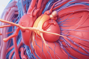
Great Vessels of the Thorax
- Ascending Aorta – arises from the left ventricle in 3rd left intercostal space, has 3 sinuses and lies in middle mediastinum. Anterior to pulmonary trunk, right lung, pleura and is posterior to left atrium, right atrium and SVC and to the left of the pulmonary trunk . Branches: Rt and Lt coronary.
- Arch of Aorta – starts at the 2 nd right costal cartilage, superior to Branches (brachiocephalic, Lt common carotid, Lt subclavian As) and Left brachiocephalic V. Inferior to division of pulmonary trunk, ligamentum arteriosum and Left main bronchus Ant to Left lung, pleura. Posterior to Trachea, esophagus and vertebral column. Branches: Brachiocephalic, Left Common Carotid, Left Subclavian, Thyroidae Ima
- Descending Aorta – starts at the Lt side of T4 vertebra Anterior Lt bronchus, Lt atrium , esophagus Vertebral column, Rt: esophagus : Lt lung & pleura and ends at the lower border of T12.
- Pulmonary Trunk – arises from Right Ventricle at the 3rd Lt costal cartilage. Anterior Left lung and pleura, ascending aorta, and Lt atrium. Distributes Right and Lt pulmonary As (caries deoxygenated blood to the lungs).N.B.:
- Ligamemtum arteriosum: fibrous band (embryological remnant) connecting arch of aorta & Lt pulmonary A causing shifting of blood from aorta
Brachiocephalic Veins
_ Begind: behind the medial end of clavicle by joining internal jugular and subclavian Veins _Short on the Right at 2.5 cm, Vertical. Relations are Righr phrenic. _Longer on left at 6cm, Obliquely horizontal Anterior- sternum, Posterior : Branches of aortic arch Inferior- arch of aorta
- SUPERIOR VENA CAVA
- *-**Begind: behind 1st Rt costal cartilage and is created by joining of 2 brachiocephalic Vs Relations are, Right: Rt phrenic. Contains B2 brachiocephalic veins and Azygos Veins
- End in: the Right atrium
Inferior vena cava
- Begins in thorax from diaphram around T8 vertebra _End in: 6th Rt costal cartilage
- PULMONARY VEINS 4 total, sup and inf Veins from each lung
- CARRIES OXXYGENATED BLOOD FROM LUNG To Left atrium
Histology of the Vascular System
-
The vascular system includes arteries, veins, and arteriovenous connections.
-
The wall of any blood vessel is formed of three layers, including the Tunica intima, Tunica media, and Tunica adventitia;
-
Arteries are classified into Large arteries, Medium sized arteries, Small arteries (arterioles), and Meta-arteriols.
| POC | Large artery [Elastic] | Medium sized artery [Muscular] | Small artery [Arteriole] |
|---|---|---|---|
| Sites | Aorta & large arteries | muscular arteries | Ends of muscular arteries |
| Wall | thick wall | thick wall | -Thin wall |
| Lumen | wide lumen | - Narrow lumen | narrowlumen. |
| Tunica intima | S. Sq. Endoth.Subendothelial CT.: rich in elasticfs. | Endothelium - Thick subendothelial CT | - Endothelium -Thin |
| Int.elasticlamina(IEL) | ill-defined [similar to elastic laminae of media. | -Thick IEL | IEL gradually disappears. |
-
Tunica media -Thick=:Fenestrated elastic membranes (40- 70 layer).with Smooth muscles in between.-EEL: ill defined -Mainly smooth muscles with many elastic fibers inbetween EEL:: may be present- One or 2 layers of smooth muscles replaced by pericytes in small arterioles, No EEL
-
Tunica adventitia -elastic fibers rich in Present -elastic fibers -ewf may be present- absent
-
Arteries that supply blood to capillaries are Called "Metarterioles" that connect terminal arterioles connect with capillaries" -Arteries that supply blood to capillaries Are surrounded by smooth muscles to act as *sphincters to control blood how to the capillaries. They carry blood from heart.
-
*-STRUCTURE OF MEDIUM SIZED VEIN LIKE MEDIUM SIZED ARTERY**With some Differences
| POC | Medium sized artery | Medium sized vein |
|---|---|---|
| 1. Wall thickness | Thick | Thin |
| 2. Lumen | Narrow Does not contain blood after death | WideContains blood after death |
| 3. Valves | Absent | Present |
| 4. Tunica Intima | -thick-foldedClear internal elastic lamina | +-Thin+-not foldedNo internal elastic lamina |
| 5. Tunica Media | Thick (main layer)-Smooth muscles with many elastic hbers.-External elastic lamina may be present | Thin.Smooth muscles withfew elastic hbers.Absent |
| 6. Tunica Adventitia | Thin.-Rich in elastic hbers | Thick (main layer)Absent |
- ""Tunica intima has No int. EL."The ""Tunica media is thin."++"Tunica adventitiais very thick & longitudinal smooth muscle""
Arteries & veins Connections And Blood Capilliaries
- I-Blood Capillaries. II- Arterio venous anastomoses.
- Diameter: about 8m.*
- Lumen: narrow.*
- Structure*: It is formed of one layer of endothelial cells. Resting on basement layer
- Types*: 1-continuous (somatic capillaries)2- fenestrated (visceral) Sinusoidal capillaries
| POC | Continuous(somatic)caps. | Fenestrated(visceral)capillaries | Sinusoidial capillaries |
|---|---|---|---|
| Diamter | 8 μm | 8 um | 5-40 μm |
| Lumen | Narrow & regular | Narrow & regular | Wide & irregular |
| Endothelium | Continuous | Fenestrated pores in kidneys | Have withoud diaphram |
| Basement Membrane | Continuous | Continuous | Non Continous |
| Preicytes | Persent | Pesent | Macrophage between endoth-Cells |
- Arteriovenous Anastomoses, These are dierct connections from arteriole Side branch that arise form an arteriole and is similair to a vanule.-Gloumrs, complacted bracnshng surrounded in a consitent structure
Physiology Arterial Blood Pressure ABP
-
Arterial pressure of blood on arterial wall maximum is SBP about 1200 normally.The minimum is distolic to 70 m hg to mHg.-Pulse pressure is 50 mmHg normally 50mmHg.-Mean systemic Arteiral Pressure Is the average of the cycle MAPis
-
Changes of age from the blood SBP systolic,DBP distotic Map arterial measures. Measuremnt-By cathter (invassive- By non sphg momerter (noninbasire
-
Physicology of the variation ABP increase to loss if elasitciy-new8/4, four years as well Femals also hve low ABP until age 45-Race cause ABP chnages as well:
-
Symphetic Emonotion increase ABP-SP ecersize too-Gravity to lower heart increase ABP, by 0.77 mmHg-Resperations cause
-
Factiors which control ABP--CO increase SV and HR. IF HR is constant so Sv decreses ABP. if SV increase diaticyly pressure
-
Each 1 to aboce heart to deceae ABP-Ealsicy buffer -volume is relation can affed Resistace- TPR too, "Nevers mechanism 1. reapud 2-Inter 3. low ""Cardiac Center: Cardio Pressore area and Center"" "Cardiothr Centre = Its heart heart, and reach sympaically."
-
Recrptors the artialy that influence the artioanly process and ABP
Studying That Suits You
Use AI to generate personalized quizzes and flashcards to suit your learning preferences.