Podcast
Questions and Answers
In the context of direct pes cavus deformity, which of the following statements most accurately characterizes the orientation of the foot and calcaneus relative to the distal tibial-fibular articulation?
In the context of direct pes cavus deformity, which of the following statements most accurately characterizes the orientation of the foot and calcaneus relative to the distal tibial-fibular articulation?
- The foot and calcaneus rotate as a unit on the distal tibial-fibular articulation via the subtalar joint, excluding the talus, contributing to compensatory supination of the calcaneal-pedal unit.
- The foot and calcaneus do not entail rotation on the distal tibial-fibular articulation; instead, the deformity is primarily uniplanar (sagittal) and does not involve the subtalar joint. (correct)
- The foot and calcaneus exhibit rotational changes as a unit at the distal tibial-fibular articulation, with the talus serving as the central axis of rotation within the sagittal plane.
- The foot and calcaneus rotate as a unit via the talocalcaneal joint, independently of the subtalar joint, leading to a uniplanar deformity primarily in the axial plane.
In assessing a patient with suspected pes cavus, which radiographic view is MOST critical for evaluating the degree of metatarsus adductus and transverse plane malalignments, especially in the context of cavovarus deformities?
In assessing a patient with suspected pes cavus, which radiographic view is MOST critical for evaluating the degree of metatarsus adductus and transverse plane malalignments, especially in the context of cavovarus deformities?
- Calcaneal axial view.
- Modified Coleman block test.
- Anteroposterior weight-bearing radiograph. (correct)
- Lateral weight-bearing radiograph.
Which statement BEST describes the relationship between the calcaneal inclination angle (CIA) and the lateral talocalcaneal angle in the context of varying degrees of pronation and supination?
Which statement BEST describes the relationship between the calcaneal inclination angle (CIA) and the lateral talocalcaneal angle in the context of varying degrees of pronation and supination?
- The lateral talocalcaneal angle is geometrically equivalent to the sum of the calcaneal inclination and talar declination angles; it tends to increase with pronation and decrease with supination. (correct)
- The lateral talocalcaneal angle is geometrically equivalent to the sum of the calcaneal inclination and talar declination angles; it decreases with pronation and increases with supination.
- The lateral talocalcaneal angle is inversely proportional to the calcaneal inclination angle, increasing as the calcaneal inclination angle decreases, irrespective of pronation or supination.
- The lateral talocalcaneal angle remains constant regardless of changes in the calcaneal inclination angle, providing a stable reference point for deformity assessment.
Which of the following statements BEST characterizes the radiographic apex location in combined (mixed) pes cavus deformity?
Which of the following statements BEST characterizes the radiographic apex location in combined (mixed) pes cavus deformity?
In the context of ancillary radiographic imaging for pes cavus deformities, what is the MOST accurate rationale for using the Saltzman and el-Khoury view, and how is it technically executed?
In the context of ancillary radiographic imaging for pes cavus deformities, what is the MOST accurate rationale for using the Saltzman and el-Khoury view, and how is it technically executed?
Which of the following statements accurately characterizes the impact of adult-acquired pes cavus on the peroneus brevis (PB) tendon, and what pathophysiological mechanisms are MOST commonly involved?
Which of the following statements accurately characterizes the impact of adult-acquired pes cavus on the peroneus brevis (PB) tendon, and what pathophysiological mechanisms are MOST commonly involved?
In the context of MRI evaluation for adult-acquired pes cavus, what is the significance of the 'magic angle artifact,' and how can it be mitigated to ensure accurate interpretation of peroneal tendon pathology?
In the context of MRI evaluation for adult-acquired pes cavus, what is the significance of the 'magic angle artifact,' and how can it be mitigated to ensure accurate interpretation of peroneal tendon pathology?
While MRI has shown an increased application to pes cavus deformities, which statement accurately reflects its limitations relative to conventional methods?
While MRI has shown an increased application to pes cavus deformities, which statement accurately reflects its limitations relative to conventional methods?
In the context of tarsal coalitions associated with pes cavus, which radiographic finding is considered a sine qua non for diagnosing a middle facet coalition, and under what specific conditions is this finding most reliable?
In the context of tarsal coalitions associated with pes cavus, which radiographic finding is considered a sine qua non for diagnosing a middle facet coalition, and under what specific conditions is this finding most reliable?
Which of the following statements accurately characterizes the rationale and methodology for using high-resolution T2* relaxation time mapping of ankle joint cartilage in patients with pes cavus?
Which of the following statements accurately characterizes the rationale and methodology for using high-resolution T2* relaxation time mapping of ankle joint cartilage in patients with pes cavus?
What are the key differentiating features depicted by 3D weight-bearing CT analysis when comparing Charcot-Marie-Tooth (CMT) cavovarus foot deformities with idiopathic cavovarus feet?
What are the key differentiating features depicted by 3D weight-bearing CT analysis when comparing Charcot-Marie-Tooth (CMT) cavovarus foot deformities with idiopathic cavovarus feet?
Given the diverse etiologies of pes cavus, which of the following statements BEST encapsulates the utility of MRI in differentiating between various underlying causes?
Given the diverse etiologies of pes cavus, which of the following statements BEST encapsulates the utility of MRI in differentiating between various underlying causes?
Which statement accurately characterizes the relationship between hindfoot alignment, ankle osteoarthritis, and subtalar joint orientation as revealed by imaging studies?
Which statement accurately characterizes the relationship between hindfoot alignment, ankle osteoarthritis, and subtalar joint orientation as revealed by imaging studies?
Under what conditions would one consider advanced multiplanar computed tomographic (CT) imaging techniques along with 3D reconstructions indispensable in the assessment of pes cavus deformity?
Under what conditions would one consider advanced multiplanar computed tomographic (CT) imaging techniques along with 3D reconstructions indispensable in the assessment of pes cavus deformity?
How does the Meary-Tomeno line, and its disruption, factor into the radiographic evaluation of Pes Cavus?
How does the Meary-Tomeno line, and its disruption, factor into the radiographic evaluation of Pes Cavus?
What role does the Harris (Harris-Beath) view play when imaging tarsal coalition?
What role does the Harris (Harris-Beath) view play when imaging tarsal coalition?
How does the average fibula position vary, between subjects of cavus and control?
How does the average fibula position vary, between subjects of cavus and control?
In pediatric pes cavus, what is the normal range for the Tibiocalcaneal angle, and it's relation to equninus?
In pediatric pes cavus, what is the normal range for the Tibiocalcaneal angle, and it's relation to equninus?
What is the position that the patient should be placed for ankle MRI, and why?
What is the position that the patient should be placed for ankle MRI, and why?
What is the Tibiocalcaneal angle generally?
What is the Tibiocalcaneal angle generally?
In the context of triplane cavovarus deformity, what is the MOST accurate description of the relative movement and positioning of the foot and calcaneus in relation to the tibial-fibular articulation?
In the context of triplane cavovarus deformity, what is the MOST accurate description of the relative movement and positioning of the foot and calcaneus in relation to the tibial-fibular articulation?
In assessing the etiology of adult-acquired pes cavus, which of the following pathophysiological mechanisms is LEAST likely to be directly evaluated and differentiated using MRI?
In assessing the etiology of adult-acquired pes cavus, which of the following pathophysiological mechanisms is LEAST likely to be directly evaluated and differentiated using MRI?
Which advanced imaging modality provides the MOST comprehensive assessment for pre-operative planning in complex triplanar cavovarus foot deformities?
Which advanced imaging modality provides the MOST comprehensive assessment for pre-operative planning in complex triplanar cavovarus foot deformities?
When evaluating a lateral weight-bearing radiograph of a patient with pes cavus, a tibiocalcaneal angle significantly exceeding the normal pediatric range (60°-90°) is MOST indicative of which underlying condition?
When evaluating a lateral weight-bearing radiograph of a patient with pes cavus, a tibiocalcaneal angle significantly exceeding the normal pediatric range (60°-90°) is MOST indicative of which underlying condition?
In the context of ancillary radiographic imaging for pes cavus deformities, which of the following statements BEST describes the primary objective for using the Saltzman and el-Khoury view?
In the context of ancillary radiographic imaging for pes cavus deformities, which of the following statements BEST describes the primary objective for using the Saltzman and el-Khoury view?
Which technical modification to traditional ankle MRI protocols is MOST critical to minimize the 'magic angle artifact' when evaluating peroneal tendon pathology in patients with adult-acquired pes cavus?
Which technical modification to traditional ankle MRI protocols is MOST critical to minimize the 'magic angle artifact' when evaluating peroneal tendon pathology in patients with adult-acquired pes cavus?
Which of the following statements BEST characterizes the clinical utility of high-resolution T2* relaxation time mapping of ankle joint cartilage in patients with pes cavus?
Which of the following statements BEST characterizes the clinical utility of high-resolution T2* relaxation time mapping of ankle joint cartilage in patients with pes cavus?
In the context of tarsal coalitions associated with pes cavus, which of the following statements accurately describes a characteristic radiographic finding?
In the context of tarsal coalitions associated with pes cavus, which of the following statements accurately describes a characteristic radiographic finding?
In instances of adult-acquired pes cavus secondary to progressive deterioration of peroneus brevis (PB) function, lateral weight-bearing radiographs may demonstrate what?
In instances of adult-acquired pes cavus secondary to progressive deterioration of peroneus brevis (PB) function, lateral weight-bearing radiographs may demonstrate what?
When assessing a suspected calcaneonavicular coalition, which of the following radiographic findings is MOST reliable on a medial oblique view?
When assessing a suspected calcaneonavicular coalition, which of the following radiographic findings is MOST reliable on a medial oblique view?
Which statement accurately reflects a limitation of Harris views in imaging subtalar joint coalitions in the presence of pes cavus deformity?
Which statement accurately reflects a limitation of Harris views in imaging subtalar joint coalitions in the presence of pes cavus deformity?
When evaluating a lateral weight-bearing radiograph of a patient with Anterior Cavus, the tibiotalar angle would demonstrate what?
When evaluating a lateral weight-bearing radiograph of a patient with Anterior Cavus, the tibiotalar angle would demonstrate what?
Which statement accurately reflects the average fibula position, between someone with pes cavus and a control?
Which statement accurately reflects the average fibula position, between someone with pes cavus and a control?
Which statement accurately reflects the range of the typical First metatarsal declination angle?
Which statement accurately reflects the range of the typical First metatarsal declination angle?
If the apex/vertex of the Meary angle is typically proximal to the Chopart joint, what type of cavus is most likely?
If the apex/vertex of the Meary angle is typically proximal to the Chopart joint, what type of cavus is most likely?
In anterior cavus, what is the general trend of the declination angles of the metatarsal bones?
In anterior cavus, what is the general trend of the declination angles of the metatarsal bones?
Which statement regarding subtalar joint in patients with ankle osteoarthritis, is most accurate?
Which statement regarding subtalar joint in patients with ankle osteoarthritis, is most accurate?
In anterior cavus, it is best to consider depression of which foot aspect being primarily involved?
In anterior cavus, it is best to consider depression of which foot aspect being primarily involved?
What is the normal range for the lateral talocalcaneal angle?
What is the normal range for the lateral talocalcaneal angle?
In a radiographic findings of secondary WB lateral radiographic findings, what might be seen?
In a radiographic findings of secondary WB lateral radiographic findings, what might be seen?
Which type of pes cavus deformity is most commonly encountered?
Which type of pes cavus deformity is most commonly encountered?
What is the cornerstone of imaging evaluation for direct pes cavus foot deformity?
What is the cornerstone of imaging evaluation for direct pes cavus foot deformity?
On a lateral radiograph, what does the apex of the Meary talo-first metatarsal angle represent?
On a lateral radiograph, what does the apex of the Meary talo-first metatarsal angle represent?
Which ancillary radiographic imaging techniques give valuable insight into deformity assessment and surgical planning?
Which ancillary radiographic imaging techniques give valuable insight into deformity assessment and surgical planning?
What do advanced multiplanar computed tomographic (CT) imaging techniques promise to improve?
What do advanced multiplanar computed tomographic (CT) imaging techniques promise to improve?
Pes cavus is characterized by which of the following?
Pes cavus is characterized by which of the following?
Calcaneal varus with external rotation of the talo-tibfibular unit is MOST characteristic of which deformity?
Calcaneal varus with external rotation of the talo-tibfibular unit is MOST characteristic of which deformity?
Where is the longitudinal talar axis directed in triplanar cavus foot deformity associated with clubfoot?
Where is the longitudinal talar axis directed in triplanar cavus foot deformity associated with clubfoot?
What does a calcaneal inclination angle (CIA) greater than 30° typically indicate?
What does a calcaneal inclination angle (CIA) greater than 30° typically indicate?
What is the normal range for the talo-first metatarsal (Meary) angle on a lateral radiograph?
What is the normal range for the talo-first metatarsal (Meary) angle on a lateral radiograph?
In anterior cavus, what general trend is observed in the declination angles of the metatarsal bones?
In anterior cavus, what general trend is observed in the declination angles of the metatarsal bones?
What radiographic finding is associated with posterior cavus deformity?
What radiographic finding is associated with posterior cavus deformity?
What is the inclination angle of the beam used for a modified Saltzman view?
What is the inclination angle of the beam used for a modified Saltzman view?
How should a patient be positioned for an ankle MRI to minimize magic angle artifact?
How should a patient be positioned for an ankle MRI to minimize magic angle artifact?
What statement is true regarding Harris views in imaging subtalar joint coalitions in the presence of pes cavus deformity?
What statement is true regarding Harris views in imaging subtalar joint coalitions in the presence of pes cavus deformity?
What is one of the primary problems in a cavus foot deformity, in regards to the Peroneus Brevis?
What is one of the primary problems in a cavus foot deformity, in regards to the Peroneus Brevis?
Which of the following best describes the radiographic appearance of the anterior calcaneal process in the presence of a calcaneonavicular coalition?
Which of the following best describes the radiographic appearance of the anterior calcaneal process in the presence of a calcaneonavicular coalition?
Which MRI sequence(s) will demonstrate increased intratendinous signal or signals leading to an erroneous false positive interpretation of tendon rupture or ruptures, due to the peroneal tendons changing direction from leg to foot around the distal tip of the fibula?
Which MRI sequence(s) will demonstrate increased intratendinous signal or signals leading to an erroneous false positive interpretation of tendon rupture or ruptures, due to the peroneal tendons changing direction from leg to foot around the distal tip of the fibula?
What is the general consensus on classifying pes cavus using weight-bearing computed tomography (WB CT)?
What is the general consensus on classifying pes cavus using weight-bearing computed tomography (WB CT)?
In the context of adult-acquired pes cavus secondary to muscle imbalance, which muscle is MOST central to structural changes?
In the context of adult-acquired pes cavus secondary to muscle imbalance, which muscle is MOST central to structural changes?
A lateral weight-bearing radiograph is not useful in the imaging evaluation of direct pes cavus foot deformity.
A lateral weight-bearing radiograph is not useful in the imaging evaluation of direct pes cavus foot deformity.
What does the apex of the Meary talo-first metatarsal angle represent on the lateral radiograph?
What does the apex of the Meary talo-first metatarsal angle represent on the lateral radiograph?
The angle formed by the intersection of the longitudinal axis of the talus and the first metatarsal is known as the ______ angle.
The angle formed by the intersection of the longitudinal axis of the talus and the first metatarsal is known as the ______ angle.
Match the radiographic findings with their corresponding tarsal coalition:
Match the radiographic findings with their corresponding tarsal coalition:
Why has the usefulness of the calcaneal-first metatarsal (Hibbs) angle been questioned?
Why has the usefulness of the calcaneal-first metatarsal (Hibbs) angle been questioned?
What is the normal range for the tibiotalar angle?
What is the normal range for the tibiotalar angle?
In cases of forefoot valgus combined with depression of the lateral forefoot pillar, compensation would result in decreased lateral TC angle and increased CIA.
In cases of forefoot valgus combined with depression of the lateral forefoot pillar, compensation would result in decreased lateral TC angle and increased CIA.
What are the two most commonly encountered tarsal coalitions?
What are the two most commonly encountered tarsal coalitions?
What is one of the most common acute musculoskeletal injuries?
What is one of the most common acute musculoskeletal injuries?
In instances of progressive deterioration of PB function, the ______ eventually, and essentially, functions unopposed, and the adult-acquired pes cavus deformity develops.
In instances of progressive deterioration of PB function, the ______ eventually, and essentially, functions unopposed, and the adult-acquired pes cavus deformity develops.
Routine ankle MRI is best accomplished with the patient in a supine position.
Routine ankle MRI is best accomplished with the patient in a supine position.
What is the typical range of normal values for the calcaneal inclination angle (CIA)?
What is the typical range of normal values for the calcaneal inclination angle (CIA)?
What is the primary imaging modality for demonstrating typical coronal plane features in subtalar joint coalition when Harris views cannot reliably image these features in subjects with pes cavus deformity?
What is the primary imaging modality for demonstrating typical coronal plane features in subtalar joint coalition when Harris views cannot reliably image these features in subjects with pes cavus deformity?
Which of the following is a typical radiographic finding in anterior cavus deformity?
Which of the following is a typical radiographic finding in anterior cavus deformity?
The subtalar joint vertical axis is used to assess the varus/valgus configuration of the ______ facet.
The subtalar joint vertical axis is used to assess the varus/valgus configuration of the ______ facet.
In triplane cavovarus deformity, rotation of the foot and calcaneus occurs on the distal tibial-fibular articulation as a unit (calcaneopedal unit) via the subtalar joint, including the talus.
In triplane cavovarus deformity, rotation of the foot and calcaneus occurs on the distal tibial-fibular articulation as a unit (calcaneopedal unit) via the subtalar joint, including the talus.
In the context of ankle sprains, what does the 'magic angle artifact' primarily affect in MRI imaging?
In the context of ankle sprains, what does the 'magic angle artifact' primarily affect in MRI imaging?
Explain the concept of the 'calcaneopedal unit' in the context of triplane cavovarus deformity, detailing which bone is excluded.
Explain the concept of the 'calcaneopedal unit' in the context of triplane cavovarus deformity, detailing which bone is excluded.
Which specific MRI technique has been used to potentially differentiate between CMT1A and CMT2A patients based on muscle involvement patterns?
Which specific MRI technique has been used to potentially differentiate between CMT1A and CMT2A patients based on muscle involvement patterns?
What is the primary characteristic that distinguishes direct pes cavus deformity from triplane pes cavovarus deformities?
What is the primary characteristic that distinguishes direct pes cavus deformity from triplane pes cavovarus deformities?
In cases of neurological cavus foot deformities, a calcaneal inclination angle (CIA) greater than 40° is pathognomonic and definitively indicates the presence of an underlying neuromuscular disorder.
In cases of neurological cavus foot deformities, a calcaneal inclination angle (CIA) greater than 40° is pathognomonic and definitively indicates the presence of an underlying neuromuscular disorder.
Explain the biomechanical rationale behind why a forefoot valgus, when combined with depression of the lateral forefoot pillar, leads to a specific compensatory pattern involving both decreased lateral talocalcaneal (TC) angle and increased calcaneal inclination angle (CIA).
Explain the biomechanical rationale behind why a forefoot valgus, when combined with depression of the lateral forefoot pillar, leads to a specific compensatory pattern involving both decreased lateral talocalcaneal (TC) angle and increased calcaneal inclination angle (CIA).
The Harris view is utilized for coronal plane imaging of the subtalar joint, but its reliability diminishes in pes cavus feet due to the ______ of the subtalar joint facets.
The Harris view is utilized for coronal plane imaging of the subtalar joint, but its reliability diminishes in pes cavus feet due to the ______ of the subtalar joint facets.
Match the following radiographic findings with their corresponding tarsal coalition:
Match the following radiographic findings with their corresponding tarsal coalition:
In the assessment of adult-acquired pes cavus secondary to peroneal brevis (PB) tendon pathology, which imaging modality is considered the 'workhorse' for evaluating the tendon's condition and integrity?
In the assessment of adult-acquired pes cavus secondary to peroneal brevis (PB) tendon pathology, which imaging modality is considered the 'workhorse' for evaluating the tendon's condition and integrity?
The 'magic angle artifact' in ankle MRI, particularly affecting the peroneal tendons, is best mitigated by performing the MRI with the patient supine and the ankle dorsiflexed.
The 'magic angle artifact' in ankle MRI, particularly affecting the peroneal tendons, is best mitigated by performing the MRI with the patient supine and the ankle dorsiflexed.
Describe how 3D weight-bearing computed tomography (WBCT) analyses differentiate cavovarus foot deformities associated with Charcot-Marie-Tooth disease (CMT) from idiopathic cavovarus feet in terms of forefoot and hindfoot alignment.
Describe how 3D weight-bearing computed tomography (WBCT) analyses differentiate cavovarus foot deformities associated with Charcot-Marie-Tooth disease (CMT) from idiopathic cavovarus feet in terms of forefoot and hindfoot alignment.
In combined (mixed) cavus deformity, the apex of the Meary angle characteristically intersects ______ relative to the Chopart and Lisfranc joints.
In combined (mixed) cavus deformity, the apex of the Meary angle characteristically intersects ______ relative to the Chopart and Lisfranc joints.
Match the imaging modality with its primary role in evaluating pes cavus deformity:
Match the imaging modality with its primary role in evaluating pes cavus deformity:
Which of the following is the most accurate description of Meary's angle in the context of pes cavus deformity radiographic assessment?
Which of the following is the most accurate description of Meary's angle in the context of pes cavus deformity radiographic assessment?
A decreased tibiotalar angle, as observed in some cavus deformities, is solely indicative of ankle joint dorsiflexion and does not reflect any underlying osseous adaptation or deformity within the ankle joint itself.
A decreased tibiotalar angle, as observed in some cavus deformities, is solely indicative of ankle joint dorsiflexion and does not reflect any underlying osseous adaptation or deformity within the ankle joint itself.
Explain the biomechanical rationale underlying the use of the modified Coleman block test in evaluating the flexibility of the first ray plantarflexion in a cavus foot, and how this influences surgical planning.
Explain the biomechanical rationale underlying the use of the modified Coleman block test in evaluating the flexibility of the first ray plantarflexion in a cavus foot, and how this influences surgical planning.
In evaluating cavus foot for potential surgical correction, advanced multiplanar computed tomographic (CT) imaging, specifically with ______, enhances accuracy in delineating intraosseous and interosseous deformities and hindfoot alignment.
In evaluating cavus foot for potential surgical correction, advanced multiplanar computed tomographic (CT) imaging, specifically with ______, enhances accuracy in delineating intraosseous and interosseous deformities and hindfoot alignment.
Match the following radiographic findings with its clinical implication in pes cavus deformity:
Match the following radiographic findings with its clinical implication in pes cavus deformity:
What underlying condition should be suspected when encountering uncommon tarsal coalitions, involving the midfoot (e.g., talonavicular, naviculocuboid, or calcaneocuboid joints) in conjunction with pes cavus?
What underlying condition should be suspected when encountering uncommon tarsal coalitions, involving the midfoot (e.g., talonavicular, naviculocuboid, or calcaneocuboid joints) in conjunction with pes cavus?
In cases of adult-acquired pes cavus secondary to peroneal brevis tendon pathology, direct surgical repair of the tendon alone is typically sufficient to correct the cavus deformity and restore normal foot biomechanics, negating the need for any additional reconstructive procedures.
In cases of adult-acquired pes cavus secondary to peroneal brevis tendon pathology, direct surgical repair of the tendon alone is typically sufficient to correct the cavus deformity and restore normal foot biomechanics, negating the need for any additional reconstructive procedures.
Explain the relationship between a rigid plantarflexed first ray and the potential for compensatory changes at the subtalar joint, and discuss how these compensatory changes might manifest radiographically. What radiographic findings would you expect?
Explain the relationship between a rigid plantarflexed first ray and the potential for compensatory changes at the subtalar joint, and discuss how these compensatory changes might manifest radiographically. What radiographic findings would you expect?
The Saltzman view estimates the moment arm between the weight-bearing axis of the leg and the contact point of the heel and is particularly useful in capturing ______ malalignments of the leg and foot.
The Saltzman view estimates the moment arm between the weight-bearing axis of the leg and the contact point of the heel and is particularly useful in capturing ______ malalignments of the leg and foot.
Match each radiographic term with its most accurate definition within the context of imaging the pes cavus foot:
Match each radiographic term with its most accurate definition within the context of imaging the pes cavus foot:
Match the following radiographic findings with their corresponding interpretations in the context of cavus foot deformities:
Match the following radiographic findings with their corresponding interpretations in the context of cavus foot deformities:
Match each of the following 'secondary' radiographic findings with its corresponding implication in assessing pes cavus:
Match each of the following 'secondary' radiographic findings with its corresponding implication in assessing pes cavus:
Match the following classifications of pes cavus deformity with their primary characteristics or associated conditions:
Match the following classifications of pes cavus deformity with their primary characteristics or associated conditions:
Match the following tarsal coalition findings seen on imaging with their corresponding anatomical implications:
Match the following tarsal coalition findings seen on imaging with their corresponding anatomical implications:
Match the following MRI findings in pes cavus with their clinical relevance:
Match the following MRI findings in pes cavus with their clinical relevance:
Match the following imaging modalities with their corresponding strengths in evaluating pes cavus and related conditions:
Match the following imaging modalities with their corresponding strengths in evaluating pes cavus and related conditions:
Match the method to correct heel varus using the oblique block test by Wicart:
Match the method to correct heel varus using the oblique block test by Wicart:
Match the primary plane of the deformity to the type of Pes Cavus:
Match the primary plane of the deformity to the type of Pes Cavus:
Match the classification of the pes cavus with the characteristics of the Meary angle:
Match the classification of the pes cavus with the characteristics of the Meary angle:
Match the normal values of First Metatarsal Declination to its characteristics:
Match the normal values of First Metatarsal Declination to its characteristics:
Match the normal values of Calcaneal Inclination Angle (CIA) to its characteristics:
Match the normal values of Calcaneal Inclination Angle (CIA) to its characteristics:
Match the method to correcting heel varus with the use of an oblique block:
Match the method to correcting heel varus with the use of an oblique block:
Match the MRI findings associated with adult-acquired pes cavus to the corresponding tendon issue:
Match the MRI findings associated with adult-acquired pes cavus to the corresponding tendon issue:
Match the angles indicated in anterior pes cavus using the provided references angles to its characteristics:
Match the angles indicated in anterior pes cavus using the provided references angles to its characteristics:
Match the following characteristic of the hindfoot-alignment view with the following descriptions:
Match the following characteristic of the hindfoot-alignment view with the following descriptions:
Match the classification to types of hindfoot mal-alignment with the following views:
Match the classification to types of hindfoot mal-alignment with the following views:
Associate the injury with conditions acquired Pes Cavus patients obtain:
Associate the injury with conditions acquired Pes Cavus patients obtain:
Match the characteristics of the tibiocalcaneal angle with the following descriptions:
Match the characteristics of the tibiocalcaneal angle with the following descriptions:
Match the type of Tarsal Coalitions with the following features:
Match the type of Tarsal Coalitions with the following features:
Associate the types with general characteristics that help categorize the plane of deformity involved in a Pes Cavus:
Associate the types with general characteristics that help categorize the plane of deformity involved in a Pes Cavus:
Flashcards
Pes Cavus
Pes Cavus
A pedal deformity with elevation of the medial and lateral longitudinal foot arches, giving a hollow/cave-like appearance.
Direct-type cavus foot deformities
Direct-type cavus foot deformities
Most commonly encountered and are primarily sagittal plane deformities.
Lateral weight-bearing radiograph
Lateral weight-bearing radiograph
Cornerstone of imaging evaluation for direct pes cavus foot deformity.
Apex of Meary talo-first metatarsal angle
Apex of Meary talo-first metatarsal angle
Signup and view all the flashcards
Ancillary radiographic imaging techniques
Ancillary radiographic imaging techniques
Signup and view all the flashcards
Multiplanar CT imaging techniques
Multiplanar CT imaging techniques
Signup and view all the flashcards
Cavovarus deformity
Cavovarus deformity
Signup and view all the flashcards
Standard WB plain radiographic imaging
Standard WB plain radiographic imaging
Signup and view all the flashcards
AP foot radiographs
AP foot radiographs
Signup and view all the flashcards
Lateral WB radiograph
Lateral WB radiograph
Signup and view all the flashcards
Calcaneal inclination angle (CIA)
Calcaneal inclination angle (CIA)
Signup and view all the flashcards
Talo-first metatarsal (Meary) angle
Talo-first metatarsal (Meary) angle
Signup and view all the flashcards
Sagittal apex of pes cavus deformity
Sagittal apex of pes cavus deformity
Signup and view all the flashcards
Calcaneal-first metatarsal (Hibbs) angle
Calcaneal-first metatarsal (Hibbs) angle
Signup and view all the flashcards
Lateral TC angle
Lateral TC angle
Signup and view all the flashcards
First metatarsal declination angle
First metatarsal declination angle
Signup and view all the flashcards
Tibiotalar angle
Tibiotalar angle
Signup and view all the flashcards
Tibiocalcaneal angle
Tibiocalcaneal angle
Signup and view all the flashcards
Anterior cavus
Anterior cavus
Signup and view all the flashcards
Posterior (hindfoot) cavus deformity
Posterior (hindfoot) cavus deformity
Signup and view all the flashcards
Hindfoot/rearfoot deformity
Hindfoot/rearfoot deformity
Signup and view all the flashcards
Tibiocalcaneal use
Tibiocalcaneal use
Signup and view all the flashcards
Anteater nose sign
Anteater nose sign
Signup and view all the flashcards
Inconspicuous/absent middle facet
Inconspicuous/absent middle facet
Signup and view all the flashcards
C-Sign
C-Sign
Signup and view all the flashcards
Lateral talar process blunting
Lateral talar process blunting
Signup and view all the flashcards
Talar neck "beaking"
Talar neck "beaking"
Signup and view all the flashcards
Cavovarus deformity result
Cavovarus deformity result
Signup and view all the flashcards
Advanced CT Imaging
Advanced CT Imaging
Signup and view all the flashcards
Saltzman view objective
Saltzman view objective
Signup and view all the flashcards
Harris (Harris-Beath) view
Harris (Harris-Beath) view
Signup and view all the flashcards
Adult acquired pes cavus workhorse
Adult acquired pes cavus workhorse
Signup and view all the flashcards
Best way to think of deformities
Best way to think of deformities
Signup and view all the flashcards
Ancillary plain radiographic views
Ancillary plain radiographic views
Signup and view all the flashcards
MRI potential ability
MRI potential ability
Signup and view all the flashcards
Tibiotalar angle and deformity
Tibiotalar angle and deformity
Signup and view all the flashcards
Apex of Meary angle reflection
Apex of Meary angle reflection
Signup and view all the flashcards
Primary problem in cavus foot deformity
Primary problem in cavus foot deformity
Signup and view all the flashcards
Best way to use MRI
Best way to use MRI
Signup and view all the flashcards
Weight-bearing CT Use
Weight-bearing CT Use
Signup and view all the flashcards
Ankle Studies
Ankle Studies
Signup and view all the flashcards
Adult acquired pes cavus MRI capability
Adult acquired pes cavus MRI capability
Signup and view all the flashcards
Direct pes cavus plane
Direct pes cavus plane
Signup and view all the flashcards
MRI use
MRI use
Signup and view all the flashcards
Meary angle apex location
Meary angle apex location
Signup and view all the flashcards
Subtalar joint alignment
Subtalar joint alignment
Signup and view all the flashcards
Total forefoot equinus compensation
Total forefoot equinus compensation
Signup and view all the flashcards
Anterior Cavus deformity finding
Anterior Cavus deformity finding
Signup and view all the flashcards
Study Notes
Key points for imaging
- Lateral weight-bearing radiographs are essential for evaluating direct pes cavus foot deformity.
- The Meary talo-first metatarsal angle apex helps in subclassifying the deformity.
- Ancillary radiographic techniques like the modified Saltzman view or Coleman block test provide insight for assessment and surgical planning.
- Advanced CT imaging along with 3D reconstructions improves precision in delineating intraosseous and interosseous deformity.
- Weight-bearing CT can aid hindfoot alignment and compensatory motions evaluation in pes cavus.
- Direct-type cavus foot deformities are most often seen and are sagittal plane deformities
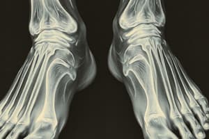
Imaging of pes cavus
- Pes cavus is a pedal deformity has a hollow or cave-like appearance.
- This appearance results from elevation of the medial and lateral longitudinal foot arches.
- Direct pes cavus is the most common form and is defined as a sagittal plane deformity. Direct pes cavus should not be confused with rarer, triplane cavovarus deformities
Triplane Cavovarus Deformity
- Triplane cavovarus deformity is distinct from uniplanar direct pes cavus which is a sagittal plane deformity.
- Cavovarus deformity involves rotation of the foot and calcaneus on the distal tibial-fibular articulation, minus the talus.
- Cavovarus deformity results in compensatory supination of the calcaneopedal unit.
- This supination causes calcaneal varus, external rotation of talo-tibfibular unit, forefoot/metatarsal equinus, and forefoot-midfoot adduction.
- Advanced modalities like weight-bearing CT scanning shows promise as potential future adjuncts.
- Plain radiographs are the cornerstone for imaging this deformity.
- Plain radiographs, along with modified specialized views, confirm clinical diagnosis, classify deformity, and aid surgical planning.
- Standard weight-bearing plain radiographic imaging includes anteroposterior, lateral, and calcaneal axial views.
- Ankle studies assess associated extrapedal causes like ankle/tibia plafond varus deformities or rotational malalignments.
- Triplane cavovarus deformity can be associated with CMT disease or clubfoot
Anteroposterior Weight-Bearing Radiographic Assessment
- Anteroposterior foot radiographs is essential to gain insight into transverse plane malalignments in cavus foot deformities.
- Anteroposterior foot radiographs help evaluate cavovarus deformities and is potentially valuable
- Metatarsus adduction can be assessed
- Forefoot adduction or displacement can be identified in clubfoot
- Degree of midfoot and subtalar joint supination (talocalcaneal and talonavicular) can be examined
- Overlap of midfoot and rearfoot bones and parallelism of talus and calcaneus can be reviewed
- Marked decrease of TC angle (<5°; normal values 25°–30°) is notable in triplanar cavus foot deformity from a clubfoot.
- The longitudinal talar axis is directed through the lateral rays in these cases.
Lateral Weight-Bearing Plain Radiographic Assessment
- Lateral weight-bearing radiograph is integral in evaluating sagittal plane deformity in direct pes cavus, and aids in clinical diagnosis and surgical planning.
- The lateral radiograph is essential in assessing sagittal plane component of triplanar cavovarus deformity.
- Multiplanar CT with 3D reconstruction of the foot and ankle are suitable for preoperative and postoperative cavovarus deformity evaluation.
Calcaneal Inclination Angle (CIA):
- Angle is a tangent to the inferior calcaneal surface and the weight-bearing reference line.
- The normal range is 25° ± 5°.
- Values over 30° indicate moderate pes cavus.
- Severe cavus often has CIAs of 40° or higher and neurologic cavus foot deformities show CIAs in this range.
Studying That Suits You
Use AI to generate personalized quizzes and flashcards to suit your learning preferences.