Podcast
Questions and Answers
In the context of pes cavus deformity, which statement most accurately differentiates between direct pes cavus and cavovarus deformity?
In the context of pes cavus deformity, which statement most accurately differentiates between direct pes cavus and cavovarus deformity?
- Cavovarus deformity exclusively occurs in conjunction with neuromuscular disorders, whereas direct pes cavus is typically idiopathic.
- Cavovarus deformity is primarily a sagittal plane deformity, whereas direct pes cavus always involves a triplane deformity with adduction.
- Direct pes cavus involves uniplanar (sagittal plane) deformity, while cavovarus deformity encompasses multiplanar rotation of the foot and calcaneus relative to the talus. (correct)
- Direct pes cavus is characterized by compensatory supination, while cavovarus deformity results from isolated plantarflexion of the first ray.
When evaluating pediatric pes cavus, which radiographic parameter offers the MOST SPECIFIC insight into underlying ankle and/or rearfoot equinus?
When evaluating pediatric pes cavus, which radiographic parameter offers the MOST SPECIFIC insight into underlying ankle and/or rearfoot equinus?
- Tibiotalar Angle
- Tibiocalcaneal Angle (correct)
- Lateral Talocalcaneal Angle
- Calcaneal Inclination Angle (CIA)
In the assessment of combined (mixed) pes cavus deformity, which statement accurately reflects the utility of the Meary angle?
In the assessment of combined (mixed) pes cavus deformity, which statement accurately reflects the utility of the Meary angle?
- The apex of the Meary angle typically intersects between the Chopart and Lisfranc joints, with an anterior apex correlating with increased metatarsal declination. (correct)
- The Meary angle is consistently normal, negating its diagnostic relevance in combined deformities.
- The apex of the Meary angle intersects proximal to the Chopart joint, exclusively indicating a calcaneonavicular coalition.
- The Meary angle is exclusively increased in the presence of marked talar declination, irrespective of the location of the apex.
Which of the following statements accurately describes the utility of weight-bearing computed tomography (WBCT) in evaluating pes cavus deformity, especially in the context of surgical planning?
Which of the following statements accurately describes the utility of weight-bearing computed tomography (WBCT) in evaluating pes cavus deformity, especially in the context of surgical planning?
What imaging modality is MOST sensitive for detecting early signs of peroneal tendon pathology associated with adult-acquired pes cavus secondary to lateral ankle sprain?
What imaging modality is MOST sensitive for detecting early signs of peroneal tendon pathology associated with adult-acquired pes cavus secondary to lateral ankle sprain?
Which of the following is the MOST accurate description of the "anteater nose sign" concerning tarsal coalitions associated with pes cavus?
Which of the following is the MOST accurate description of the "anteater nose sign" concerning tarsal coalitions associated with pes cavus?
What is the MOST specific radiographic finding indicative of a middle facet tarsal coalition in a patient with pes cavus?
What is the MOST specific radiographic finding indicative of a middle facet tarsal coalition in a patient with pes cavus?
In evaluating potential intraosseous or interosseous deformity contributing to pes cavus, what is the MOST significant advantage of advanced multiplanar computed tomographic (CT) imaging and three-dimensional (3D) reconstructions?
In evaluating potential intraosseous or interosseous deformity contributing to pes cavus, what is the MOST significant advantage of advanced multiplanar computed tomographic (CT) imaging and three-dimensional (3D) reconstructions?
Which statement accurately describes the imaging technique best suited to assess hindfoot alignment, specifically coronal plane tibiocalcaneal alignment in the context of pes cavus?
Which statement accurately describes the imaging technique best suited to assess hindfoot alignment, specifically coronal plane tibiocalcaneal alignment in the context of pes cavus?
How does the "Magic Angle Artifact" MOST significantly impact MRI evaluation of peroneal tendons in patients with pes cavus and lateral ankle instability?
How does the "Magic Angle Artifact" MOST significantly impact MRI evaluation of peroneal tendons in patients with pes cavus and lateral ankle instability?
In assessing muscle involvement using MRI in cavovarus deformity secondary to Charcot-Marie-Tooth disease (CMT), which finding MOST accurately differentiates between CMT1A and CMT2A subtypes?
In assessing muscle involvement using MRI in cavovarus deformity secondary to Charcot-Marie-Tooth disease (CMT), which finding MOST accurately differentiates between CMT1A and CMT2A subtypes?
When evaluating pes cavus, especially focusing on Meary's angle, which statement BEST describes the significance of this measurement?
When evaluating pes cavus, especially focusing on Meary's angle, which statement BEST describes the significance of this measurement?
What is the MOST accurate method for ensuring the reliability of hindfoot alignment assessments when using radiographic techniques, considering inherent limitations related to leg rotation and subject posture?
What is the MOST accurate method for ensuring the reliability of hindfoot alignment assessments when using radiographic techniques, considering inherent limitations related to leg rotation and subject posture?
Which of the following statements accurately reflects the diagnostic significance of T2* relaxation time mapping when assessing ankle joint cartilage in pes cavus?
Which of the following statements accurately reflects the diagnostic significance of T2* relaxation time mapping when assessing ankle joint cartilage in pes cavus?
In cases of suspected tarsal coalition in patients with pes cavus, medial oblique radiographs are particularly useful for visualizing which specific anatomical structure?
In cases of suspected tarsal coalition in patients with pes cavus, medial oblique radiographs are particularly useful for visualizing which specific anatomical structure?
Which factor is MOST crucial in distinguishing between compensatory alignment and true deformity when evaluating hindfoot alignment?
Which factor is MOST crucial in distinguishing between compensatory alignment and true deformity when evaluating hindfoot alignment?
Which of the following statements accurately describes the PRIMARY advantage of using 7-Tesla MRI over lower-field MRI in evaluating intrinsic foot muscles in pes cavus?
Which of the following statements accurately describes the PRIMARY advantage of using 7-Tesla MRI over lower-field MRI in evaluating intrinsic foot muscles in pes cavus?
When assessing the clinical significance of the lateral talocalcaneal angle, which factor is MOST critical in its interpretation regarding clubfoot correction or pes cavus subclassification?
When assessing the clinical significance of the lateral talocalcaneal angle, which factor is MOST critical in its interpretation regarding clubfoot correction or pes cavus subclassification?
In the context of radiographic imaging of pes cavus, what is the MOST accurate description of the C-sign and its clinical significance?
In the context of radiographic imaging of pes cavus, what is the MOST accurate description of the C-sign and its clinical significance?
Which statement accurately describes the potential utility of ultrahigh-resolution MRI in evaluating subtle abnormalities in foot and ankle alignment within cavovarus deformities?
Which statement accurately describes the potential utility of ultrahigh-resolution MRI in evaluating subtle abnormalities in foot and ankle alignment within cavovarus deformities?
What is the MOST common type of pes cavus deformity encountered?
What is the MOST common type of pes cavus deformity encountered?
Which radiographic view is considered the cornerstone for imaging evaluation of direct pes cavus foot deformity?
Which radiographic view is considered the cornerstone for imaging evaluation of direct pes cavus foot deformity?
What does the apex of the Meary talo-first metatarsal angle on a lateral radiograph represent in the context of pes cavus deformity?
What does the apex of the Meary talo-first metatarsal angle on a lateral radiograph represent in the context of pes cavus deformity?
In evaluating pes cavus deformity, what information can be gained from ancillary radiographic imaging techniques like the modified Saltzman view or the modified Coleman block test?
In evaluating pes cavus deformity, what information can be gained from ancillary radiographic imaging techniques like the modified Saltzman view or the modified Coleman block test?
What is the PRIMARY advantage of using multiplanar computed tomographic (CT) imaging with 3D reconstructions in evaluating pes cavus deformity?
What is the PRIMARY advantage of using multiplanar computed tomographic (CT) imaging with 3D reconstructions in evaluating pes cavus deformity?
In the context of radiographic interpretation, what does an increased calcaneal inclination angle (CIA) typically indicate?
In the context of radiographic interpretation, what does an increased calcaneal inclination angle (CIA) typically indicate?
Which of the following conditions is MOST frequently associated with forefoot-driven hindfoot varus in anterior cavus deformity?
Which of the following conditions is MOST frequently associated with forefoot-driven hindfoot varus in anterior cavus deformity?
What is the MOST likely compensatory mechanism in the subtalar joint when there is a sagittal plane depression of the medial forefoot column?
What is the MOST likely compensatory mechanism in the subtalar joint when there is a sagittal plane depression of the medial forefoot column?
In radiographic assessment, which condition is suggested by a tibiocalcaneal angle that tends to decrease with increasing values of calcaneal pitch/CIA?
In radiographic assessment, which condition is suggested by a tibiocalcaneal angle that tends to decrease with increasing values of calcaneal pitch/CIA?
What radiographic finding is indicative of a calcaneonavicular coalition?
What radiographic finding is indicative of a calcaneonavicular coalition?
What is the PRIMARY role of MRI in evaluating adult-acquired pes cavus secondary to an ankle sprain?
What is the PRIMARY role of MRI in evaluating adult-acquired pes cavus secondary to an ankle sprain?
Why is it important to plantarflex the ankle by about 20 degrees during an ankle MRI when evaluating for peroneal tendon pathology?
Why is it important to plantarflex the ankle by about 20 degrees during an ankle MRI when evaluating for peroneal tendon pathology?
In the context of combined (mixed) cavus deformity, where is the typical intersection point of the apex of the Meary angle?
In the context of combined (mixed) cavus deformity, where is the typical intersection point of the apex of the Meary angle?
What key information is provided from weight-bearing CT scans that cannot be obtained from standard radiographs in evaluating pes cavus?
What key information is provided from weight-bearing CT scans that cannot be obtained from standard radiographs in evaluating pes cavus?
How does the presence of high-arched/cavus feet affect the reliability of Harris views (Harris-Beath view) in imaging tarsal coalitions?
How does the presence of high-arched/cavus feet affect the reliability of Harris views (Harris-Beath view) in imaging tarsal coalitions?
In the context of using MRI to evaluate muscle involvement in pes cavus due to Charcot-Marie-Tooth disease (CMT), what is a key difference in muscle involvement patterns between CMT1A and CMT2A patients?
In the context of using MRI to evaluate muscle involvement in pes cavus due to Charcot-Marie-Tooth disease (CMT), what is a key difference in muscle involvement patterns between CMT1A and CMT2A patients?
What is the MOST significant limitation of hindfoot alignment studies that assess coronal plane alignment of the calcaneus relative to the tibia?
What is the MOST significant limitation of hindfoot alignment studies that assess coronal plane alignment of the calcaneus relative to the tibia?
In cavovarus foot deformities, which statement accurately describes how muscle imbalance significantly contributes to the mentioned deformity?
In cavovarus foot deformities, which statement accurately describes how muscle imbalance significantly contributes to the mentioned deformity?
While lateral weigthbearing radiographs are accurate in the assessing of direct cavus deformities, for accuracy, what additional method is needed when subclassifying a complex triplanar deformity?
While lateral weigthbearing radiographs are accurate in the assessing of direct cavus deformities, for accuracy, what additional method is needed when subclassifying a complex triplanar deformity?
If a patient presents with a decrease in subtalar joint motion, which deformity is most likely to occur?
If a patient presents with a decrease in subtalar joint motion, which deformity is most likely to occur?
In assessing a direct pes cavus deformity, what is the PRIMARY focus when using a lateral weight-bearing radiograph?
In assessing a direct pes cavus deformity, what is the PRIMARY focus when using a lateral weight-bearing radiograph?
What is the TYPICAL radiographic presentation of neurologic cavus foot deformities regarding the calcaneal inclination angle (CIA)?
What is the TYPICAL radiographic presentation of neurologic cavus foot deformities regarding the calcaneal inclination angle (CIA)?
In radiographic analysis of pes cavus, an increased first metatarsal declination angle typically indicates what specific type of cavus deformity?
In radiographic analysis of pes cavus, an increased first metatarsal declination angle typically indicates what specific type of cavus deformity?
Concerning the tibiocalcaneal angle in the pediatric population, what condition is generally associated with increased angles?
Concerning the tibiocalcaneal angle in the pediatric population, what condition is generally associated with increased angles?
Which radiographic finding is MOST indicative of a calcaneonavicular coalition?
Which radiographic finding is MOST indicative of a calcaneonavicular coalition?
In the context of anterior cavus deformity, what is the PRIMARY characteristic?
In the context of anterior cavus deformity, what is the PRIMARY characteristic?
What is a COMMON clinical presentation in instances where the forefoot demonstrates valgus relative to the rearfoot?
What is a COMMON clinical presentation in instances where the forefoot demonstrates valgus relative to the rearfoot?
Following an acute lateral ankle sprain, what muscle imbalance is MOST likely to contribute to a resultant cavus foot deformity?
Following an acute lateral ankle sprain, what muscle imbalance is MOST likely to contribute to a resultant cavus foot deformity?
What specific information does multiplanar CT scanning add to the evaluation of triplanar cavovarus deformity, beyond what plain film radiographs provide?
What specific information does multiplanar CT scanning add to the evaluation of triplanar cavovarus deformity, beyond what plain film radiographs provide?
In assessing for tarsal coalitions, particularly when using the Harris view (Harris-Beath view), what factor limits the reliability of this view in patients with high-arched/cavus feet?
In assessing for tarsal coalitions, particularly when using the Harris view (Harris-Beath view), what factor limits the reliability of this view in patients with high-arched/cavus feet?
Why is an ankle MRI typically performed with the foot plantarflexed by about 20 degrees when evaluating for peroneal tendon pathology?
Why is an ankle MRI typically performed with the foot plantarflexed by about 20 degrees when evaluating for peroneal tendon pathology?
What is the MOST LIKELY underlying etiology if AP ankle views reveal a ball-and-socket ankle joint deformity in a pes cavus presentation?
What is the MOST LIKELY underlying etiology if AP ankle views reveal a ball-and-socket ankle joint deformity in a pes cavus presentation?
In the context of hindfoot alignment, what is the primary limitation of studies assessing coronal plane alignment of the calcaneus relative to the tibia?
In the context of hindfoot alignment, what is the primary limitation of studies assessing coronal plane alignment of the calcaneus relative to the tibia?
What does the apex of the Meary talo-first metatarsal angle represent, and why is it clinically significant in the subclassification of pes cavus?
What does the apex of the Meary talo-first metatarsal angle represent, and why is it clinically significant in the subclassification of pes cavus?
How might MRI demonstrate altered bone marrow edema patterns and a predilection for lateral stress reactions or fractures in the context of pes cavus/cavovarus deformity?
How might MRI demonstrate altered bone marrow edema patterns and a predilection for lateral stress reactions or fractures in the context of pes cavus/cavovarus deformity?
Which statement accurately describes the influence of the subtalar joint in instances of varus ankle/varus plafond?
Which statement accurately describes the influence of the subtalar joint in instances of varus ankle/varus plafond?
A patient presents with radiographic findings of a Meary angle of nearly 0°, increased CIA, and increased tibiotalar angle. What type of pes cavus deformity is MOST likely?
A patient presents with radiographic findings of a Meary angle of nearly 0°, increased CIA, and increased tibiotalar angle. What type of pes cavus deformity is MOST likely?
In a combined (mixed) cavus deformity, if the apex of the Meary angle intersects somewhere between the Chopart and Lisfranc joints, what component of the deformity is generally reflected?
In a combined (mixed) cavus deformity, if the apex of the Meary angle intersects somewhere between the Chopart and Lisfranc joints, what component of the deformity is generally reflected?
In extremely severe cases of pes cavus, plain film lateral radiographs may not be sensitive enough to evaluate the condition. According to the experts, advanced 3D weight bearing CT and MRI studies are beginning to show a previously unseen relationship in the rearfoot. That previously unseen condition is:
In extremely severe cases of pes cavus, plain film lateral radiographs may not be sensitive enough to evaluate the condition. According to the experts, advanced 3D weight bearing CT and MRI studies are beginning to show a previously unseen relationship in the rearfoot. That previously unseen condition is:
Weight-bearing CT (WBCT) allows for a more detailed assessment of pes cavus deformity. Beyond identifying different types of pes cavus and evaluating compensatory rearfoot motions, what additional analysis does WBCT facilitate?
Weight-bearing CT (WBCT) allows for a more detailed assessment of pes cavus deformity. Beyond identifying different types of pes cavus and evaluating compensatory rearfoot motions, what additional analysis does WBCT facilitate?
Which type of pes cavus deformity is most commonly encountered?
Which type of pes cavus deformity is most commonly encountered?
A lateral weight-bearing radiograph is NOT a key component in imaging evaluation of a direct pes cavus foot deformity.
A lateral weight-bearing radiograph is NOT a key component in imaging evaluation of a direct pes cavus foot deformity.
What radiographic angle represents the pinnacle of the cavus deformity and aids in its subclassification?
What radiographic angle represents the pinnacle of the cavus deformity and aids in its subclassification?
The apex of the Meary talo-first metatarsal angle assists in ___________ of the deformity.
The apex of the Meary talo-first metatarsal angle assists in ___________ of the deformity.
Match the following radiographic views with their primary diagnostic use in pes cavus evaluation:
Match the following radiographic views with their primary diagnostic use in pes cavus evaluation:
Which advanced imaging technique shows promise in improving the precision and accuracy of delineating intraosseous and interosseous deformity in pes cavus?
Which advanced imaging technique shows promise in improving the precision and accuracy of delineating intraosseous and interosseous deformity in pes cavus?
The calcaneal inclination angle (CIA) is formed by the intersection of the longitudinal axis of the talus and the first metatarsal.
The calcaneal inclination angle (CIA) is formed by the intersection of the longitudinal axis of the talus and the first metatarsal.
What is the normal range for the calcaneal inclination angle (CIA)?
What is the normal range for the calcaneal inclination angle (CIA)?
Values greater than ______ degrees for the calcaneal inclination angle (CIA) are indicative of moderate pes cavus/increase in pitch.
Values greater than ______ degrees for the calcaneal inclination angle (CIA) are indicative of moderate pes cavus/increase in pitch.
Match the radiographic finding with its description in pes cavus:
Match the radiographic finding with its description in pes cavus:
In anterior cavus deformity, which of the following is a common radiographic finding?
In anterior cavus deformity, which of the following is a common radiographic finding?
The usefulness of the Calcaneal-first metatarsal (Hibbs) angle has not been questioned in that it does not aid in the determination of the apex of the deformity.
The usefulness of the Calcaneal-first metatarsal (Hibbs) angle has not been questioned in that it does not aid in the determination of the apex of the deformity.
What is the primary imaging modality for demonstrating typical coronal plane features in subtalar joint coalition when Harris views cannot reliably image subtalar joint features in subjects with pes cavus deformity?
What is the primary imaging modality for demonstrating typical coronal plane features in subtalar joint coalition when Harris views cannot reliably image subtalar joint features in subjects with pes cavus deformity?
The ________ angle is formed by the intersection of the longitudinal axes of the calcaneus and first metatarsal bones.
The ________ angle is formed by the intersection of the longitudinal axes of the calcaneus and first metatarsal bones.
Match the muscle group with its associated cavus deformity pattern:
Match the muscle group with its associated cavus deformity pattern:
Which of the following is NOT a typical finding in a secondary weight-bearing lateral radiographic assessment associated with pes cavus deformity?
Which of the following is NOT a typical finding in a secondary weight-bearing lateral radiographic assessment associated with pes cavus deformity?
The tibiocalcaneal angle generally increases with increasing values of calcaneal pitch/CIA in pes cavus deformities.
The tibiocalcaneal angle generally increases with increasing values of calcaneal pitch/CIA in pes cavus deformities.
What specific imaging feature, observable via MRI, can demonstrate altered patterns of bone marrow edema in pes cavus/cavovarus deformity?
What specific imaging feature, observable via MRI, can demonstrate altered patterns of bone marrow edema in pes cavus/cavovarus deformity?
In the context of adult-acquired pes cavus secondary to chronic sequelae of a peroneus brevis (PB) injury, imaging begins with weight-bearing plain film imaging of the foot and ankle, AP and lateral of the foot, axial views of the _________, and AP mortise and lateral of the ankle.
In the context of adult-acquired pes cavus secondary to chronic sequelae of a peroneus brevis (PB) injury, imaging begins with weight-bearing plain film imaging of the foot and ankle, AP and lateral of the foot, axial views of the _________, and AP mortise and lateral of the ankle.
Match the following parameters of pes cavus with their typical values or characteristics:
Match the following parameters of pes cavus with their typical values or characteristics:
Which of the following is considered the cornerstone of imaging evaluation for direct pes cavus foot deformity?
Which of the following is considered the cornerstone of imaging evaluation for direct pes cavus foot deformity?
Direct-type cavus foot deformities are most commonly triplane deformities.
Direct-type cavus foot deformities are most commonly triplane deformities.
What angle, when assessed on a lateral radiograph, represents the pinnacle of the cavus deformity?
What angle, when assessed on a lateral radiograph, represents the pinnacle of the cavus deformity?
The calcaneal inclination angle is formed by a tangent to the inferior calcaneal surface and the ______ reference line.
The calcaneal inclination angle is formed by a tangent to the inferior calcaneal surface and the ______ reference line.
Match the following radiographic views with their primary use in evaluating pes cavus:
Match the following radiographic views with their primary use in evaluating pes cavus:
What does the term 'pes cavus' refer to?
What does the term 'pes cavus' refer to?
The Meary angle is formed by the intersection of the calcaneal and talar longitudinal axes.
The Meary angle is formed by the intersection of the calcaneal and talar longitudinal axes.
In the context of imaging pes cavus, what is the significance of multiplanar CT scanning?
In the context of imaging pes cavus, what is the significance of multiplanar CT scanning?
Severe pes cavus deformities may demonstrate decreased ______ angles approaching 90 degrees.
Severe pes cavus deformities may demonstrate decreased ______ angles approaching 90 degrees.
In anterior cavus deformity, which of the following muscles is typically weakened?
In anterior cavus deformity, which of the following muscles is typically weakened?
In the evaluation of flatfoot, loss of visualization of the middle facet is a sin qua non for a middle facet coalition.
In the evaluation of flatfoot, loss of visualization of the middle facet is a sin qua non for a middle facet coalition.
What radiographic finding is indicative of CN coalition?
What radiographic finding is indicative of CN coalition?
Typical findings demonstrated utilizing coronal plane features in subtalar joint coalition includes dysmorphic sustentaculum tali, altered middle facet angulation (``drunken ______'' sign), presence of incorporated os sustentacula, and the degree of posterior facet involvement in the coalition.
Typical findings demonstrated utilizing coronal plane features in subtalar joint coalition includes dysmorphic sustentaculum tali, altered middle facet angulation (``drunken ______'' sign), presence of incorporated os sustentacula, and the degree of posterior facet involvement in the coalition.
What is the most common acute musculoskeletal injury of the body?
What is the most common acute musculoskeletal injury of the body?
MRI is the standard imaging technique for direct cavus deformities.
MRI is the standard imaging technique for direct cavus deformities.
What is the significance of the "bullet-hole" sinus tarsi in radiographic findings related to pes cavus?
What is the significance of the "bullet-hole" sinus tarsi in radiographic findings related to pes cavus?
Anterior cavus is primarily a sagittal plane deformity associated with excessive plantarflexion of either the first ______ or the forefoot equinus
Anterior cavus is primarily a sagittal plane deformity associated with excessive plantarflexion of either the first ______ or the forefoot equinus
Which of the following statements regarding the use of subtalar joint vertical axis for assessing varus/valgus configuration of the subtalar joint posterior facet is true?
Which of the following statements regarding the use of subtalar joint vertical axis for assessing varus/valgus configuration of the subtalar joint posterior facet is true?
The tibiocalcaneal angle typically decreases with increasing values of calcaneal pitch and/or with ankle dorsiflexion.
The tibiocalcaneal angle typically decreases with increasing values of calcaneal pitch and/or with ankle dorsiflexion.
What type of imaging can cause the 'magic angle artifact'?
What type of imaging can cause the 'magic angle artifact'?
Flashcards
Pes Cavus
Pes Cavus
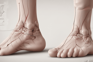
A foot deformity with a high arch, giving a hollow or cave-like appearance.
Direct Pes Cavus
Direct Pes Cavus
Direct pes cavus is primarily a sagittal plane deformity and is the most common type of cavus foot.
Meary Talo-First Metatarsal Angle
Meary Talo-First Metatarsal Angle
An angle measured on a lateral radiograph, formed by the intersection of the talus and first metatarsal axes; assesses cavus deformity.
Calcaneal Inclination Angle (CIA)
Calcaneal Inclination Angle (CIA)
Signup and view all the flashcards
Cavovarus Deformity
Cavovarus Deformity
Signup and view all the flashcards
Modified Coleman Block Test
Modified Coleman Block Test
Signup and view all the flashcards
Modified Saltzman View
Modified Saltzman View
Signup and view all the flashcards
Imaging for Pes Cavus
Imaging for Pes Cavus
Signup and view all the flashcards
AP Foot Radiographs
AP Foot Radiographs
Signup and view all the flashcards
Standard Plain Radiographs
Standard Plain Radiographs
Signup and view all the flashcards
Calcaneal-First Metatarsal (Hibbs) Angle
Calcaneal-First Metatarsal (Hibbs) Angle
Signup and view all the flashcards
Tibiotalar Angle
Tibiotalar Angle
Signup and view all the flashcards
Anteater Nose Sign
Anteater Nose Sign
Signup and view all the flashcards
Peroneus Brevis (PB) Acute Injuries
Peroneus Brevis (PB) Acute Injuries
Signup and view all the flashcards
MRI in pes cavus/cavovarus deformity
MRI in pes cavus/cavovarus deformity
Signup and view all the flashcards
Apex of Meary angle.
Apex of Meary angle.
Signup and view all the flashcards
Cavovarus deformity details
Cavovarus deformity details
Signup and view all the flashcards
AP radiograph benefit
AP radiograph benefit
Signup and view all the flashcards
Tibiocalcaneal angle significance
Tibiocalcaneal angle significance
Signup and view all the flashcards
Anterior Cavus
Anterior Cavus
Signup and view all the flashcards
Cause of Anteater nose sign
Cause of Anteater nose sign
Signup and view all the flashcards
Inconspicuous middle facet
Inconspicuous middle facet
Signup and view all the flashcards
Peroneus Brevis (PB) injuries
Peroneus Brevis (PB) injuries
Signup and view all the flashcards
CT scanning for tarsal coalition
CT scanning for tarsal coalition
Signup and view all the flashcards
Lateral Radiographs
Lateral Radiographs
Signup and view all the flashcards
Calcaneal Inclination Angle
Calcaneal Inclination Angle
Signup and view all the flashcards
Talo-first metatarsal (Meary) angle
Talo-first metatarsal (Meary) angle
Signup and view all the flashcards
Lateral TC Angle
Lateral TC Angle
Signup and view all the flashcards
First Metatarsal Declination Angle
First Metatarsal Declination Angle
Signup and view all the flashcards
Classifying Pes Cavus
Classifying Pes Cavus
Signup and view all the flashcards
Increased Lateral TC Angle
Increased Lateral TC Angle
Signup and view all the flashcards
Ancillary Radiographic views
Ancillary Radiographic views
Signup and view all the flashcards
Uncommon Tarsal Coalitions
Uncommon Tarsal Coalitions
Signup and view all the flashcards
Lateral Weight-Bearing Radiograph
Lateral Weight-Bearing Radiograph
Signup and view all the flashcards
Tibiocalcaneal Angle
Tibiocalcaneal Angle
Signup and view all the flashcards
Secondary Pes Cavus Radiographic Signs
Secondary Pes Cavus Radiographic Signs
Signup and view all the flashcards
Ankle Sprain
Ankle Sprain
Signup and view all the flashcards
Cavus Foot Muscle Imbalance
Cavus Foot Muscle Imbalance
Signup and view all the flashcards
MRI for Pes Cavus
MRI for Pes Cavus
Signup and view all the flashcards
Medial Oblique Imaging
Medial Oblique Imaging
Signup and view all the flashcards
Triplane Cavovarus Deformity
Triplane Cavovarus Deformity
Signup and view all the flashcards
Tibiocalcaneal Angle Increase
Tibiocalcaneal Angle Increase
Signup and view all the flashcards
Horizontal Talus
Horizontal Talus
Signup and view all the flashcards
Talar Neck “Beaking”
Talar Neck “Beaking”
Signup and view all the flashcards
Peroneal Tendon Deformations
Peroneal Tendon Deformations
Signup and view all the flashcards
Study Notes
- Direct-type cavus foot deformities are most commonly encountered and are primarily sagittal plane deformities
- Direct deformities should be delineated from rarer triplane pes cavovarus deformities
- The lateral weight-bearing radiograph is the cornerstone of imaging evaluation of direct pes cavus foot deformity
- Ancillary radiographic imaging techniques give valuable insight into deformity assessment and surgical planning, like the modified Saltzman view or the modified Coleman block test
- Advanced multiplanar computed tomographic (CT) imaging techniques along with 3-dimensional reconstructions promise to improve precision and accuracy in delineating the presence and extent of intraosseous and interosseous deformity
- Weight-bearing CT can facilitate hindfoot alignment and compensatory motions in pes cavus deformity
- Pes cavus is perceived as a pedal deformity with elevation of the medial and lateral longitudinal foot arches, imparting a cave-like appearance
- Lateral weight-bearing radiograph is integral to the evaluative process for the sagittal plane deformity seen in direct pes cavus, whether for confirming clinical diagnosis, classifying the deformity, or planning surgical correction
- The lateral radiograph is essential in assessing the sagittal plane component of the triplanar cavovarus deformity
- Multiplanar CT scanning with 3D reconstruction appears better suited for preoperative and postoperative evaluation of cavovarus deformity
- Primary WB lateral radiographic determinations comprise the main measurements and/or determinations in pes cavus deformity
- Calcaneal inclination angle (CIA) (aka calcaneal “pitch” angle) is formed by a tangent to the inferior calcaneal surface and the WB reference line. 25° ± 5° is normal, greater than 30° indicates moderate pes cavus/increase in pitch. Severe cavus often demonstrates excessive CIAs of 40° or greater
- A talo-first metatarsal (Meary) angle is formed by the intersection of longitudinal axes, ranging 0°-5° normally. As first metatarsal declination increases, the Meary angle generally increases, with values greater than 15°-18° in pes cavus
- Sagittal apex of pes cavus deformity is determined by where the apex of the Meary talo-first metatarsal angle is positioned, and is a key guideline used in helping to subclassify pes cavus deformities
- A calcaneal-first metatarsal (Hibbs) angle is formed by the intersection of longitudinal axes of the calcaneus and first metatarsal bones. Hibb angles less than 150° are indicative of pes cavus, possibly approaching 90°
- A lateral TC angle is formed by the intersection of the calcaneal and talar longitudinal axes. Normal values are 35°-50°, tending to increase with pronation and decrease with supination, classifically used to evaluate clubfoot correction
- First metatarsal declination angle is formed by the longitudinal axis of the first metatarsal and the WB reference line. Normal values range from 15°-23°, and angles commonly increase beyond 25° in cavus deformity
- Tibiotalar angle is formed by the longitudinal axes of the talus and tibia, which normally measures 110°, and decreases with cavus deformity associated with ankle joint dorsiflexion and vice versa
- Tibiocalcaneal angle is formed by a tangent to the inferior calcaneal surface and the longitudinal axis of the tibia, ranging 60°-90° normally in pediatric cases
- Increased angles are generally associated with ankle and/or rearfoot equinus, and tends to decrease with increasing values of calcaneal pitch/CIA and/or with ankle dorsiflexion
- Anterior cavus ("forefoot driven hindfoot varus") is primarily a sagittal plane deformity associated with excessive plantarflexion of either the first ray or the forefoot equinus
- General radiographic findings for anterior cavus deformity include increased declination angles of the metatarsal bones
Studying That Suits You
Use AI to generate personalized quizzes and flashcards to suit your learning preferences.