Podcast
Questions and Answers
Which of the following best describes pathogenesis in the context of pathology?
Which of the following best describes pathogenesis in the context of pathology?
- The signs and symptoms of a disease, providing insight into its clinical presentation.
- The structural alterations in cells or tissues that are characteristic of a disease.
- The initiating cause of a disease, such as genetic mutations or infectious agents.
- The sequence of molecular, biochemical, and cellular events that lead to the development of a disease. (correct)
Which of the following is the MOST accurate distinction between necrosis and apoptosis?
Which of the following is the MOST accurate distinction between necrosis and apoptosis?
- Necrosis is often accompanied by inflammation, while apoptosis typically does not induce inflammation. (correct)
- Necrosis affects individual cells, while apoptosis affects large groups of cells.
- Necrosis involves cell shrinkage, while apoptosis involves cell swelling.
- Necrosis is always physiologic, while apoptosis is always pathologic.
In the context of tissue repair, what condition would MOST LIKELY result in scar formation rather than regeneration?
In the context of tissue repair, what condition would MOST LIKELY result in scar formation rather than regeneration?
- Mild injury to the liver, where cells are capable of proliferation.
- Damage limited to the epithelial layer of the skin.
- Extensive damage involving both the epithelium and underlying connective tissue. (correct)
- A superficial abrasion that does not penetrate the basement membrane.
A patient presents with a persistent non-healing ulcer on their foot. Microscopic examination reveals granulation tissue. What is the MOST accurate description of granulation tissue's role in wound healing?
A patient presents with a persistent non-healing ulcer on their foot. Microscopic examination reveals granulation tissue. What is the MOST accurate description of granulation tissue's role in wound healing?
What is the underlying mechanism behind ischemia leading to coagulative necrosis?
What is the underlying mechanism behind ischemia leading to coagulative necrosis?
Why are teratogenic effects MOST detrimental during the embryonic period (3-9 weeks)?
Why are teratogenic effects MOST detrimental during the embryonic period (3-9 weeks)?
What is the MOST likely cause of wet gangrene?
What is the MOST likely cause of wet gangrene?
Which of the following is considered an example of adaptation?
Which of the following is considered an example of adaptation?
What is the key difference between stable cells and labile cells in terms of proliferative capacity?
What is the key difference between stable cells and labile cells in terms of proliferative capacity?
Which of the following is likely to result in gangrenous necrosis?
Which of the following is likely to result in gangrenous necrosis?
Flashcards
Pathology
Pathology
The study of structural, biochemical, and functional changes in cells, tissues, and organs underlying disease.
Etiology
Etiology
The initiating cause of a disease, which can be genetic or environmental.
Pathogenesis
Pathogenesis
The sequence of molecular, biochemical, and cellular events leading to the development of a disease.
Morphologic changes
Morphologic changes
Signup and view all the flashcards
Clinical Manifestations
Clinical Manifestations
Signup and view all the flashcards
Homeostasis
Homeostasis
Signup and view all the flashcards
Adaptation
Adaptation
Signup and view all the flashcards
Cell Death relevance
Cell Death relevance
Signup and view all the flashcards
Necrosis Cell Size
Necrosis Cell Size
Signup and view all the flashcards
Irreversible injuries
Irreversible injuries
Signup and view all the flashcards
Study Notes
Pathology
- Pathology studies structural, biochemical, and functional changes in cells, tissues, and organs underlying disease.
- It aims to explain the reasons behind a patient's signs and symptoms.
- Pathology provides the scientific basis for clinical care and therapy.
- Four key aspects of a disease are etiology, pathogenesis, morphologic changes, and clinical manifestations.
Etiology
- Etiology refers to the initiating cause of a disease.
- The two main categories of etiology are genetic and environmental.
- Genetic causes include mutations and gene variants.
- Environmental factors include infectious, nutritional, chemical, and physical causes.
- Multifactorial diseases involve a combination of genetic predisposition and environmental trigger.
- An example of a multifactorial disease is a smoker born with genetic risk for cancer having a higher chance of developing it.
Pathogenesis
- Pathogenesis is the sequence of molecular, biochemical, and cellular events that lead to disease development.
- It explains how underlying etiologies cause the morphologic and clinical signs of the disease.
Morphologic Changes
- Morphologic changes involve structural alterations in cells or tissues characteristic of a disease.
- They aid in diagnosing the etiologic process.
Clinical Manifestations
- Clinical manifestations are the signs and symptoms of a disease.
Cellular Response
- Normal cells can handle physiological demands and maintain a steady state of homeostasis.
- Adaptation refers to reversible functional and structural responses to changes in physiological states.
- Examples of adaptation include pregnancy and exposure to pathogenic stimuli.
- Cells achieve a new steady state to survive and function.
- After the stressor is removed, the cell can return to its original state.
- Exceeding adaptive limits, exposure to damaging insults, nutrient deprivation, or genetic mutations affecting cellular function leads to disturbed cell injury.
Causes of Cell Injury
- Oxygen deprivation can cause hypoxia or ischemia, such as in a stroke or heart attack.
- Physical agents include traumatic injuries or temperature extremes, like frostbite, burns, or electrical injuries.
- Chemical agents and drugs include toxins and certain medications.
- Infectious agents, like COVID-19, can cause cell injury.
- Immunologic agents can damage the body while fighting off infection.
- Genetic abnormalities can lead to cells more susceptible to damage.
- Nutritional imbalances can also make cells more susceptible to damage.
- Cell injury often has multiple causes acting in combination.
Cell Aging
- Cell aging involves a progressive decline in cellular function and viability.
- It results from accumulated cellular damage, reduced capacity to divide (replicative senescence), impaired DNA repair, and defective protein homeostasis.
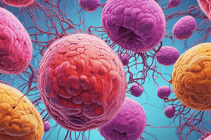
Cell Death
- Cell death occurs when further progressive injury leads to a point of no return.
- It can also be an essential process in embryogenesis, organ development, and homeostasis.
- Cell death removes damaged, unneeded, and aged cells and is considered normal.
- Necrosis is enlarged (swelling) cell size, disrupted plasma membrane, enzymatic digestion and usually pathologic.
- Apoptosis is reduced (shrinkage) cell size, fragmentation into a nucleosome-size fragments, intact plasma membrane, release of apoptotic bodies and often physiologic.
Patterns of Tissue Necrosis
- Tissue necrosis occurs when a large group of cells die.
Coagulative Necrosis
- Coagulative necrosis preserves dead tissue architecture for a few days.
- It is caused by ischemia, due to a blood vessel obstruction, leading to an infarct.
- An infarct is a localized area of coagulative necrosis.
- The pattern is generally found in all organs except the brain.
Liquefactive Necrosis
- Liquefactive necrosis does not preserve dead tissue architecture.
- Digestion of dead cells transforms the tissue into a viscous liquid (pus).
- Necrotic material (pus) is frequently creamy yellow because of leukocytes.
- This pattern occurs in the brain and focal bacterial or fungal infections.
- Microbes stimulate leukocyte accumulation and enzyme liberation from these cells.
Gangrenous Necrosis
- Gangrenous necrosis refers to coagulative necrosis of the limbs, typically the lower extremities, due to lost blood supply.
- Generally considered coagulative necrosis involving multiple tissue planes.
- Wet gangrene involves superimposed bacterial infection leading to liquefactive necrosis due to degradative enzymes, bacteria, and attracted leukocytes.
- Wet gangrene is frequently seen in diabetic feet.
- Untreated wet gangrene can result in ascending infection and may require amputation.
Fibrinoid Necrosis
- Fibrinoid necrosis is observed in vascular damage from immune reactions.
- It occurs when antigen-antibody complexes deposit in artery walls causing plasma proteins to leak out of the vessel
- The result is an amorphous, bright pink deposit.
Caseous Necrosis
- Caseous necrosis is associated with tuberculous infection.
- It presents as a cheese-like, friable white appearance.
Fat Necrosis
- Fat necrosis involves focal fat destruction resulting from activated pancreatic lipases/enzymes entering the peritonal cavity.
- Grossly visible chalky white areas (fat saponification) result from fatty acid and calcium combination.
Tissue Repair
- Tissue repair, also known as healing, involves restoring tissue architecture and function after injury.
- This process is critical for survival.
- The inflammatory response eliminates dangers and initiates repair.
- Tissue repair occurs through regeneration or scarring.
Regeneration
- Regeneration restores normal cells.
- It occurs after mild injuries that damage the epithelium but not the underlying tissue and resolution occurs.
Scarring
- Scarring consists of connective tissue deposition.
- It occurs due to more severe injury with damage to the connective tissue and repaired by scar formation.
Cell and Tissue Regeneration
- Cell and tissue regeneration involves cell proliferation and development of mature cells from tissue stem cells, driven by growth factors.
- Restoration of normal tissue structure occurs only if residual tissue is structurally intact and undamaged.
Connective Tissue Deposition
- Connective tissue deposition occurs when repair cannot be accomplished by regeneration alone
- Replacement of injured cells with connective tissue leading to a scar or a combination of regeneration of some residual cells and scar
- After injury, a hemostatic plug of platelets forms, stopping bleeding and providing a scaffold for fibrin deposition.
- Inflammatory cells enter the wound in the next 48 hours to eliminate microbes and clear debris.
- In the days that follow epithelial, vascular, and fibroblast cells proliferate and migrate to close the wound.
- Fibroblasts lay down collagen fibers.
- Over weeks, cells form granulation tissue, filling the injury site.
- Over months, collagen slowly replaces tissue forming a stable fibrous scar. Scar tissue results in fibrosis and injury, like stroke and heart attack.
Proliferative Capacity of Cells
- Tissue regeneration or scar formation depends on proliferative capacity.
Labile Tissues
- Labile tissues' cells are continuously lost and replaced by maturation of stem cells
- Labile tissues can readily regenerate if the stem cell pool remains
- Examples include blood cells in the bone marrow, skin epithelium, and mucous membranes
Stable Tissues
- Stable tissues have limited proliferative activity in their normal state but can divide in response to injury or loss
- Most solid tissues, except heart and brain, are stable tissues
Permanent Tissues
- Permanent tissues are considered nonproliferative and postnatal
Cerebrovascular Disease (Stroke)
- Cerebrovascular disease involves an altered blood flow to the brain, leading to tissue infarction
- It can be ischemic or hemorrhagic.
- Ischemic stroke is caused by an occlusive vascular disease, usually an embolism or thrombosis.
- Hemorrhagic stroke is caused by hypertension-induced blood vessel weakening.
- Genetic predisposition to aneurysm formation and tissue damage.
- If tissue perfusion is not achieved, the affected brain part undergoes irreversible injury and liquefaction necrosis.
- Stroke is usually characterized by memory deficits, visuospatial issues, judgment, personality, motor skills, and language difficulties.
Congenital Anomalies
- Anatomic defects are present at birth with genetic or environmental causes.
- Those born with anomalies might represent a less serious developemental failure in early embryogenesis.
Trisomy 21 (Down Syndrome)
- Trisomy 21 is a the most common of syndromes affecting cells with 47 chromosomes with an extra copy of chromosome 21 as a result of meiotic nondisjunction.
- Incidence increases with increasing maternal age.
- Diagnostic clinical features (usually evident even at birth) include flat facial profile, oblique palpebral fissures, and epicanthic folds.
- Over 40 year olds develop neuropathic changes like Alzheimer's.
- Leads to severe Intellectual Disability.
Studying That Suits You
Use AI to generate personalized quizzes and flashcards to suit your learning preferences.