Podcast
Questions and Answers
What ovarian volume measurement is typically associated with Polycystic Ovarian Syndrome (PCOS)?
What ovarian volume measurement is typically associated with Polycystic Ovarian Syndrome (PCOS)?
- 3.2 - 5.1 cm
- 2 - 20 cm
- Greater than 10 cm (correct)
- Less than 3 cm
Which of the following is a characteristic sonographic finding associated with ovarian torsion?
Which of the following is a characteristic sonographic finding associated with ovarian torsion?
- Presence of multiple small follicles
- Decreased ovarian size
- Increased arterial flow
- Absent color flow (correct)
What is the approximate five-year survival rate for ovarian cancer when detected and treated at an early stage?
What is the approximate five-year survival rate for ovarian cancer when detected and treated at an early stage?
- 30-50%
- 65%
- 80% (correct)
- 5%
Which of the following is a common clinical sign associated with Polycystic Ovarian Syndrome (PCOS)?
Which of the following is a common clinical sign associated with Polycystic Ovarian Syndrome (PCOS)?
For a simple ovarian cyst, at what size does it typically warrant follow-up?
For a simple ovarian cyst, at what size does it typically warrant follow-up?
Which ovarian tumor is known for containing hair, teeth, and bones?
Which ovarian tumor is known for containing hair, teeth, and bones?
During which stage of clot development in a hemorrhagic cyst might it mimic a solid mass on ultrasound with acoustic enhancement?
During which stage of clot development in a hemorrhagic cyst might it mimic a solid mass on ultrasound with acoustic enhancement?
Which of the following Doppler findings would be most concerning for ovarian malignancy?
Which of the following Doppler findings would be most concerning for ovarian malignancy?
What is the typical sonographic appearance of a follicular cyst?
What is the typical sonographic appearance of a follicular cyst?
Which of the following laboratory findings is often elevated in women with ovarian cancer?
Which of the following laboratory findings is often elevated in women with ovarian cancer?
What is the most common benign cystic tumor of the ovary?
What is the most common benign cystic tumor of the ovary?
A corpus luteum cyst is most likely to secrete which hormone?
A corpus luteum cyst is most likely to secrete which hormone?
Which type of ovarian cyst is often associated with infertility treatment and high levels of hCG?
Which type of ovarian cyst is often associated with infertility treatment and high levels of hCG?
What is the most common germ cell tumor of the ovary?
What is the most common germ cell tumor of the ovary?
What is the term for ovarian torsion in which the right side mimics appendicitis?
What is the term for ovarian torsion in which the right side mimics appendicitis?
Which type of ovarian tumor may be associated with Meigs' syndrome?
Which type of ovarian tumor may be associated with Meigs' syndrome?
Which sonographic characteristic helps differentiate mucinous from serous cystadenomas?
Which sonographic characteristic helps differentiate mucinous from serous cystadenomas?
Which of the following is considered a risk factor for ovarian cancer?
Which of the following is considered a risk factor for ovarian cancer?
What is the origin of epithelial ovarian tumors?
What is the origin of epithelial ovarian tumors?
A unilateral, estrogen-producing tumor in a postmenopausal woman is most likely which of the following?
A unilateral, estrogen-producing tumor in a postmenopausal woman is most likely which of the following?
Which of the following is a typical sonographic appearance of a fibroma?
Which of the following is a typical sonographic appearance of a fibroma?
In ovarian cancer staging, which stage indicates that the cancer has spread beyond the abdomen?
In ovarian cancer staging, which stage indicates that the cancer has spread beyond the abdomen?
What is the most common malignant tumor of the ovary?
What is the most common malignant tumor of the ovary?
In the context of the ovaries, where do paraovarian cysts arise?
In the context of the ovaries, where do paraovarian cysts arise?
What is the primary mechanism by which ovarian torsion causes harm?
What is the primary mechanism by which ovarian torsion causes harm?
What is the significance of the 'ring of fire' sign seen in a corpus luteum cyst?
What is the significance of the 'ring of fire' sign seen in a corpus luteum cyst?
Which ovarian syndrome is characterized by swollen and painful ovaries, often as a result of fertility treatments?
Which ovarian syndrome is characterized by swollen and painful ovaries, often as a result of fertility treatments?
What is the typical age range for women diagnosed with mucinous cystadenoma?
What is the typical age range for women diagnosed with mucinous cystadenoma?
Which of the following characteristics is associated with granulosa cell tumors?
Which of the following characteristics is associated with granulosa cell tumors?
What is a Krukenberg tumor?
What is a Krukenberg tumor?
Which of the following is considered one of the clinical signs of PCOS (Polycystic Ovarian Syndrome)?
Which of the following is considered one of the clinical signs of PCOS (Polycystic Ovarian Syndrome)?
What is the approximate normal ovarian volume?
What is the approximate normal ovarian volume?
A largely solid, unilateral ovarian mass found on a woman younger than 30 may be which of the following?
A largely solid, unilateral ovarian mass found on a woman younger than 30 may be which of the following?
A 52 year old woman presents with a unilateral ovarian mass on ultrasound. She has previously gone through menopause. Which of the following ovarian tumors is she most likely to have?
A 52 year old woman presents with a unilateral ovarian mass on ultrasound. She has previously gone through menopause. Which of the following ovarian tumors is she most likely to have?
Which of the following ovarian abnormalities is most readily characterized by the presence of ascites?
Which of the following ovarian abnormalities is most readily characterized by the presence of ascites?
Which of the following tumors arises from the sex cord of ovarian stroma?
Which of the following tumors arises from the sex cord of ovarian stroma?
Which of the following sonographic features are useful for diagnosis of malignancy?
Which of the following sonographic features are useful for diagnosis of malignancy?
How can you definitively tell the difference between Tubo-ovarian abscess, ovarian torsion, hemorrhagic cysts, and ectopic pregnancy?
How can you definitively tell the difference between Tubo-ovarian abscess, ovarian torsion, hemorrhagic cysts, and ectopic pregnancy?
Which functional ovarian cyst occurs following ovulation?
Which functional ovarian cyst occurs following ovulation?
Which functional ovarian cyst secretes progesterone?
Which functional ovarian cyst secretes progesterone?
Flashcards
Ovaries
Ovaries
Paired, almond-shaped organs in the female pelvis that produce eggs and hormones.
Physiologic Cysts
Physiologic Cysts
Cysts that arise as a normal part of the menstrual cycle.
Follicular cyst
Follicular cyst
A common type of functional ovarian cyst that forms when the dominant follicle fails to rupture.
Corpus Luteum Cyst
Corpus Luteum Cyst
Signup and view all the flashcards
Hemorrhagic Cyst
Hemorrhagic Cyst
Signup and view all the flashcards
Theca Lutein Cyst
Theca Lutein Cyst
Signup and view all the flashcards
Ovarian Hyperstimulation Syndrome (OHS)
Ovarian Hyperstimulation Syndrome (OHS)
Signup and view all the flashcards
Polycystic Ovarian Syndrome (PCOS)
Polycystic Ovarian Syndrome (PCOS)
Signup and view all the flashcards
Par(a)ovarian Cyst
Par(a)ovarian Cyst
Signup and view all the flashcards
Ovarian Torsion
Ovarian Torsion
Signup and view all the flashcards
Cystic Teratoma
Cystic Teratoma
Signup and view all the flashcards
Epithelial Tumor Types
Epithelial Tumor Types
Signup and view all the flashcards
Mucinous Cystadenoma
Mucinous Cystadenoma
Signup and view all the flashcards
Serous Cystadenoma
Serous Cystadenoma
Signup and view all the flashcards
Brenner Tumor
Brenner Tumor
Signup and view all the flashcards
Stromal Tumors
Stromal Tumors
Signup and view all the flashcards
Thecoma
Thecoma
Signup and view all the flashcards
Sertoli-Leydig Tumor
Sertoli-Leydig Tumor
Signup and view all the flashcards
Ovarian Malignancy
Ovarian Malignancy
Signup and view all the flashcards
CA 125
CA 125
Signup and view all the flashcards
Treatment of Ovarian Cancer
Treatment of Ovarian Cancer
Signup and view all the flashcards
Sonographic Screening
Sonographic Screening
Signup and view all the flashcards
Cancer Staging
Cancer Staging
Signup and view all the flashcards
Serous Cystadenocarcinoma
Serous Cystadenocarcinoma
Signup and view all the flashcards
Mucinous Cystadenocarcinoma
Mucinous Cystadenocarcinoma
Signup and view all the flashcards
Most common Germ cell Tumor
Most common Germ cell Tumor
Signup and view all the flashcards
Dysgerminoma
Dysgerminoma
Signup and view all the flashcards
Endodermal Sinus Tumor
Endodermal Sinus Tumor
Signup and view all the flashcards
Granulosa Cell Tumor
Granulosa Cell Tumor
Signup and view all the flashcards
Krukenberg Tumor
Krukenberg Tumor
Signup and view all the flashcards
Always USE DOPPLER
Always USE DOPPLER
Signup and view all the flashcards
Study Notes
Ovaries
- Paired and almond-shaped organs
- Internal iliac arteries lie posterior to the ovaries and it's a good landmark
- The homogeneous echotexture consists of an echogenic medulla and multiple anechoic follicles around the cortex
- Ovarian appearance varies with age and menstrual cycle
- Standard ovarian size is 3.5 x 2.0 x 1.5 cm
- Standard ovarian volume is 3.2 - 5.1 cm
- Ovarian volume calculation formula: 0.523 x length x width x height
Physiologic Cysts
- Functional cysts include:
- Follicular cysts
- Corpus luteum cysts
- Hemorrhagic cysts
- Theca lutein cysts
Functional Ovarian Cysts
- Function of ovary is to produce the dominant follicle
- A dominant follicle measures 3 mm to 24 mm
- Corpus Luteum can be 1 to 10 cm
- Physiologic ovarian enlargement
- Cysts less than 3 cm are considered Within Normal Limits (WNL) if simple, anechoic, and thin-walled
- Cysts greater than 5 cm need follow up
- Hormonal therapy suppresses cysts
- Surgery is considered for cysts > 6 cm persisting for 8 weeks
Follicular Cyst
- Follicular cysts occur due to the non-rupture of a dominant follicle
- Follicular cysts occur due to the failure of an immature follicle to undergo atresia
- They range from 2-20cm
- They are an incidental finding, usually unilateral and asymptomatic
- On ultrasound, they appear anechoic, unilocular, and thin-walled
Corpus Luteum Cyst
- Corpus Luteum Cysts occur following ovulation
- Corpus Luteum Cyst secrete PROGESTERONE
- No treatment is required
- Usually less than 4 cm and Unilateral
- They are prone to hemorrhage and rupture
- If pregnancy occurs, it secretes progesterone for up to 16 weeks
- On ultrasound, the can appear as simple or complex, thick walled.
Hemorrhagic Cyst
- This is bleeding into a cyst during the follicular or, more commonly, the corpus luteal stage
- Sonographic appearance varies as clot develops
- Acute: Hyperechoic, mimicking a solid mass but has acoustic enhancement
- Subacute: Internal echoes become more complex and echogenic
- This is unilateral and can cause sudden onset of pelvic pain
Theca Lutein Cyst
- These are the largest of the functional cysts, ranging from 3-20 cm
- This results from high levels of hCG
- Often found with Molar pregnancy which is about -30% of the time
- Can be caused by infertility treatment (Clomid/Pergonal)
- usually Bilateral, multi-loculated cystic masses
Ovarian Hyperstimulation Syndrome (OHS)
- This is often a Complication of ovulation induction
- Ovaries are swollen and painful
- This is an Iatrogenic cause, usually due to fertility treatments
- Cases are typically Bilateral
- Severe cases can be associated with ascites, pleural effusion and large painful cysts
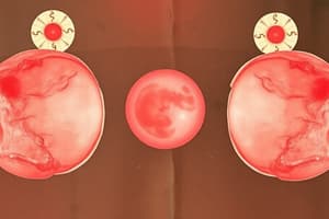
Polycystic Ovarian Syndrome (PCOS)
- AKA Stein-Leventhal Syndrome
- Endocrine disorder (imbalance of LH, FSH and testosterone)
- Chronic anovulation and the clinical signs include
- Infertility
- Obesity
- Oligomenorrhea
- Hirsutism (dark hair on face or chest of woman)
- Have more male characteristics; deeper voice, smaller breasts, more muscle mass
- Younger women in their teens to 20's will often have this
- It's Diagnosis based on clinical signs and blood tests
- The sonographic appearance includes
- They appear two to five times normal size
- Increased number of follicles (peripheral cyst)
- More than 19 follicular cysts
- Always bilateral
- Ovarian volume > 10cm3
Par(a)ovarian Cyst
- 10% of adnexal simple cysts are Paraovarian
- These arise from the broad ligament, NOT the ovary
- Located separate from the ovary, within the broad ligament
- Wolffian duct remnants
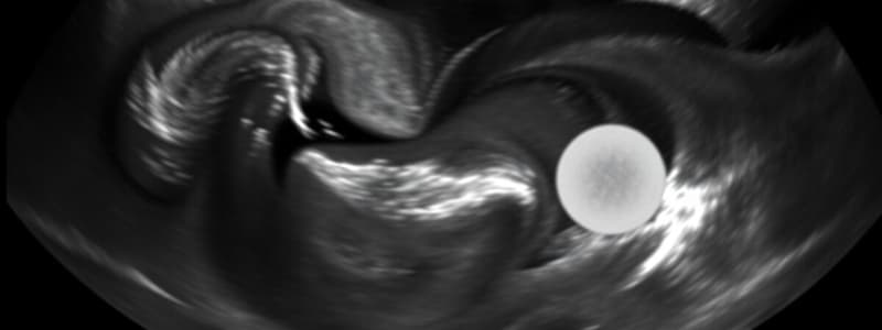
Ovarian Torsion
- This is Caused by partial or complete rotation of the ovary on its axis
- This Restricts blood flow
- Clinical symptoms inlcude acute and severe unilateral pain
- Right side pain can mimic appendicitis
- Sonographic Appearance:
- Enlarged ovary
- Absent or dampened arterial flow on color or pulsed doppler; always compare to other side (should be the same)
- Usually occurs in childhood and adolescence, during pregnancy, or in a patient with a history of a large ovarian cyst
- Accounts for 3% of GYN emergency surgeries
- Ovarian death occurs quickly
Benign Ovarian Tumors
- 80% of all Ovarian masses are Benign
Cystic Teratoma
- Also known as Dermoid Cyst
- Most Common Germ cell Tumor
- Found in Women of childbearing age
- Composed of hair, teeth, bones, & fat
- Complex mass may have:
- Calcifications
- Fat/fluid levels
- Diffusely echogenic with shadows
- "Tip of the iceberg” appearance
- Dermoid plug
- May mimic bowel
- high Risk for torsion
Epithelial Tumors
- Most Common Types
- Mucinous Cystadenoma
- Serous Cystadenoma
- Less Common Type
- Brenner Tumor
Mucinous Cystadenoma
- Most common benign cystic tumor of ovary
- Tumor lined with mucinous elements of the endo cervix
- 25% of benign ovarian neoplasms
- Occurs in patients ages 30 - 50 years
- 85% are benign
- Large 15 to 50 cm (weighing up to 100 lbs)
- Unilateral
- Ultrasound shows numerous, thick-walled septations, multiloculated cysts
- Clinical symptoms: Pressure, pain, increased abdominal girth
Serous Cystadenoma
- Second most common benign tumor of ovary
- Unilateral
- Smaller than mucinous
- Multiloculated cysts with thin septations
- Clinical: Pressure and bloating
- Sonographic Appearance:
- Anechoic
- Large, usually unilocular
- Thin wall septations
Brenner Tumor
- (Epithelial/Transitional Cells)
- Found in Women 40–80 yrs; avg 50's
- Unilateral
- Incidental finding
- It is Rare
- Ultrasound Appearance:
- Small, hypoechoic, solid mass, may have small cystic spaces, may have calcifications
Stromal Tumors
- Sex cord of ovarian stroma
- These include Fibroma, Thecoma and Sertoli-Leydig tumor
- Fibroma
- Unilateral
- Postmenopausal women
- Microscopic to melon size
- Homogeneous, hypoechoic mass with posterior attenuation, shadowing
- Larger tumors are prone to torsion (pressure and pain)
- When tumor is over 5 cm, it can be associated with Meigs' Syndrome (Ascites and Pleural Effusion)
Thecoma
- Unilateral
- Estrogen Producing
- Rare
- Postmenopausal women
- Ultrasound:
- Solid hypoechoic mass with posterior shadow
- Possible thick endometrium due to estrogen secretion
Steroli-Leydig Tumor
- AKA Arrhenoblastoma/Androblastoma
- Very rare
- Unilateral
- Usually in Women <30 years
- Androgen (testosterone) producing
- Masculinization symptoms 50%
- 5-15cm in size
- 20% may become malignant
- Ultrasound Findings
- Solid, echogenic mass
Ovarian Malignancy
- 8th leading cause of cancer death in women worldwide
- 5-year survival rate is 30-50%
- 15,000 women die annually in US
- 207,000 women die globally
- 80% are women > 60
Ovarian Cancer: Lab Tests
- CA 125
- A biological tumor marker that is elevated in the blood of most women with ovarian cancer
- 80% ovarian cancers result in high levels
- Non-specific to ovarian cancer
- Insensitive to mucinous and germ cell tumor
- In any patient with a suspicious ovarian mass, always consider additional testing for ascites and mets in peritoneum, lymph nodes, liver, pleural space
Risk Factors For Ovarian Cancer
- Age (women over 50)
- Nulliparity
- Early menses or late menopause
- Family history of ovarian or breast cancer
- BRCA 1 or 2 gene
Ovarian Cancer Clinical Signs and Symptoms
- Known as silent killer
- Vague complaints (early on)
- Abdominal pain/swelling, Frequent urination
- Increasing abdominal girth & vaginal bleeding
- Poor prognosis due to late diagnosis
- 65% of patients have stage III or IV at time of diagnosis
Treatment
- Early detection has an 80% five-year survival rate
- Late detection has 5% five-year survival rate
- Multifaceted treatment methods include surgery, chemotherapy, radiation
Sonographic Screening
- It's useful to use Endovaginal imaging with CA125 tool
- Check the ovarian
- Size/Volume, Echogenicity, Symmetry
- Arterial flow resistance: most ALL cancer has low resistant flow patterns
- Best to screen Days 3-7 of menstrual cycle due to changes in ovarian arteries' waveforms
Cancer Staging
- Stage I, Limited to ovary
- Stage II, Limited to pelvis (uterus, fallopian tubes)
- Stage III, Limited to abdomen (colon, retroperitoneal)
- Stage IV, Spread beyond abdomen (liver)
Epithelial Cell Tumor
- Arise from surface that covers ovary
- 80 – 90 % of ovarian malignancies
Serous Cystadenocarcinoma
- It accounts for about 80% of ovarian cancers
- Most common malignant tumor
- Found Bilaterally 50% of the time
- Peri- and postmenopausal women
- Sonographic appearance:
- Multilocular
- Multiple papillary projections/septations
- Calcification, Ascites
- Smaller than Mucinous
Mucinous Cystadenocarcinoma
- Rare 5-10% of tumors
- Occurs in women ages 40-70 years
- Can become very large and is likely to rupture
- Can cause Pseudomyxoma peritonei
- Tumor capsule ruptures spreading cells into peritoneal cavity filling it with gelatinous material (cancer cells)
- Appears as complex ascites, fluid with septations
- Sonographic appearance:
- Multiloculated cystic lesions measuring 15-30 cm
- Contain echogenic material
- Similar to Serous
- Ascites with possible septations
Germ Cell Neoplasms
- Derived from germ cells of the embryonic gonads
Immature Teratoma
- Very rare and rapidly growing
- Occurs in the first two decades of life (girls ages 10-20 years)
- Unilateral and creates Elevated AFP (Alpha Fetoprotein)
- Sonographic appearance:
- Looks the Same as mature teratoma (dermoid cyst)
Dysgerminoma
- A malignant germ cell tumor originating from germ cells of ovary
- Occurs in women under 30 and it
- Highly malignant
- Appears Unilateral
- Sonographic appearance:
- Multilobulated hyperechoic solid mass with areas of hemorrhage and necrosis
- Usually unilateral
Endodermal Sinus (Yolk Sac) Tumor
- 2nd most common germ cell malignancy
- Occurs in young adults (20-30 years)
- Unilateral and variable in size
- Highly malignant
- Metastasizes to surrounding organs
- Increased AFP & LDH
- Mostly solid mass with necrosis
Sex Cord Tumor
- Stromal Tumor arise from connective tissue
Granulosa Cell Tumor
- Most common hormone-active estrogenic tumor
- Mimics Graafian Follicle
- Occurs in the range of Ages 50-55, more common after menopause
- Commonly produce estrogen which causes bleeding
- Possible thickened endometrium due to estrogen influence
- Unilateral
- May lead to Meigs' syndrome
- Sonographic appearance:
- Small tumors which are Predominantly solid and Similar to adenoma or fibroids
- Large tumors are Multiloculated/cystic and Similar to cystadenomas
- Can have Thickened endometrium
Metastases to Ovary
- Arise from Breast, GI, Uterus and Lymph
- Krukenberg Tumor indicates the metastasis is from the Primary GI tract and is
- Typically Bilateral
- Sonographic findings:
- Bilateral solid hypoechoic or complex solid masses
- Possible ascites
Key points to remember
- The more sonographically complex the tumor, the more likely it is malignant
- Malignancies rely heavily on BLOOD flow to sustain their spread
- Solid adnexal masses have many differential diagnoses including pedunculated fibroid, dermoid, fibroma, thecoma, granulosa cell tumor, Brenner tumor, or metastasis, get a BIOPSY to verify
- Tubo-ovarian abscess, ovarian torsion, hemorrhagic cysts, and ectopic pregnancy may appear as solid adnexal masses. How can you tell?
- Always use DOPPLER on any SOLID APPEARING MASS!!!! COLOR, POWER, & PULSED DOPPLER
Studying That Suits You
Use AI to generate personalized quizzes and flashcards to suit your learning preferences.