Podcast
Questions and Answers
What is the primary purpose of biomicroscopy, as stated in the provided material?
What is the primary purpose of biomicroscopy, as stated in the provided material?
- To evaluate the drainage system of the eye.
- To measure eye pressure.
- To assess the front health of the eyes. (correct)
- To assess the posterior health of the eyes.
During gonioscopy, expressing that there are no bubbles or lashes trapped underneath the lens is crucial.
During gonioscopy, expressing that there are no bubbles or lashes trapped underneath the lens is crucial.
True (A)
When assessing the superior quadrant during gonioscopy, what specific information should be stated regarding the visible structures?
When assessing the superior quadrant during gonioscopy, what specific information should be stated regarding the visible structures?
The most posterior visible structure is...
When using Goldmann Applanation Tonometry, what range of IOP readings indicates a normal pressure?
When using Goldmann Applanation Tonometry, what range of IOP readings indicates a normal pressure?
During binocular indirect ophthalmoscopy, the practitioner should strive to obtain a clear image by filling the condensing lens as much as possible without the ______ in view.
During binocular indirect ophthalmoscopy, the practitioner should strive to obtain a clear image by filling the condensing lens as much as possible without the ______ in view.
What symptoms are associated with Demodex Blepharitis?
What symptoms are associated with Demodex Blepharitis?
A chalazion is typically painful and tender due to an acute infection.
A chalazion is typically painful and tender due to an acute infection.
According to the document, what is a key initial step in managing preseptal cellulitis?
According to the document, what is a key initial step in managing preseptal cellulitis?
Match the conditions with their respective chief complaints:
Match the conditions with their respective chief complaints:
A chief complaint of a red spot with no pain or vision change might indicate which condition?
A chief complaint of a red spot with no pain or vision change might indicate which condition?
Dry eye disease is only caused by reduced tear production.
Dry eye disease is only caused by reduced tear production.
According to the document, the classic finding during a slit lamp examination for blepharitis involves inflammation and redness at ______ margins.
According to the document, the classic finding during a slit lamp examination for blepharitis involves inflammation and redness at ______ margins.
What is the chief complaint associated with vitreous opacities, such as floaters?
What is the chief complaint associated with vitreous opacities, such as floaters?
Blood tests, typically a rheumatoid factor and ANA, are required when assessing for episcleritis.
Blood tests, typically a rheumatoid factor and ANA, are required when assessing for episcleritis.
In cases of severe or recurrent scleritis, what is a topical medication option?
In cases of severe or recurrent scleritis, what is a topical medication option?
Match the glaucoma types from the provided material with a description of what is occurring based on pathophysiology.
Match the glaucoma types from the provided material with a description of what is occurring based on pathophysiology.
What is the most common symptom among patients with thyroid eye disease (TED)?
What is the most common symptom among patients with thyroid eye disease (TED)?
If bacterial conjunctivitis does not improve with topical antibiotic treatment, a conjunctival scraping to identify the causative organism is not needed.
If bacterial conjunctivitis does not improve with topical antibiotic treatment, a conjunctival scraping to identify the causative organism is not needed.
With a chief complaint of blurred vision, a family history of glaucoma, and a closed anterior chamber angle detected via gonioscopy, a patient most likely has acute ______ glaucoma.
With a chief complaint of blurred vision, a family history of glaucoma, and a closed anterior chamber angle detected via gonioscopy, a patient most likely has acute ______ glaucoma.
According to the overview information, which of the items below should you NOT perform with pre-test information from Clinical station?
According to the overview information, which of the items below should you NOT perform with pre-test information from Clinical station?
Ocular Hypertension has obvious vision changes
Ocular Hypertension has obvious vision changes
Slit lamp examination to differentiate episcleritis from scleritis, what test can be conducted?
Slit lamp examination to differentiate episcleritis from scleritis, what test can be conducted?
What class of medications is okay to be used for anterior uveitis?
What class of medications is okay to be used for anterior uveitis?
During retinoscopy, an examiner must ensure that the lens used obtains a clear image, filling the condensing lens as much as possible without obstructing the ______
During retinoscopy, an examiner must ensure that the lens used obtains a clear image, filling the condensing lens as much as possible without obstructing the ______
According to the document, to diagnose and characterize the following disease, multiple sclerosis, what is the most likely test to be?
According to the document, to diagnose and characterize the following disease, multiple sclerosis, what is the most likely test to be?
It is important to note that after administering both the anesthetic drop and the fluorescein dye, that tear film qualities should not be noted until after tonometry has been performed.
It is important to note that after administering both the anesthetic drop and the fluorescein dye, that tear film qualities should not be noted until after tonometry has been performed.
If a subconjunctival hemorrhage is recurrent or unexplained what optional tool or test can be performed?
If a subconjunctival hemorrhage is recurrent or unexplained what optional tool or test can be performed?
If the history is significant for diabetes as well as having a chief complaint about yellow bumps this is suggestive of what?
If the history is significant for diabetes as well as having a chief complaint about yellow bumps this is suggestive of what?
During the initial work up for a patient, the history of stroke is not needed to be reviewed or added.
During the initial work up for a patient, the history of stroke is not needed to be reviewed or added.
Steroid-induced glaucoma occurs when prolonged steroid use causes increased IOP, leading to optic nerve ______ if untreated.
Steroid-induced glaucoma occurs when prolonged steroid use causes increased IOP, leading to optic nerve ______ if untreated.
What type of medications should be avoided in angle recession or traumatic glaucoma due to structural concerns?
What type of medications should be avoided in angle recession or traumatic glaucoma due to structural concerns?
If there is a pupillary block, a laser peripheral iridotomy (LPI) should never be used
If there is a pupillary block, a laser peripheral iridotomy (LPI) should never be used
If central vision loss, distorted central vision, difficulty reading or seeing fine details, describe the chief complaint as?
If central vision loss, distorted central vision, difficulty reading or seeing fine details, describe the chief complaint as?
Match the following disease's required ancillary test.
Match the following disease's required ancillary test.
A chief complaint has a sudden onset of flashes of light, new floaters, a shadow or curtain over part of the visual field indicates?
A chief complaint has a sudden onset of flashes of light, new floaters, a shadow or curtain over part of the visual field indicates?
Explaind that asteroid hyalosis is an age-related condition where calcium and phosphate crystal deposits accumulate in the vitreous humor, often resutling from an infection .
Explaind that asteroid hyalosis is an age-related condition where calcium and phosphate crystal deposits accumulate in the vitreous humor, often resutling from an infection .
*insert the word that best suits- Chief complaint: eye pain ___________ upon waking?
*insert the word that best suits- Chief complaint: eye pain ___________ upon waking?
All of the following is relevant for a case history/recent history part is not important for...
All of the following is relevant for a case history/recent history part is not important for...
true or false to assess ulcer size and depth the slit lamp exam is not helpful or required.
true or false to assess ulcer size and depth the slit lamp exam is not helpful or required.
There is a foreign body in the eye, what should be ordered?
There is a foreign body in the eye, what should be ordered?
Match the diagnosis name with the correct description
Match the diagnosis name with the correct description
Flashcards
General Rule for Exam
General Rule for Exam
Disinfect ocular instruments before use and maintain hygiene.
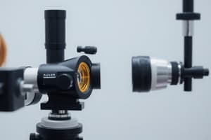
Biomicroscopy Purpose
Biomicroscopy Purpose
Assess front eye health by examining lids, conjunctiva, cornea, etc.
Goldmann Tonometry
Goldmann Tonometry
Measures eye pressure; patient looks at a fixation target
Binocular Indirect Ophthalmoscopy
Binocular Indirect Ophthalmoscopy
Signup and view all the flashcards
Dilated fundus exam
Dilated fundus exam
Signup and view all the flashcards
Blepharitis
Blepharitis
Signup and view all the flashcards
Demodex Blepharitis
Demodex Blepharitis
Signup and view all the flashcards
Ocular Rosacea
Ocular Rosacea
Signup and view all the flashcards
Chalazion
Chalazion
Signup and view all the flashcards
Hordeolum
Hordeolum
Signup and view all the flashcards
Preseptal Cellulitis
Preseptal Cellulitis
Signup and view all the flashcards
Orbital Cellulitis
Orbital Cellulitis
Signup and view all the flashcards
Bacterial Conjunctivitis
Bacterial Conjunctivitis
Signup and view all the flashcards
Viral Conjunctivitis
Viral Conjunctivitis
Signup and view all the flashcards
Allergic Conjunctivitis
Allergic Conjunctivitis
Signup and view all the flashcards
Subconjunctival Hemorrhage
Subconjunctival Hemorrhage
Signup and view all the flashcards
Pinguecula/Pterygium
Pinguecula/Pterygium
Signup and view all the flashcards
Dry Eye Disease (DED)
Dry Eye Disease (DED)
Signup and view all the flashcards
Episcleritis
Episcleritis
Signup and view all the flashcards
Scleritis
Scleritis
Signup and view all the flashcards
Recurrent Corneal Erosions (RCE)
Recurrent Corneal Erosions (RCE)
Signup and view all the flashcards
Herpes Simplex Keratitis
Herpes Simplex Keratitis
Signup and view all the flashcards
Herpes Zoster Ophthalmicus (HZO)
Herpes Zoster Ophthalmicus (HZO)
Signup and view all the flashcards
Corneal Ulcer (Bacterial Keratitis)
Corneal Ulcer (Bacterial Keratitis)
Signup and view all the flashcards
Foreign Body
Foreign Body
Signup and view all the flashcards
Fuch's Endothelial Dystrophy
Fuch's Endothelial Dystrophy
Signup and view all the flashcards
Anterior Uveitis
Anterior Uveitis
Signup and view all the flashcards
cateracts
cateracts
Signup and view all the flashcards
Xanthelasma
Xanthelasma
Signup and view all the flashcards
Non-Proliferative Diabetic Retinopathy (NPDR)
Non-Proliferative Diabetic Retinopathy (NPDR)
Signup and view all the flashcards
Hypertensive Retinopathy
Hypertensive Retinopathy
Signup and view all the flashcards
Age-Related Macular Degeneration (AMD)
Age-Related Macular Degeneration (AMD)
Signup and view all the flashcards
Central Serous Chorioretinopathy (CSCR)
Central Serous Chorioretinopathy (CSCR)
Signup and view all the flashcards
Epiretinal Membrane
Epiretinal Membrane
Signup and view all the flashcards
Macular Hole
Macular Hole
Signup and view all the flashcards
Retinal Vein Occlusion (RVO)
Retinal Vein Occlusion (RVO)
Signup and view all the flashcards
Retinal Arterial Occlusion (RAO)
Retinal Arterial Occlusion (RAO)
Signup and view all the flashcards
Ocular Ischemic Syndrome
Ocular Ischemic Syndrome
Signup and view all the flashcards
Posterior Vireous Detachment (PVD)
Posterior Vireous Detachment (PVD)
Signup and view all the flashcards
Retinal Detachment
Retinal Detachment
Signup and view all the flashcards
Study Notes
Exam Logistics and Preparation
- Exam cycle duration at each station is 15 minutes, observational cycle is 3 min
- At the anterior segment and posterior segments skills stations, disinfect the slit lamp, focus it, and organize necessary equipment before patient interactions
- During patient encounters, connect the device to the docking station, take notes on the note board, and use hand sanitizer.
Anterior Segment Evaluation
- Proper disinfection of ocular instruments and hygiene must be maintained
- Best practice efficiency is to start temporally during slit lamp use
Biomicroscopy Procedure
- Assess the front health of the eyes using this process
- Instruct patients where to look throughout the procedure
Clinical Steps
- Use 10x magnification, low illumination, diffuse beam, 0 degrees to examine the eyelids and lashes
- Confirm upper and lower lids and lashes are clean
- Palpebral conjunctiva should be healthy and pink with lower lid eversion
- Check that inferior bulbar conjunctiva and sclera are white and quiet
- Verify that lower lid margin is clear and lower puncta are open with good apposition to the globe
- Verify that nasal and temporal bulbar conjunctiva and sclera are white and quiet
- Check that upper lid margin is clear and upper puncta are open with good apposition to the globe
- Confirm that superior palpebral conjunctiva is pink and healthy with upper lid eversion
- Use 16x mag, medium-high illumination with 3-4mm parallelpiped full height with variable angle technique
- Assessing superior and central cornea using parallelpiped and optic sections confirms that the epithelium, stroma, and endothelium are clear
- Use 16x mag, high illumination, 0.5mm optic section full height, 60-degree locked parameter to assess temporal anterior chamber angle
- Employ 16x mag, high illumination, conical beam (1mm x 1.5mm rectangle, 45 degrees to examine angle with room lights off
- Typically allow 15 seconds for dark adaptation to prevent cells or flares in the anterior chamber
- Utilize the parameters 16x mag, medium illumination, 3-4mm parallelpiped full height, 0 degrees to find iris conditions flat and avascular with a good pupillary response
- Employ 10x mag, medium illumination, 3-4mm parallelpiped, height equal to pupil size, 0 degrees to confirm that iris has no transillumination defects
Gonioscopy Information
- Assess the drainage system using procedure
- Instruct the patient of where to look throughout the procedure
Clinical Steps
- Use 16x mag, wide parallelpiped outside of the pupil, perpendicular to the flat mirror surface
- Inquire about allergies or pregnancy before instilling an anesthetic drop
- Add cushioning solution to the gonio lens
- Check for bubbles beneath the gonio lens or eyelashes trapped
- Identify inferior, superior, and nasal post visible structures as well as iris approach is
Goldmann Applanation Tonometry Information
- This procedure checks a patient's eye pressure
- Instruct the patient of where to look throughout the procedure
Clinical Steps
- With 10x mag, high illumination, diffuse beam, cobalt blue filter, and light housing on the temporal side, ask the patient
- Confirm allergies and pregnancy before instilling an anesthetic-fluorescein drop
- Make sure the cornea and tear film are clear before tonometry, use fixation target during the procedure
- Record results (mmHg) and time of reading.
- With normal readings (10-21 mmHg) recommend the patient have their eye pressure checked annually
- With high reading (>21 mmHg) tell the patient that pressure reading is higher than expected and recommend them return to clinic to have their eye pressure rechecked
Posterior Segment Evaluation
- Proper disinfection of ocular instruments and hygiene must be maintained
Binocular Indirect Ophthalmoscopy Info
- Procedure allows assessment of posterior health
- Instruct the patient of where to look throughout the procedure
Clinical Steps
- Get a clear image filling the condensing lens without the arcades in view and ask
- Ask preceptor if they have a view
- Posterior health with peripheral intact is a normal finding
Information About Dilated Fundus Exam
- Tool to assess lens, vitreous substance, and retina health after dilation
- Instruct the patient of where to look throughout the procedure when performing
Clinical Steps
- Anterior and posterior lens should be clear.
- Lens should have no defects w/ retroillumination (set to max)
- Anterior and posterior vitreous should be clear
- Optic nerve should have distinct margins and healthy rim tissue
- Normal C/D ratio is horizontal and vertical
- All arcades A/V ratio should be 2/3 without crossings
- Fovea should be flat and avascular with a +/- foveal reflex
Patient Encounter Station Details
- Essential information including patient demographics, chief complaints, current medications list, and symptom reviews
- Assessment data such as pretest info, refractive data, and anterior/posterior segment information
- Ancilary tests are not alterable in the system
- Evaluations to use to make written/verbalized assessments and plans
Studying That Suits You
Use AI to generate personalized quizzes and flashcards to suit your learning preferences.