Podcast
Questions and Answers
What structure persists as the urachus in adults?
What structure persists as the urachus in adults?
- Notochord
- Median umbilical ligament (correct)
- Neural tube
- Cranial nerve ganglia
Which developmental process involves the formation of the neural plate and the neural tube?
Which developmental process involves the formation of the neural plate and the neural tube?
- Organogenesis
- Somitogenesis
- Neurulation (correct)
- Gastrulation
What is the role of the notochord in the development of the neural plate?
What is the role of the notochord in the development of the neural plate?
- It induces the thickening of the overlying ectoderm. (correct)
- It generates mesodermal tissues.
- It serves as a blood vessel conduit.
- It forms the neural tube directly.
What is formed when the neural folds fuse?
What is formed when the neural folds fuse?
Neuroectodermal cells that lose their epithelial characteristics and detach from the neural folds form what structure?
Neuroectodermal cells that lose their epithelial characteristics and detach from the neural folds form what structure?
When does the process of neurulation complete?
When does the process of neurulation complete?
Which structure serves as the primary source for the sensory ganglia of spinal and cranial nerves?
Which structure serves as the primary source for the sensory ganglia of spinal and cranial nerves?
What major developmental change occurs to the size of the neural plate during development?
What major developmental change occurs to the size of the neural plate during development?
What is the primary signaling function of the notochord during embryonic development?
What is the primary signaling function of the notochord during embryonic development?
During which developmental stage does the notochord detach from the endoderm of the umbilical vesicle?
During which developmental stage does the notochord detach from the endoderm of the umbilical vesicle?
What structure does the notochord contribute to as it degenerates?
What structure does the notochord contribute to as it degenerates?
What role does the allantois play during embryonic development?
What role does the allantois play during embryonic development?
What is the significance of the notochord in axial development?
What is the significance of the notochord in axial development?
How does the notochord influence the formation of the neural plate?
How does the notochord influence the formation of the neural plate?
What occurs during the invagination of cells from the primitive pit in relation to the notochord?
What occurs during the invagination of cells from the primitive pit in relation to the notochord?
What connects the notochordal canal to the umbilical vesicle during initial development?
What connects the notochordal canal to the umbilical vesicle during initial development?
What is the primary function of somites during embryogenesis?
What is the primary function of somites during embryogenesis?
How do somites form in relation to the growth of the embryonic structure?
How do somites form in relation to the growth of the embryonic structure?
What are the two layers formed from the lateral mesoderm due to the intraembryonic coelom?
What are the two layers formed from the lateral mesoderm due to the intraembryonic coelom?
What is the embryonic structure that serves as the precursor to the axial skeleton?
What is the embryonic structure that serves as the precursor to the axial skeleton?
In what stage does the intraembryonic coelom start to take shape within the embryo?
In what stage does the intraembryonic coelom start to take shape within the embryo?
Which major body cavities are formed from the division of the intraembryonic coelom?
Which major body cavities are formed from the division of the intraembryonic coelom?
What is the initial source of nutrition for the embryo during early development?
What is the initial source of nutrition for the embryo during early development?
Which structure forms as a segmental block of mesodermal tissue during the development phase?
Which structure forms as a segmental block of mesodermal tissue during the development phase?
What is the primary role of the primitive streak in embryonic development?
What is the primary role of the primitive streak in embryonic development?
The notochordal process is formed by the migration of which type of cells?
The notochordal process is formed by the migration of which type of cells?
What forms at the cranial end of the primitive streak?
What forms at the cranial end of the primitive streak?
Which layer of the embryonic disc does the primitive streak appear on?
Which layer of the embryonic disc does the primitive streak appear on?
By the end of which week does the primitive streak typically disappear?
By the end of which week does the primitive streak typically disappear?
What embryonic structure does the notochord contribute to as it degenerates?
What embryonic structure does the notochord contribute to as it degenerates?
What does mesenchyme give rise to in the embryo?
What does mesenchyme give rise to in the embryo?
The prechordal plate is significant for signaling the development of which structures?
The prechordal plate is significant for signaling the development of which structures?
What process occurs at the beginning of the third week of embryonic development?
What process occurs at the beginning of the third week of embryonic development?
Which germ layer is primarily responsible for forming the epidermis and nervous system?
Which germ layer is primarily responsible for forming the epidermis and nervous system?
What is one of the first indications of pregnancy related to the development of the embryo?
What is one of the first indications of pregnancy related to the development of the embryo?
During the process of gastrulation, the bilaminar embryonic disk is converted into what structure?
During the process of gastrulation, the bilaminar embryonic disk is converted into what structure?
What significant feature develops during the third week of embryonic development?
What significant feature develops during the third week of embryonic development?
Which of these structures is derived from the embryonic endoderm?
Which of these structures is derived from the embryonic endoderm?
What characterizes the rapid development of the embryo during the third week?
What characterizes the rapid development of the embryo during the third week?
What is the primary outcome of the differentiation of the three germ layers in the embryo?
What is the primary outcome of the differentiation of the three germ layers in the embryo?
What sequence describes the development of somites during embryogenesis?
What sequence describes the development of somites during embryogenesis?
Which of the following is NOT a function of somites?
Which of the following is NOT a function of somites?
What is the composition of the somatic mesoderm?
What is the composition of the somatic mesoderm?
What major body cavity is formed from the division of the intraembryonic coelom?
What major body cavity is formed from the division of the intraembryonic coelom?
How many pairs of somites are typically present by the end of the fifth week of embryonic development?
How many pairs of somites are typically present by the end of the fifth week of embryonic development?
What does the term 'splanchnopleure' refer to in embryonic development?
What does the term 'splanchnopleure' refer to in embryonic development?
What initiates the formation of blood vessels during early embryonic development?
What initiates the formation of blood vessels during early embryonic development?
What structure forms a continuous layer with the ectoderm in the developing embryo?
What structure forms a continuous layer with the ectoderm in the developing embryo?
What is the adult representation of the urachus?
What is the adult representation of the urachus?
During which week does the closure of the neural tube complete?
During which week does the closure of the neural tube complete?
Which structure is essential for the thickening of the embryonic ectoderm to form the neural plate?
Which structure is essential for the thickening of the embryonic ectoderm to form the neural plate?
What is the process called that involves the formation of new blood vessels from existing ones?
What is the process called that involves the formation of new blood vessels from existing ones?
What happens to the neural folds at the end of the third week?
What happens to the neural folds at the end of the third week?
When does the embryonic blood circulation begin and the heart starts beating?
When does the embryonic blood circulation begin and the heart starts beating?
What cellular transition do neural crest cells undergo as the neural folds fuse?
What cellular transition do neural crest cells undergo as the neural folds fuse?
What primarily drives the development of embryonic blood vessels?
What primarily drives the development of embryonic blood vessels?
What does the neural crest give rise to?
What does the neural crest give rise to?
What type of cells aggregate to form blood islands during the vascular development?
What type of cells aggregate to form blood islands during the vascular development?
Which of the following structures develops first in the formation of the cardiovascular system?
Which of the following structures develops first in the formation of the cardiovascular system?
What is the initiator of the formation of the neural groove?
What is the initiator of the formation of the neural groove?
Where does the separation of the neural crest occur during development?
Where does the separation of the neural crest occur during development?
What type of villi has visible blood vessels and connects with the embryonic heart?
What type of villi has visible blood vessels and connects with the embryonic heart?
During which week does hematopoiesis, the formation of blood, begin?
During which week does hematopoiesis, the formation of blood, begin?
What type of cells differentiate into the muscular and connective tissue components of blood vessels?
What type of cells differentiate into the muscular and connective tissue components of blood vessels?
Flashcards are hidden until you start studying
Study Notes
Notochord Development
- The notochord is a rod-like structure that forms from notochordal precursor cells induced by signals from the primitive streak.
- The notochordal process elongates by invagination of cells from the primitive pit.
- An indentation forms and extends into the notochordal process, forming the notochordal canal.
- The floor of the notochordal process fuses with the underlying embryonic endoderm, leading to the degeneration of the fused layers.
- Openings form connecting the notochordal canal with the umbilical vesicle.
- As the openings in the floor of the notochordal process become confluent, the floor disappears, forming the notochordal plate.
- The notochordal plate cells proliferate and undergo infolding, creating the definitive notochord.
- The proximal part of the notochordal canal temporarily persists as the neurenteric canal, forming a communication between the amniotic cavity and the umbilical vesicle.
- The neurenteric canal eventually obliterates as development progresses.
- The notochord detaches from the endoderm of the umbilical vesicle, becoming a continuous layer.
- The notochord extends from the oropharyngeal membrane to the primitive node.
- As it degenerates, the notochord contributes to the formation of the nucleus pulposus of each intervertebral disc.
Functions of the Notochord
- The notochord defines the primordial longitudinal axis of the embryo, providing rigidity and serving as the initial scaffold for body structure.
- The notochord acts as a primary signaling center, inducing the overlying embryonic ectoderm to thicken and form the neural plate, the primordium of the CNS.
- The notochord sends signals necessary for the development of axial musculoskeletal structures and the CNS.
Allantois
- The allantois appears as a small outpouching from the caudal wall of the umbilical vesicle around day 16 of embryonic development.
- The allantois extends into the connecting stalk.
- The allantoic mesoderm expands beneath the chorion and forms blood vessels that eventually supply the placenta.
- The proximal part of the allantoic diverticulum persists as a stalk called the urachus, which extends from the bladder to the umbilical region.
- In adults, the urachus is represented by the median umbilical ligament.
- The blood vessels of the allantoic stalk develop into the umbilical arteries.
- The intraembryonic part of the umbilical veins has a separate origin from the allantois.
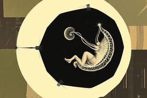
Neurulation
- Neurulation is the process of forming the neural plate, neural folds, and closure of the folds to form the neural tube.
- Neurulation is completed by the end of the fourth week when the caudal neuropore closes.
- The notochord induces the overlying embryonic ectoderm to thicken, forming the neural plate.
- The neural plate consists of thickened epithelial cells called neuroectoderm, which gives rise to the CNS.
- The neural plate initially matches the length of the notochord but eventually extends cranially to the oropharyngeal membrane.
- Around the 18th day, the neural plate begins to invaginate along its central axis, creating the neural groove flanked by neural folds.
- The neural folds, prominent at the cranial end, are the first indicators of brain development.
- By the end of the third week, the neural folds begin to fuse, transforming the neural plate into the neural tube, the primordium of the brain vesicles and spinal cord.
- The neural tube detaches from the surface ectoderm as the neural folds meet.
- As the neural folds fuse, neural crest cells undergo an epithelial-to-mesenchymal transition and migrate away.
- The surface ectoderm fuses over the neural tube, later differentiating into the epidermis.
- Neurulation is completed during the fourth week.
Neural Crest
- As the neural folds fuse to form the neural tube, some neuroectodermal cells along the inner margin of each fold detach from neighboring cells.
- These cells form a flattened, irregular mass called the neural crest, located between the neural tube and the overlying surface ectoderm.
- The neural crest soon separates into right and left parts, shifting to the dorsolateral aspects of the neural tube.
- Neural crest cells give rise to the sensory ganglia of spinal and cranial nerves.
Development of Somites
- Somites are segmented blocks of mesodermal tissue that form along the sides of the neural tube during embryogenesis.
- Somites develop from the paraxial mesoderm, derived from the primitive node.
- The paraxial mesoderm is continuous laterally with the intermediate mesoderm and thins out into the lateral mesoderm.
- Toward the end of the third week, the paraxial mesoderm begins to differentiate, condense, and divide into paired cuboidal bodies called somites.
- Somites form in a craniocaudal sequence, developing from the head region downward along the body.
- During the somite period (days 20 to 30), about 38 pairs of somites form.
- By the end of the fifth week, there are 42 to 44 pairs of somites.
- The number and appearance of somites are used as criteria to determine the age of the embryo.
Functions of Somites
- Somites contribute to the development of:
- Most of the axial skeleton (bones of the head and trunk)
- Associated musculature
- The adjacent dermis of the skin
- Motor axons from the spinal cord innervate the muscle cells in the somites, guided by specific pathways.
Development of Intraembryonic Coelom
- The intraembryonic coelom begins as isolated coelomic spaces in the lateral intraembryonic mesoderm and cardiogenic mesoderm.
- These spaces merge to form a single horseshoe-shaped cavity, the intraembryonic coelom.
- The intraembryonic coelom divides the lateral mesoderm into two layers:
- Somatic (Parietal) Layer: Located beneath the ectoderm, continuous with the extraembryonic mesoderm covering the amnion.
- Splanchnic (Visceral) Layer: Located adjacent to the endoderm, continuous with the extraembryonic mesoderm covering the umbilical vesicle.
- The somatic mesoderm and the overlying ectoderm form the embryonic body wall (somatopleure).
- The splanchnic mesoderm and the underlying endoderm form the embryonic gut (splanchnopleure).
- During the second month, the intraembryonic coelom divides into three major body cavities:
- Pericardial Cavity
- Pleural Cavities
- Peritoneal Cavity
Early Development of Cardiovascular System
- Initially, the embryo obtains nutrition from maternal blood through diffusion via the extraembryonic coelom and umbilical vesicle.
- Blood vessels begin to form in the extraembryonic mesoderm of the umbilical vesicle, connecting stalk, and chorion.
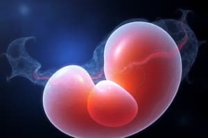
Third Week of Embryo Development
- The beginning of the third week marks the period of gastrulation.
- This period is characterized by the development of the primitive streak, notochord, and three germ layers.
- The third week corresponds to 5 weeks after the first day of the last normal menstrual period.
- Cessation of menstruation is often the first indication of pregnancy.
Gastrulation
- Process by which the three germ layers are established in embryos.
- Bilaminar embryonic disc transitions into a trilaminar embryonic disc.
- Involves significant cell shape changes, rearrangement, movement, and alterations in adhesive properties.
- Beginning of morphogenesis (development of body form).
- Most significant event during the third week.
- The embryo is referred to as a gastrula.
Germ Layer Derivatives
- Ectoderm: Epidermis, nervous system (central and peripheral), eyes, internal ears, neural crest cells, head connective tissues.
- Endoderm: Epithelial linings of respiratory and digestive tracts, glands, liver, pancreas.
- Mesoderm: Skeletal muscles, blood cells, blood vessel linings, smooth muscle, serosal linings of body cavities, reproductive and excretory systems, cardiovascular system.
Primitive Streak
- First morphological sign of gastrulation.
- Appears on the epiblast surface of the bilaminar embryonic disc.
- Thickened linear band of epiblast.
- Crucial for establishing the embryo's craniocaudal axis (cranial and caudal ends, dorsal and ventral surfaces, right and left sides).
- Elongates as cells are added to its caudal end.
- Forms the primitive node at its cranial end.
- Develops a groove (primitive groove) and a small depression (primitive pit).
- As cells move between the epiblast and hypoblast layers, they form mesenchyme.
- Produces extraembryonic mesoderm covering the yolk sac and amnion.
- Diminishes in size and disappears by the end of the fourth week.
Notochord Formation
- Critical structure for signaling the formation of central nervous system and axial skeleton.
- Mesenchymal cells migrate through the primitive streak and acquire mesodermal cell fates.
- Cells migrate cranially from the primitive node and pit, forming the notochordal process.
- Notochordal process develops a lumen, becoming the notochordal canal.
- Notochordal process continues to grow cranially between the ectoderm and endoderm, reaching the prechordal plate.
- Prechordal plate is crucial for signaling cranial structure development.
- Mesenchymal cells from the primitive streak and notochordal process migrate laterally and cranially, contributing to the formation of the cardiogenic mesoderm.
Neurulation
- Process of forming the neural plate, neural folds, and neural tube.
- Completed by the end of the fourth week.
- Notochord induces the overlying embryonic ectoderm to thicken, forming the neural plate.
- Neural plate consists of thickened epithelial cells (neuroectoderm), which gives rise to the CNS.
- Neural plate begins to invaginate, creating the neural groove and neural folds.
- Neural folds begin to fuse, transforming the neural plate into the neural tube.
- Neural tube detaches from the surface ectoderm.
- Neural crest cells detach from the folds, forming a flattened, irregular mass.
- Neural crest cells migrate and differentiate into sensory ganglia of spinal and cranial nerves.
Development of Somites
- Segmented blocks of mesodermal tissue.
- Form along the sides of the neural tube.
- Develop from the paraxial mesoderm.
- Form in a craniocaudal sequence.
- About 38 pairs of somites form during the somite period (days 20 to 30).
- Number and appearance of somites are used to determine the age of the embryo.
Functions of Somites
- Most of the axial skeleton (bones of the head and trunk).
- Associated musculature.
- Adjacent dermis of the skin.
- Motor axons from the spinal cord innervate the muscle cells in the somites.
Development of Intraembryonic Coelom
- Initially isolated coelomic spaces in the lateral intraembryonic mesoderm and cardiogenic mesoderm.
- Spaces merge to form a single horseshoe-shaped cavity.
- Divides the lateral mesoderm into two layers:
- Somatic (Parietal) Layer: beneath the ectoderm.
- Splanchnic (Visceral) Layer: adjacent to the endoderm.
- Somatic mesoderm and overlying ectoderm form the embryonic body wall.
- Splanchnic mesoderm and underlying endoderm form the embryonic gut.
- Divides into three major body cavities during the second month:
- Pericardial Cavity
- Pleural Cavities
- Peritoneal Cavity
Early Development of Cardiovascular System
- Nutrition initially obtained by diffusion through the extraembryonic coelom and umbilical vesicle.
- Blood vessels form in the extraembryonic mesoderm.
- Embryonic blood vessels develop to deliver oxygen and nutrients.
Development of the Vascular System
- Vasculogenesis: Formation of new vascular channels by the assembly of angioblasts.
- Angiogenesis: Formation of new blood vessels by budding and branching from existing vessels.
- Blood Islands: Clusters of angioblasts that differentiate into endothelial cells and form vascular channels.
- Mesenchymal cells differentiate into muscular and connective tissue components of blood vessels.
- Blood cells first develop from specialized endothelial cells.
- Hematopoiesis begins in the fifth week.
- Heart and great vessels develop from mesenchymal cells in the cardiogenic area.
- Paired endocardial heart tubes form and fuse into the primordial heart tube.
- Heart tube connects with blood vessels, forming a primitive cardiovascular system.
- Blood circulation begins by the end of the third week.
- Heart starts beating on the 21st or 22nd day.
- Cardiovascular system is the first functional organ system.
- Heartbeat detectable by Doppler ultrasonography during the fourth week.
Development of Chorionic Villi
- Primary chorionic villi appear at the end of the second week and branch.
- Mesenchyme grows into these villi, forming secondary chorionic villi.
- Mesenchymal cells differentiate into capillaries and blood cells, forming tertiary chorionic villi with visible blood vessels.
- Capillaries in the villi fuse to form arteriocapillary networks, connecting with the embryonic heart.
Studying That Suits You
Use AI to generate personalized quizzes and flashcards to suit your learning preferences.