Podcast
Questions and Answers
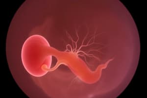
What is the first step in the process of neurulation?
What is the first step in the process of neurulation?
- Formation of the neural groove
- Formation of the neural plate (correct)
- Formation of the neural crest
- Migration of neural crest cells
What structure is formed by the neural tube during neurulation?
What structure is formed by the neural tube during neurulation?
- Heart
- Spinal cord (correct)
- Dorsal root ganglia
- Facial bones
Which of the following best describes the process of epithelial-mesenchymal transition (EMT) in neural crest cells?
Which of the following best describes the process of epithelial-mesenchymal transition (EMT) in neural crest cells?
- It is the transition from a mesenchymal state to an epithelial state.
- It allows neural crest cells to become tightly connected to one another.
- It enables neural crest cells to transition to a migratory state, moving freely. (correct)
- It prevents neural crest cells from migrating.
Which of the following structures is NOT formed by migrating neural crest cells? (Select one)
Which of the following structures is NOT formed by migrating neural crest cells? (Select one)
Which of the following is a function of trunk neural crest cells?
Which of the following is a function of trunk neural crest cells?
Animal neural crest cells contribute to the development of which of the following? (Select all that apply)
Animal neural crest cells contribute to the development of which of the following? (Select all that apply)
Which structure is responsible for hormone secretion and is derived from neural crest cells?
Which structure is responsible for hormone secretion and is derived from neural crest cells?
During neurulation, what forms at the edges of the neural folds?
During neurulation, what forms at the edges of the neural folds?
What is the function of the dorsal root ganglia formed by migrating neural crest cells?
What is the function of the dorsal root ganglia formed by migrating neural crest cells?
Which structure is NOT directly formed by trunk neural crest cells?
Which structure is NOT directly formed by trunk neural crest cells?
What is the primary role of the enteric ganglia formed by neural crest cells?
What is the primary role of the enteric ganglia formed by neural crest cells?
Which process allows neural crest cells to migrate from the neural tube to other areas of the body?
Which process allows neural crest cells to migrate from the neural tube to other areas of the body?
Neural crest cells are important for the development of which of the following structures?
Neural crest cells are important for the development of which of the following structures?
What is the initial location where neural crest cells form during neurulation?
What is the initial location where neural crest cells form during neurulation?
Which transition allows neural crest cells to migrate away from the neural tube?
Which transition allows neural crest cells to migrate away from the neural tube?
Which of the following pathways is followed by neural crest cells that migrate to form melanocytes?
Which of the following pathways is followed by neural crest cells that migrate to form melanocytes?
Which of the following structures is formed by the ventral migration of trunk neural crest cells?
Which of the following structures is formed by the ventral migration of trunk neural crest cells?
Which of the following structures is involved in regulating autonomic functions, particularly in the abdomen, and is derived from ventral trunk neural crest cells?
Which of the following structures is involved in regulating autonomic functions, particularly in the abdomen, and is derived from ventral trunk neural crest cells?
The adrenal medulla, which plays a role in the body's response to stress, is formed by which type of neural crest cell migration?
The adrenal medulla, which plays a role in the body's response to stress, is formed by which type of neural crest cell migration?
Which of the following structures does NOT form from ventral trunk neural crest cell migration?
Which of the following structures does NOT form from ventral trunk neural crest cell migration?
Ventral trunk neural crest cells contribute to the formation of which part of the nervous system?
Ventral trunk neural crest cells contribute to the formation of which part of the nervous system?
Neural crest cells are derived from which layer of the embryo?
Neural crest cells are derived from which layer of the embryo?
Which of the following structures is derived from the cranial neural crest cells and contributes to the development of teeth?
Which of the following structures is derived from the cranial neural crest cells and contributes to the development of teeth?
Which of the following is formed by the dorsal migration of trunk neural crest cells?
Which of the following is formed by the dorsal migration of trunk neural crest cells?
Which structure is formed from the ventral migration of trunk neural crest cells?
Which structure is formed from the ventral migration of trunk neural crest cells?
Trunk neural crest cells contribute to the formation of all of the following EXCEPT:
Trunk neural crest cells contribute to the formation of all of the following EXCEPT:
Cranial neural crest cells contribute to the development of which of the following?
Cranial neural crest cells contribute to the development of which of the following?
Cranial neural crest cells contribute to the formation of which of the following structures in the heart?
Cranial neural crest cells contribute to the formation of which of the following structures in the heart?
Which of the following structures are derived from the cranial neural crest cells in the pharyngeal arches?
Which of the following structures are derived from the cranial neural crest cells in the pharyngeal arches?
The enteric nervous system is formed from which type of neural crest cells?
The enteric nervous system is formed from which type of neural crest cells?
Odontoblasts, which form teeth, are derived from?
Odontoblasts, which form teeth, are derived from?
Which of the following is NOT a derivative of cranial neural crest cells?
Which of the following is NOT a derivative of cranial neural crest cells?
Cranial neural crest cells contribute to the formation of which of the following in the face and neck?
Cranial neural crest cells contribute to the formation of which of the following in the face and neck?
Which of the following cells, derived from cranial neural crest, play a crucial role in the development of teeth?
Which of the following cells, derived from cranial neural crest, play a crucial role in the development of teeth?
Cranial neural crest cells contribute to the formation of which structure related to the nervous system?
Cranial neural crest cells contribute to the formation of which structure related to the nervous system?
Which of the following cranial neural crest derivatives plays a key role in the formation of facial structures?
Which of the following cranial neural crest derivatives plays a key role in the formation of facial structures?
Cranial neural crest cells contribute to the formation of which of the following structures in the nervous system?
Cranial neural crest cells contribute to the formation of which of the following structures in the nervous system?
Which of the following structures is formed by cranial neural crest cells and provides support and protection to the central nervous system?
Which of the following structures is formed by cranial neural crest cells and provides support and protection to the central nervous system?
The mesenchyme of the pharyngeal arches derived from cranial neural crest cells contributes to the development of which of the following?
The mesenchyme of the pharyngeal arches derived from cranial neural crest cells contributes to the development of which of the following?
Neural crest cells are particularly vulnerable to damage from substances like alcohol or retinoic acid because they:
Neural crest cells are particularly vulnerable to damage from substances like alcohol or retinoic acid because they:
Errors in the migration of neural crest cells can result in:
Errors in the migration of neural crest cells can result in:
Which of the following congenital defects is commonly associated with neural crest cell migration errors?
Which of the following congenital defects is commonly associated with neural crest cell migration errors?
Hirschsprung's disease is primarily caused by:
Hirschsprung's disease is primarily caused by:
Which part of the colon is most commonly affected in Hirschsprung's disease?
Which part of the colon is most commonly affected in Hirschsprung's disease?
Which of the following is a symptom of Hirschsprung's disease?
Which of the following is a symptom of Hirschsprung's disease?
The connective tissue development of the head is derived from which of the following sources?
The connective tissue development of the head is derived from which of the following sources?
In which week does eye development begin with the formation of optic grooves from the neural tube?
In which week does eye development begin with the formation of optic grooves from the neural tube?
The optic grooves, which give rise to the optic vesicles, appear in which part of the neural tube?
The optic grooves, which give rise to the optic vesicles, appear in which part of the neural tube?
What forms when the optic grooves extend and evaginate from the neural tube?
What forms when the optic grooves extend and evaginate from the neural tube?
During the formation of the lens vesicle, the lens placodes: (Select all that apply)
During the formation of the lens vesicle, the lens placodes: (Select all that apply)
Which structure remains connected to the neural tube via the optic stalk during early eye development?
Which structure remains connected to the neural tube via the optic stalk during early eye development?
The anterior cells of the lens vesicle become cuboidal and form which part of the lens?
The anterior cells of the lens vesicle become cuboidal and form which part of the lens?
The anterior cells of the lens vesicle become cuboidal and form which part of the lens?
The anterior cells of the lens vesicle become cuboidal and form which part of the lens?
What do the inner and outer layers of the optic cup form?
What do the inner and outer layers of the optic cup form?
The hyaloid artery passes through which structure to reach the developing eye?
The hyaloid artery passes through which structure to reach the developing eye?
Which of the following is true about optic nerve development?
Which of the following is true about optic nerve development?
Raised intracranial pressure may lead to the appearance of which condition at the point where the optic nerve leaves the retina?
Raised intracranial pressure may lead to the appearance of which condition at the point where the optic nerve leaves the retina?
The pigmented layer of the retina is derived from which layer of the optic cup?
The pigmented layer of the retina is derived from which layer of the optic cup?
The optic nerve fibers grow into the inner layer of the optic stalk and form the optic nerve. Which of the following structures supplies the blood to the developing retina?
The optic nerve fibers grow into the inner layer of the optic stalk and form the optic nerve. Which of the following structures supplies the blood to the developing retina?
Which of the following occurs around the 7th week of eye development?
Which of the following occurs around the 7th week of eye development?
The optic stalk is responsible for connecting the optic vesicle to the neural tube. What does the optic stalk eventually develop into?
The optic stalk is responsible for connecting the optic vesicle to the neural tube. What does the optic stalk eventually develop into?
The optic nerve is myelinated by which type of glial cells?
The optic nerve is myelinated by which type of glial cells?
Which structure contributes to the formation of the retina and the optic nerve?
Which structure contributes to the formation of the retina and the optic nerve?
Which condition may result from abnormalities during the closure of the choroid fissure?
Which condition may result from abnormalities during the closure of the choroid fissure?
The sclera and choroid are derived from which embryonic layer?
The sclera and choroid are derived from which embryonic layer?
Which structure is formed by mesodermal condensation around the optic cup?
Which structure is formed by mesodermal condensation around the optic cup?
What is the role of the primary vitreous body in early eye development?
What is the role of the primary vitreous body in early eye development?
Which artery runs through the primary vitreous body during early eye development?
Which artery runs through the primary vitreous body during early eye development?
The mesoderm forms which of the following structures in the developing eye?
The mesoderm forms which of the following structures in the developing eye?
What forms between the lens and the surface ectoderm during eye development?
What forms between the lens and the surface ectoderm during eye development?
The anterior chamber forms from which structure during eye development?
The anterior chamber forms from which structure during eye development?
Which structure forms the inner layer of the cornea during eye development?
Which structure forms the inner layer of the cornea during eye development?
What forms the outer layer of the iris during eye development?
What forms the outer layer of the iris during eye development?
The pupil forms from which structure during eye development?
The pupil forms from which structure during eye development?
Which structure forms the inner layer of the cornea during eye development?
Which structure forms the inner layer of the cornea during eye development?
What forms the outer layer of the iris during eye development?
What forms the outer layer of the iris during eye development?
Which embryonic layer contributes to the formation of both the iris and pupil?
Which embryonic layer contributes to the formation of both the iris and pupil?
The eyelids develop from which type of tissue during eye development?
The eyelids develop from which type of tissue during eye development?
The iris is formed by the combination of which two components?
The iris is formed by the combination of which two components?
Which of the following structures is NOT directly involved in the formation of the anterior chamber of the eye?
Which of the following structures is NOT directly involved in the formation of the anterior chamber of the eye?
The choroid fissure in the developing eye is important for:
The choroid fissure in the developing eye is important for:
Which of the following best describes the mesodermal contribution to the iris?
a) The entire iris is derived from the neural tube.
b) The iris is formed by a combination of neuroectoderm and mesoderm.
c) Mesoderm only forms the muscles of the iris.
d) The iris is derived from surface ectoderm only.
Which of the following best describes the mesodermal contribution to the iris? a) The entire iris is derived from the neural tube. b) The iris is formed by a combination of neuroectoderm and mesoderm. c) Mesoderm only forms the muscles of the iris. d) The iris is derived from surface ectoderm only.
The eyelids develop around which week of embryonic development?
The eyelids develop around which week of embryonic development?
The mesodermal tissue growing between the lens and surface ectoderm helps form which of the following structures?
The mesodermal tissue growing between the lens and surface ectoderm helps form which of the following structures?
Which tissue is primarily responsible for forming the retina, optic nerve, and the posterior iris during eye development?
Which tissue is primarily responsible for forming the retina, optic nerve, and the posterior iris during eye development?
The lens of the eye and corneal epithelium are derived from which tissue layer?
The lens of the eye and corneal epithelium are derived from which tissue layer?
The primary vitreous body, fibrous (sclera), and vascular (choroid) coats of the eye are mainly formed by which tissue?
The primary vitreous body, fibrous (sclera), and vascular (choroid) coats of the eye are mainly formed by which tissue?
The ciliary ganglion and mesenchymal structures of the eye are derived from which tissue?
The ciliary ganglion and mesenchymal structures of the eye are derived from which tissue?
Which of the following contributes to the formation of the fibrous sclera and vascular choroid?
Which of the following contributes to the formation of the fibrous sclera and vascular choroid?
What is the primary cause of coloboma (cleft) in the developing eye?
What is the primary cause of coloboma (cleft) in the developing eye?
The severity of coloboma can vary. Which part(s) of the eye can it affect in severe cases?
The severity of coloboma can vary. Which part(s) of the eye can it affect in severe cases?
Congenital cataracts are most commonly associated with which intrauterine condition?
Congenital cataracts are most commonly associated with which intrauterine condition?
Mesenchyme contributes to the formation of which eye structures?
Mesenchyme contributes to the formation of which eye structures?
Flashcards
Neurulation first step
Neurulation first step
Formation of the neural plate, the initial stage during development of the nervous system.
Neural tube's structure
Neural tube's structure
Forms the spinal cord, the main communication channel for the body.
EMT in neural crest cells
EMT in neural crest cells
Allows neural crest cells to change from an epithelial to a mesenchymal state, enabling migration.
Neural crest cell structure NOT formed by epidermis
Neural crest cell structure NOT formed by epidermis
Signup and view all the flashcards
Trunk neural crest cells function
Trunk neural crest cells function
Signup and view all the flashcards
Animal neural crest development
Animal neural crest development
Signup and view all the flashcards
Adrenal medulla origin
Adrenal medulla origin
Signup and view all the flashcards
Neural folds and crest cells
Neural folds and crest cells
Signup and view all the flashcards
Dorsal root ganglia function
Dorsal root ganglia function
Signup and view all the flashcards
Trunk neural crest NOT forming
Trunk neural crest NOT forming
Signup and view all the flashcards
Enteric ganglia primary role
Enteric ganglia primary role
Signup and view all the flashcards
Neural crest migration method
Neural crest migration method
Signup and view all the flashcards
Neural crest cell structure
Neural crest cell structure
Signup and view all the flashcards
Initial neural crest location
Initial neural crest location
Signup and view all the flashcards
Migration enabling transition
Migration enabling transition
Signup and view all the flashcards
Melanocyte migration path
Melanocyte migration path
Signup and view all the flashcards
Trunk neural crest ventral migration
Trunk neural crest ventral migration
Signup and view all the flashcards
Pre-aortic ganglia function
Pre-aortic ganglia function
Signup and view all the flashcards
Adrenal medulla migration
Adrenal medulla migration
Signup and view all the flashcards
NOT ventral trunk crest derived
NOT ventral trunk crest derived
Signup and view all the flashcards
Sympathetic Nervous System Formation
Sympathetic Nervous System Formation
Signup and view all the flashcards
Neural crest embryonic origin
Neural crest embryonic origin
Signup and view all the flashcards
Odontoblasts origin
Odontoblasts origin
Signup and view all the flashcards
Dorsal migration of trunk crest
Dorsal migration of trunk crest
Signup and view all the flashcards
Sympathetic ganglia derivation
Sympathetic ganglia derivation
Signup and view all the flashcards