Podcast
Questions and Answers
What is the primary basis for all fields of radiology, including neuroradiology?
What is the primary basis for all fields of radiology, including neuroradiology?
- Technological advancements
- Anatomy (correct)
- Visible symptoms
- Patient history
Which advanced imaging modality is used primarily for evaluating vascular conditions of the brain?
Which advanced imaging modality is used primarily for evaluating vascular conditions of the brain?
- Ultrasound
- CT Myelography
- Plain X-Ray
- CT Angiography (correct)
What is the main limitation of using Skull X-Ray for neurological assessments?
What is the main limitation of using Skull X-Ray for neurological assessments?
- It does not provide information on soft tissue. (correct)
- It requires patients to remain still during imaging.
- It is only effective for evaluating blood flow.
- It cannot identify fractures in the skull.
Which MRI technique is used to assess nerve pathways in the brain and spinal cord?
Which MRI technique is used to assess nerve pathways in the brain and spinal cord?
In what scenario would CT Myelography be primarily indicated?
In what scenario would CT Myelography be primarily indicated?
Which imaging modality is noted for being highly sensitive to early lesions and metastasis?
Which imaging modality is noted for being highly sensitive to early lesions and metastasis?
What is the primary purpose of using Transcranial Doppler in imaging?
What is the primary purpose of using Transcranial Doppler in imaging?
What advantage does MR Spectroscopy provide in neuroradiology?
What advantage does MR Spectroscopy provide in neuroradiology?
What does the anteroposterior (PA) view best assess?
What does the anteroposterior (PA) view best assess?
Which view is specifically recommended for assessing trauma to the occipital region in children?
Which view is specifically recommended for assessing trauma to the occipital region in children?
What is an important characteristic of the lateral view in skull radiography?
What is an important characteristic of the lateral view in skull radiography?
Which view is utilized to evaluate the orbits and sinuses, particularly the frontal and ethmoid sinuses?
Which view is utilized to evaluate the orbits and sinuses, particularly the frontal and ethmoid sinuses?
What abnormality might indicate an increase in intracranial pressure on a skull radiography?
What abnormality might indicate an increase in intracranial pressure on a skull radiography?
In which skull radiography view is the X-ray beam angled to assess specific structures?
In which skull radiography view is the X-ray beam angled to assess specific structures?
What condition could lead to macrocephaly as seen in skull radiography?
What condition could lead to macrocephaly as seen in skull radiography?
Which condition does the basal view generally assess?
Which condition does the basal view generally assess?
What is the primary function of the plexus choroideus found within the ventricles?
What is the primary function of the plexus choroideus found within the ventricles?
How is the third ventricle connected to the lateral ventricles?
How is the third ventricle connected to the lateral ventricles?
What is a characteristic of MRI compared to CT scans?
What is a characteristic of MRI compared to CT scans?
What causes hydrocephalus in the ventricles?
What causes hydrocephalus in the ventricles?
In the context of strokes, what is the main distinction between hemorrhagic and ischemic conditions?
In the context of strokes, what is the main distinction between hemorrhagic and ischemic conditions?
Which condition is characterized by multiple bone fractures and is a congenital disorder?
Which condition is characterized by multiple bone fractures and is a congenital disorder?
Which structure separates the cerebellum from the cerebrum?
Which structure separates the cerebellum from the cerebrum?
What is a significant feature of hypophosphatasia?
What is a significant feature of hypophosphatasia?
What is the appropriate imaging technique for evaluating acute ischemic stroke within the first hour?
What is the appropriate imaging technique for evaluating acute ischemic stroke within the first hour?
Which factor is crucial for determining the extent of a tumor using MRI?
Which factor is crucial for determining the extent of a tumor using MRI?
Which structure is NOT part of the skull base?
Which structure is NOT part of the skull base?
What is the primary function of sutures in the skull?
What is the primary function of sutures in the skull?
Which part of the skull is associated with the pituitary gland?
Which part of the skull is associated with the pituitary gland?
What is the significance of the fontanella in neonates?
What is the significance of the fontanella in neonates?
In osteopetrosis, what is the fundamental defect causing the condition?
In osteopetrosis, what is the fundamental defect causing the condition?
Which of the following is true regarding the anterior fontanella in infants?
Which of the following is true regarding the anterior fontanella in infants?
Flashcards are hidden until you start studying
Study Notes
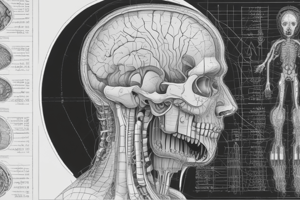
Neuroradiology Overview
- Neuroradiology is a subspecialty of radiology focused on imaging neurological cases.
- A strong understanding of anatomy is crucial for identifying lesions and their extent.
- Various imaging modalities are employed based on clinical indication for effectiveness and efficiency.
Imaging Modalities
-
Conventional Imaging
- Utilizes plain X-ray for the skull and spine.
-
Advanced Imaging
- CT Scan:
- CT Angiography assesses vascular conditions of the brain and spinal cord.
- Perfusion CT is used in early stroke assessment.
- CT Myelography studies the spinal cord condition.
- MRI:
- MRA/MRV focuses on vascular structures.
- Diffusion Tensor MRI maps nerve pathways in the brain and spinal cord.
- Perfusion MRI differentiates blood flow reductions, indicating strokes.
- MR Spectroscopy evaluates metabolites in tumors/infections to distinguish malignancy from benign conditions.
- fMRI identifies vital brain centers.
- Fluoroscopy: Evaluates vascular conditions through angiography.
- Ultrasonography:
- Head Ultrasound is primarily used for infants to check intracranial conditions.
- Transcranial Doppler assesses cerebral vascular conditions in both infants and adults.
- Nuclear Medicine:
- Combined with CT/MRI, such as PET Scan, highlighting lesions in early or metastatic stages.
- CT Scan:
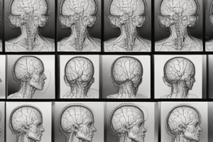
Skull Radiography
-
Skull X-Ray:
- Effective for assessing bony structures and initial evaluations.
- Limited for brain internal conditions; CT or MRI is preferred.
- Findings include calcifications, pituitary fossa enlargement, and bone lesions.
-
Indications for Skull Radiography:
- Suspected skeletal abnormalities, infections, or malignancies.
-
Standard X-Ray Positions:
- PA/AP (Anteroposterior view): Best for frontal and parietal bones.
- Lateral view: Evaluates occipital, parietal bones, and sella turcica.
- Towne’s view: Useful for assessing occipital trauma.
- Caldwell’s view: Assesses orbits and sinuses.
- Waters view and several other specialized views focus on different skull aspects.
Skull and Bone Conditions
- Skull comprises cranial and facial bones:
- Protects the brain and houses major arteries, veins, and nerves through foramina.
- Features cranial fossae: anterior, middle, posterior, and pituitary fossa.
- Sutures and Fontanelles:
- Sutures connect cranial bones, such as sagittal, coronal, and lambdoid sutures.
- Fontanelles allow for growth in infants; can be assessed using ultrasound for abnormalities.
Neurological Imaging Insights
- The ventricular system contains choroid plexus producing cerebrospinal fluid (CSF) and includes lateral, third, and fourth ventricles.
- Hydrocephalus results from excessive production or impaired absorption of CSF.
- Tentorium Cerebelli separates cerebellum from cerebrum.
Stroke Types
- Hemorrhagic Stroke results from blood vessel rupture.
- Ischemic Stroke occurs due to vascular obstruction, leading to tissue death (hypodense lesions).
MRI Applications
- MRI uses magnetic fields, safe for children and pregnant women, to detail soft tissue like the brain.
- Best for acute conditions; effective in evaluating stroke within a critical timing window.
- Can delineate tumor boundaries using contrast and assess vascular malformations (AVM).
Ultrasound Use in Neonates
- Head ultrasound is beneficial for infants with open fontanelles to evaluate for bleeding without exposing them to high radiation from X-rays.
Studying That Suits You
Use AI to generate personalized quizzes and flashcards to suit your learning preferences.