Podcast
Questions and Answers
What would be the most immediate consequence if a neuron's sodium-potassium pump ceased to function?
What would be the most immediate consequence if a neuron's sodium-potassium pump ceased to function?
- The neuron would be unable to synthesize ATP.
- The resting membrane potential would gradually dissipate. (correct)
- Neurotransmitter release at the synapse would be immediately halted.
- Action potentials would become prolonged due to unchecked potassium efflux.
The absolute refractory period primarily results from the inactivation of voltage-gated potassium channels, preventing any possibility of sodium influx.
The absolute refractory period primarily results from the inactivation of voltage-gated potassium channels, preventing any possibility of sodium influx.
False (B)
How would demyelination of axons affect the propagation of action potentials, and why?
How would demyelination of axons affect the propagation of action potentials, and why?
Demyelination causes slower or blocked action potential propagation due to the loss of saltatory conduction.
The opening of ligand-gated ion channels at a synapse typically results in a(n) ______.
The opening of ligand-gated ion channels at a synapse typically results in a(n) ______.
Match each glial cell type with its primary function in the nervous system:
Match each glial cell type with its primary function in the nervous system:
Which of the following best describes the role of the axon hillock in neuronal signaling?
Which of the following best describes the role of the axon hillock in neuronal signaling?
An excitatory postsynaptic potential (EPSP) always leads to the generation of an action potential in the postsynaptic neuron.
An excitatory postsynaptic potential (EPSP) always leads to the generation of an action potential in the postsynaptic neuron.
What is the primary mechanism by which local anesthetics, such as lidocaine, block pain signals?
What is the primary mechanism by which local anesthetics, such as lidocaine, block pain signals?
The process by which neurotransmitters are released from the presynaptic neuron into the synaptic cleft is known as ______.
The process by which neurotransmitters are released from the presynaptic neuron into the synaptic cleft is known as ______.
Match the following ions with their relative concentrations inside and outside a neuron at resting membrane potential:
Match the following ions with their relative concentrations inside and outside a neuron at resting membrane potential:
Which of the following mechanisms contributes most directly to the hyperpolarization phase of an action potential?
Which of the following mechanisms contributes most directly to the hyperpolarization phase of an action potential?
The myelin sheath increases the capacitance of the axon membrane, leading to slower action potential propagation.
The myelin sheath increases the capacitance of the axon membrane, leading to slower action potential propagation.
How does the concentration gradient of calcium ions across the presynaptic membrane contribute to neurotransmitter release?
How does the concentration gradient of calcium ions across the presynaptic membrane contribute to neurotransmitter release?
The unidirectional propagation of action potentials along the axon is ensured by the ______ of voltage-gated sodium channels.
The unidirectional propagation of action potentials along the axon is ensured by the ______ of voltage-gated sodium channels.
Match the following neuronal structures with their respective functions in signal transmission:
Match the following neuronal structures with their respective functions in signal transmission:
What would happen to neurotransmitter release if a toxin blocked voltage-dependent calcium channels in the presynaptic terminal?
What would happen to neurotransmitter release if a toxin blocked voltage-dependent calcium channels in the presynaptic terminal?
The threshold for initiating an action potential is the point at which potassium influx exceeds sodium efflux.
The threshold for initiating an action potential is the point at which potassium influx exceeds sodium efflux.
How does the structure of the myelin sheath contribute to the increased speed of action potential propagation in myelinated axons?
How does the structure of the myelin sheath contribute to the increased speed of action potential propagation in myelinated axons?
In the context of neurophysiology, the term '______' refers to the process by which a neuron becomes less negative inside relative to its resting state.
In the context of neurophysiology, the term '______' refers to the process by which a neuron becomes less negative inside relative to its resting state.
Match each phase of the action potential with its characteristic ion channel activity:
Match each phase of the action potential with its characteristic ion channel activity:
Which of the following glial cells is responsible for the myelination of axons in the central nervous system?
Which of the following glial cells is responsible for the myelination of axons in the central nervous system?
An inhibitory postsynaptic potential (IPSP) results from the influx of sodium ions into the postsynaptic neuron.
An inhibitory postsynaptic potential (IPSP) results from the influx of sodium ions into the postsynaptic neuron.
What is the role of the Na+/K+ ATPase pump in maintaining the resting membrane potential, and how does it achieve this?
What is the role of the Na+/K+ ATPase pump in maintaining the resting membrane potential, and how does it achieve this?
Saltatory conduction is characterized by action potentials 'jumping' between ______ along myelinated axons.
Saltatory conduction is characterized by action potentials 'jumping' between ______ along myelinated axons.
Match each neurotransmitter with its primary effect on postsynaptic neurons:
Match each neurotransmitter with its primary effect on postsynaptic neurons:
Which of the following is NOT a mechanism by which neurotransmitters are cleared from the synaptic cleft?
Which of the following is NOT a mechanism by which neurotransmitters are cleared from the synaptic cleft?
Increasing the extracellular concentration of potassium ions would hyperpolarize a neuron.
Increasing the extracellular concentration of potassium ions would hyperpolarize a neuron.
How does the diameter of an axon affect the speed of action potential propagation, and why?
How does the diameter of an axon affect the speed of action potential propagation, and why?
The ______ period is a time during which a stronger-than-normal stimulus is required to elicit an action potential.
The ______ period is a time during which a stronger-than-normal stimulus is required to elicit an action potential.
Match each type of ion channel with its primary mode of activation:
Match each type of ion channel with its primary mode of activation:
What is the role of SNARE proteins in neurotransmitter release?
What is the role of SNARE proteins in neurotransmitter release?
The action potential always travels bidirectionally from the point of initiation.
The action potential always travels bidirectionally from the point of initiation.
Describe the role of astrocytes in regulating the extracellular environment of neurons.
Describe the role of astrocytes in regulating the extracellular environment of neurons.
The conversion of an electrical signal to a chemical signal occurs at the ______ of a neuron.
The conversion of an electrical signal to a chemical signal occurs at the ______ of a neuron.
Match each ion channel with its primary role during an action potential:
Match each ion channel with its primary role during an action potential:
What is the primary effect of myelin on the time constant (τ) of an axon?
What is the primary effect of myelin on the time constant (τ) of an axon?
Neurotransmitters are synthesized exclusively in the cell body and then transported to the synaptic terminals.
Neurotransmitters are synthesized exclusively in the cell body and then transported to the synaptic terminals.
Explain how the balance of EPSPs and IPSPs at the axon hillock determines whether a neuron will fire an action potential.
Explain how the balance of EPSPs and IPSPs at the axon hillock determines whether a neuron will fire an action potential.
The process by which the membrane potential returns to its resting value after depolarization is called ______.
The process by which the membrane potential returns to its resting value after depolarization is called ______.
Match the following neurophysiological terms with correct example values:
Match the following neurophysiological terms with correct example values:
Which of the following best explains the role of the Na+/K+ ATPase pump in maintaining the resting membrane potential?
Which of the following best explains the role of the Na+/K+ ATPase pump in maintaining the resting membrane potential?
Nodes of Ranvier are critical for saltatory conduction because they contain a high density of voltage-gated ion channels that regenerate the action potential as it propagates along the axon.
Nodes of Ranvier are critical for saltatory conduction because they contain a high density of voltage-gated ion channels that regenerate the action potential as it propagates along the axon.
How would a drug that selectively blocks voltage-gated potassium channels affect the repolarization phase of an action potential?
How would a drug that selectively blocks voltage-gated potassium channels affect the repolarization phase of an action potential?
The period during which a neuron cannot fire another action potential, regardless of the stimulus strength, is known as the ______ refractory period.
The period during which a neuron cannot fire another action potential, regardless of the stimulus strength, is known as the ______ refractory period.
Match each phase of the action potential with the primary ion channel activity:
Match each phase of the action potential with the primary ion channel activity:
Which of the following is the most accurate description of how the myelin sheath increases the speed of action potential propagation?
Which of the following is the most accurate description of how the myelin sheath increases the speed of action potential propagation?
Inhibitory postsynaptic potentials (IPSPs) result from the influx of positive ions into the postsynaptic neuron, bringing the membrane potential closer to the threshold for firing an action potential.
Inhibitory postsynaptic potentials (IPSPs) result from the influx of positive ions into the postsynaptic neuron, bringing the membrane potential closer to the threshold for firing an action potential.
How does the function of K+ leak channels contribute to the negative resting membrane potential of a neuron?
How does the function of K+ leak channels contribute to the negative resting membrane potential of a neuron?
At the synapse, the conversion of an electrical signal into a chemical signal involves the release of ______ from synaptic vesicles.
At the synapse, the conversion of an electrical signal into a chemical signal involves the release of ______ from synaptic vesicles.
Match each component of a neuron with its primary function:
Match each component of a neuron with its primary function:
Which of the following is a critical function of the myelin sheath?
Which of the following is a critical function of the myelin sheath?
The relative refractory period occurs because voltage-gated sodium channels are still inactivated, making it impossible for the neuron to fire another action potential, regardless of stimulus strength.
The relative refractory period occurs because voltage-gated sodium channels are still inactivated, making it impossible for the neuron to fire another action potential, regardless of stimulus strength.
During the repolarization phase of an action potential, what specific event causes the membrane potential to return towards its resting negative value?
During the repolarization phase of an action potential, what specific event causes the membrane potential to return towards its resting negative value?
The concentration gradient for sodium ions (Na+) is maintained by the ______, which keeps the concentration of Na+ higher outside the cell than inside.
The concentration gradient for sodium ions (Na+) is maintained by the ______, which keeps the concentration of Na+ higher outside the cell than inside.
Match each phase of the action potential with its corresponding change in membrane potential:
Match each phase of the action potential with its corresponding change in membrane potential:
What is the primary role of the axon hillock in a neuron?
What is the primary role of the axon hillock in a neuron?
Saltatory conduction occurs only in unmyelinated axons, where the action potential travels continuously along the axon membrane.
Saltatory conduction occurs only in unmyelinated axons, where the action potential travels continuously along the axon membrane.
How do excitatory postsynaptic potentials (EPSPs) contribute to the generation of an action potential?
How do excitatory postsynaptic potentials (EPSPs) contribute to the generation of an action potential?
The small structures at the end of the axon that release neurotransmitters to communicate with other neurons or muscle cells are called ______.
The small structures at the end of the axon that release neurotransmitters to communicate with other neurons or muscle cells are called ______.
Which of the following best describes the function of voltage-gated sodium channels during an action potential?
Which of the following best describes the function of voltage-gated sodium channels during an action potential?
Flashcards
Cell Body (Soma)
Cell Body (Soma)
Contains the nucleus and organelles; integrates incoming signals.
Dendrites
Dendrites
Branch-like extensions that receive signals from other neurons at synapses; carry signals to the cell body.
Axon
Axon
A long fiber that transmits electrical signals away from the cell body.
Axon Hillock
Axon Hillock
Signup and view all the flashcards
Synaptic Endings (Terminals)
Synaptic Endings (Terminals)
Signup and view all the flashcards
Myelin Sheath
Myelin Sheath
Signup and view all the flashcards
Nodes of Ranvier
Nodes of Ranvier
Signup and view all the flashcards
Active Transporters
Active Transporters
Signup and view all the flashcards
Na+/K+ Pump
Na+/K+ Pump
Signup and view all the flashcards
Ion Channels
Ion Channels
Signup and view all the flashcards
Leak Channels
Leak Channels
Signup and view all the flashcards
Voltage-Gated Channels
Voltage-Gated Channels
Signup and view all the flashcards
Membrane Potential
Membrane Potential
Signup and view all the flashcards
Resting Membrane Potential
Resting Membrane Potential
Signup and view all the flashcards
Na+/K+ ATPase
Na+/K+ ATPase
Signup and view all the flashcards
K+ Leak Channels
K+ Leak Channels
Signup and view all the flashcards
Negatively Charged Proteins
Negatively Charged Proteins
Signup and view all the flashcards
Action Potential
Action Potential
Signup and view all the flashcards
Resting State (-70 mV)
Resting State (-70 mV)
Signup and view all the flashcards
Depolarization (Rising Phase)
Depolarization (Rising Phase)
Signup and view all the flashcards
Repolarization (Falling Phase)
Repolarization (Falling Phase)
Signup and view all the flashcards
Hyperpolarization
Hyperpolarization
Signup and view all the flashcards
Restoring the Resting Potential
Restoring the Resting Potential
Signup and view all the flashcards
Absolute Refractory Period
Absolute Refractory Period
Signup and view all the flashcards
Relative Refractory Period
Relative Refractory Period
Signup and view all the flashcards
Myelinated Axons
Myelinated Axons
Signup and view all the flashcards
Synapses
Synapses
Signup and view all the flashcards
Neurotransmitters
Neurotransmitters
Signup and view all the flashcards
EPSP
EPSP
Signup and view all the flashcards
IPSP
IPSP
Signup and view all the flashcards
Cell body (Soma)
Cell body (Soma)
Signup and view all the flashcards
Signals from Other Neurons
Signals from Other Neurons
Signup and view all the flashcards
Axon Hillock
Axon Hillock
Signup and view all the flashcards
Synaptic Endings (Terminals)
Synaptic Endings (Terminals)
Signup and view all the flashcards
Myelin Sheath
Myelin Sheath
Signup and view all the flashcards
Nodes of Ranvier
Nodes of Ranvier
Signup and view all the flashcards
Ion Transporters
Ion Transporters
Signup and view all the flashcards
Ion Channels
Ion Channels
Signup and view all the flashcards
Resting Membrane Potential
Resting Membrane Potential
Signup and view all the flashcards
Action Potential
Action Potential
Signup and view all the flashcards
Study Notes
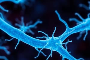
- Neurons are specialized cells transmitting electrical and chemical signals throughout the nervous system.
- Their structure facilitates rapid communication with other neurons, muscles, and glands.
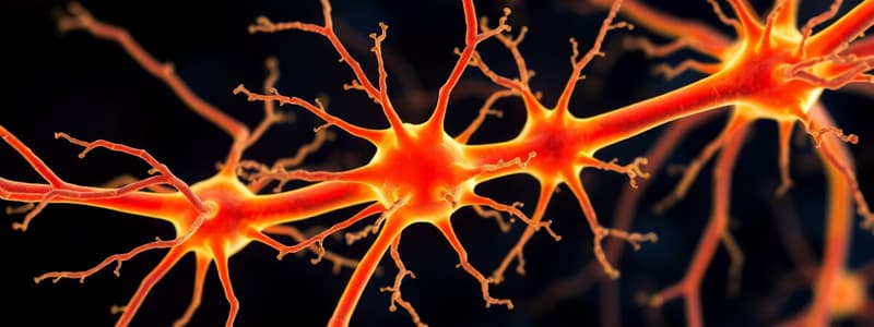
Key Components of a Neuron:
- Cell Body (Soma):
- Contains the nucleus and organelles.
- Processes incoming signals and maintains cell function.
- Acts as the control center of the neuron, integrating information from dendrites.
- Ensures neuron functions properly and decides whether to send an electrical impulse down the axon.
- This links to action potential generation, which happens if the signals received are strong enough.
- Dendrites:
- Branch-like extensions from the cell body.
- Collect information from synapses with other neurons.
- Carry signals towards the cell body.
- Receive chemical signals from other neurons at synapses.
- These signals can be excitatory or inhibitory, affecting whether the neuron will fire an action potential.
- Axon:
- A long fiber that transmits electrical signals away from the cell body.
- Can be very long, for example, the sciatic nerve extends from the spinal cord to the foot.
- Essential for carrying signals over long distances in the body.
- The electrical impulse (action potential) moves down the axon toward the synaptic terminals.
- Axon Hillock:
- The junction between the cell body and the axon.
- The site where action potentials are initiated.
- Crucial because it determines whether the neuron reaches the "threshold" required to generate an action potential.
- If enough positive charge builds up, an action potential is triggered.
- Synaptic Endings (Terminals):
- Small structures at the end of the axon.
- Release neurotransmitters to communicate with other neurons or muscles.
- Electrical signals get converted into chemical signals (neurotransmitters).
- This process allows neurons to pass messages to other cells, which links to synapse function.
- Myelin Sheath (Produced by Schwann Cells in the PNS, Oligodendrocytes in the CNS):
- A fatty insulating layer around the axon.
- Increases signal transmission speed through saltatory conduction (signal jumps between gaps in myelin).
- Dramatically increases the speed of nerve impulses.
- Diseases like multiple sclerosis degrade myelin, causing slower nerve transmission and movement issues.
- Nodes of Ranvier:
- Gaps between myelin where action potentials "jump" from node to node.
- Speeds up signal transmission.
- These nodes allow saltatory conduction, making impulses travel much faster compared to unmyelinated neurons.
- This links to the factors that affect conduction speed, such as myelination and axon diameter.
How Ions Move Across Membranes & Membrane Potentials
- Neurons function using electrical signals, which depend on ion movement across membranes.
- The plasma membrane is made of lipids, therefore charged ions cannot pass through directly.
- Instead, they move through ion transporters and channels.
Types of Ion Transporters & Channels:
- Active Transporters (e.g., Na+/K+ Pump):
- Move ions against their concentration gradient using ATP.
- Pumps 3 Na+ out and 2 K+ in, creating a charge difference.
- Critical for maintaining the resting potential (-70 mV).
- Keeps sodium outside and potassium inside, creating an electrochemical gradient.
- Links to how resting potentials are generated.
- Ion Channels (Passive Transport):
- Allow ions to move down their concentration gradient.
- Can be leak channels (always open, e.g., K+ leak channels) or voltage-gated channels (open/close based on membrane voltage).
- Allow the neuron to respond to changes in charge, leading to action potentials.
- Voltage-gated sodium and potassium channels play a key role in action potential generation and propagation.
Membrane Potential & How It Arises
- Membrane Potential:
- The difference in electrical charge between the inside and outside of the neuron
- Measured in millivolts (mV).
- Resting membrane potential: ~-70 mV (inside is more negative than outside).
- Exists because of ion gradients (mainly Na+ and K+), maintained by the Na+/K+ pump and ion leak channels.
How is the Resting Potential Generated?
- Na+/K+ ATPase (10%):
- Pumps 3 Na+ out and 2 K+ in, creating a charge imbalance.
- K+ Leak Channels (70%):
- Potassium leaks out, making the inside more negative.
- Negatively Charged Proteins Inside the Cell (20%):
- Large anions (proteins) remain inside, contributing to negativity.
- Resting potential allows neurons to respond quickly to stimuli by generating action potentials.
Action Potentials: A Step-by-Step Deep Dive
- Action potential: Neurons send electrical signals along their axons to communicate with other neurons, muscles, or glands.
- It happens due to rapid changes in the membrane potential caused by the movement of ions (Na⁺ and K⁺) through voltage-gated ion channels.
Resting State (-70 mV)
- At rest, the inside of the neuron is more negative compared to the outside
- Na⁺/K⁺ pump (Sodium-Potassium ATPase):
- Pumps 3 Na⁺ out and 2 K⁺ in, using ATP.
- This creates a concentration gradient where:
- More Na⁺ is outside the cell.
- More K⁺ is inside the cell.
- Voltage-gated Na⁺ and K⁺ channels are closed.
- The membrane potential is at -70 mV, meaning the inside is 70 mV more negative than the outside.
- The neuron is polarized, meaning there is a difference in charge across the membrane.
Depolarization (Rising Phase)
- A stimulus (such as a neurotransmitter) binds to receptors on the neuron, causing some Na⁺ channels to open.
- If the stimulus is strong enough to bring the membrane to threshold (-55 mV):
- Voltage-gated Na⁺ channels fully open.
- Na⁺ rushes into the cell, following:
- Its concentration gradient (high outside → low inside).
- Its electrical gradient (inside is negative, attracting positive Na⁺).
- As Na⁺ enters, the inside of the neuron becomes less negative, and the membrane potential rapidly increases to +30 mV.
- If the stimulus does not reach -55 mV, nothing happens (no action potential).
- If it does, an action potential always occurs (all-or-nothing principle).
Repolarization (Falling Phase)
- At +30 mV, the neuron must return to its resting state.
- Voltage-gated Na⁺ channels inactivate (they close temporarily and cannot reopen immediately).
- Voltage-gated K⁺ channels open, allowing K⁺ to leave the cell.
- Since K⁺ is positively charged, its exit makes the inside of the cell more negative again.
- The membrane potential drops back towards -70 mV.
- The closing of Na⁺ channels prevents the action potential from moving backward.
Hyperpolarization
- K⁺ channels stay open too long, allowing too much K⁺ to leave.
- This overcorrects the membrane potential, making it more negative than the resting state (about -80 mV instead of -70 mV).
- This is why a neuron cannot fire again immediately—it’s harder to reach threshold when it’s extra negative.
Restoring the Resting Potential (-70 mV)
- Voltage-gated K⁺ channels close.
- The Na⁺/K⁺ pump starts working again, pumping Na⁺ out and K⁺ in, restoring the original ion distribution.
- The neuron is now ready for another action potential.
Refractory Periods: Preventing Overlapping Signals
- Refractory period prevents the neuron from firing again immediately after an action potential
- Absolute Refractory Period:
- The neuron cannot fire another action potential because Na⁺ channels are inactivated.
- Lasts until repolarization is complete.
- Relative Refractory Period:
- The neuron can fire another action potential, but it requires a stronger stimulus.
- Occurs during hyperpolarization (-80 mV), when the membrane potential is extra negative.
- Ensures action potentials travel in one direction and do not overlap.
Ion Movements in Each Phase
- Resting State:
- Na+ Channels:Closed
- K+ Channels:Closed
- Main Ion Movement: None (Na+/K+ pump active)
- Membrane Potential:-70 mV
- Depolarization:
- Na+ Channels: Open (Na+ enters)
- K+ Channels: Closed
- Main Ion Movement:Na⁺ enters cell
- Membrane Potential: -55 mV → +30 mV
- Repolarization:
- Na+ Channels: Inactivated (Closed)
- K+ Channels: Open (K+ exits)
- Main Ion Movement: K⁺ leaves cell
- Membrane Potential:+30 mV → -70 mV
- Hyperpolarization:
- Na+ Channels: Closed
- K+ Channels: Open (K+ still exits)
- Main Ion Movement:K⁺ continues to leave
- Membrane Potential:-80 mV
- Return to Resting:
- Na+ Channels: Closed
- K+ Channels: Closed
- Main Ion Movement:Na+/K+ pump restores balance
- Membrane Potential: -70 mV
Key Takeaways
- Action potentials allow neurons to communicate via electrical impulses.
- Na⁺ influx causes depolarization (+30 mV), K⁺ efflux causes repolarization (-70 mV).
- Refractory periods prevent backwards signals and ensure one-way travel.
- The Na⁺/K⁺ pump restores ion balance after hyperpolarization.
- More Sodium (Na⁺) is Outside than Potassium (K⁺) is Inside:
- The Na⁺/K⁺ pump pumps out 3 Na⁺ ions for every 2 K⁺ ions it brings in.
- This means the outside of the cell has more positive charges than the inside.
- Since more positive charge is leaving than entering, the inside of the cell remains negative compared to the outside.
- Large Negative Proteins Inside the Cell:
- Inside the neuron, there are large negatively charged molecules (proteins, DNA, phosphates) that cannot leave the cell.
- These contribute to the inside being negative even though K⁺ is there.
What Happens During Repolarization?
- At +30 mV, the neuron needs to return to resting (-70 mV)
- Na⁺ Channels Inactivate (Close):
- No more Na⁺ can enter the cell.
- K⁺ Channels Open:
- Voltage-gated K⁺ channels open, and K⁺ exits the cell.
- K⁺ is moving down its concentration gradient (high inside → low outside).
- K⁺ is positively charged, so as it leaves, the inside of the cell becomes more negative again.
Why Does the Closing of Na⁺ Channels Prevent Backward Movement?
- When an action potential moves along the axon, it only moves forward because:
- The Na⁺ channels that just opened will now inactivate and cannot reopen right away.
- This prevents the action potential from going backward and ensures that it moves in one direction along the neuron.
Key Takeaways
- The inside of the neuron is negative at rest because of the Na⁺/K⁺ pump and large negative molecules.
- During repolarization, K⁺ exits the cell, making the inside more negative again.
- Na⁺ channels inactivate after they open, preventing the signal from going backward.
Propagation of Action Potential
- Once an action potential starts at the axon hillock, it travels along the axon.
- In unmyelinated axons, the impulse moves continuously.
- In myelinated axons, the impulse "jumps" between Nodes of Ranvier (saltatory conduction), making it faster.
- Factors like myelination and axon diameter affect conduction speed.
- Myelinated neurons conduct impulses much faster than unmyelinated ones.
Synapses & Neurotransmitter Release
- At the synapse, electrical signals are converted into chemical signals. Neurotransmitters (NTs) are released from synaptic vesicles.
- NTs bind to receptors on the postsynaptic neuron, causing either:
- Excitatory Postsynaptic Potential (EPSP) → depolarization.
- Inhibitory Postsynaptic Potential (IPSP) → hyperpolarization.
- The balance of EPSPs and IPSPs determines whether the neuron fires an action potential.
- This process underlies learning, memory, and many brain functions
How All Parts Work Together
- Neurons are specialized cells designed to transmit information rapidly throughout the nervous system.
- The structure of the neuron enables these functions, with each part playing a distinct role in processing and transmitting electrical and chemical signals
- The cell body is located centrally, and it acts as the control center of the neuron.
- It's where the nucleus and most of the organelles are found.
- The soma integrates all the incoming signals from the dendrites and determines whether the neuron should generate an action potential.
- The soma processes these signals and maintains cell function (like energy production and protein synthesis).
- The action potential only happens if the signal is strong enough to reach a specific threshold.
- Once the soma receives signals from the dendrites, it decides whether these signals are enough to cause an action potential at the axon hillock.
- The soma contains ion channels and pumps to maintain the resting potential, ensuring the neuron is ready for action when needed.
- Dendrites are tree-like extensions that protrude from the soma.
- They are the "input" regions of the neuron
- Dendrites receive incoming chemical signals from other neurons.
- These signals are either excitatory (encouraging the neuron to fire) or inhibitory (discouraging the neuron from firing).
- The sum of these signals determines whether an action potential is triggered
- Signals from other neurons arrive at the dendrites, and if enough excitatory signals come through, the neuron becomes depolarize.
- This leads to the generation of an action potential that will travel down the axon.
- If inhibitory signals predominate, the neuron will not fire an action potential.
- The combination of excitatory and inhibitory input from various neurons is crucial for processing complex information, such as learning or decision-making.
- The axon is a long, thin extension of the neuron that can extend from the soma all the way to distant target cells (other neurons, muscles, or glands).
- Axons can be very long
- The axon transmits electrical signals (action potentials) away from the soma toward the synaptic endings.
- It's responsible for long-distance communication within the nervous system.
- Once the soma decides an action potential is needed, the signal travels down the axon.
- The axon hillock is the key junction where this decision is made, determining whether the neuron will fire or remain inactive.
- The myelin sheath, which surrounds the axon, dramatically speeds up this signal transmission
- The Axon Hillock is located at the junction where the axon meets the soma.:
- The axon hillock is the decision point where the neuron "decides" whether to fire an action potential
- The electrical signal from the soma accumulates at the axon hillock, and if it is strong enough (reaching a certain threshold), an action potential is initiated.
- It acts as a gatekeeper that determines whether or not the signal is strong enough to send an impulse down the axon.
- It’s crucial for the all-or-nothing principle of action potentials
- The Synaptic Endings (Terminals) are small structures at the end of the axon, where the axon branches into multiple smaller branches.
- They come into close contact with other neurons or muscle cells.
- the electrical signal (action potential) is converted into a chemical signal in the form of neurotransmitters
- These neurotransmitters cross the synapse (the gap between two neurons) and bind to receptors on the next cell, passing on the signal.
- Once the action potential travels down the axon, it reaches the synaptic endings, where the signal is transferred to the next neuron or muscle via neurotransmitter release.
- This is where the electrical signal becomes a chemical one, allowing communication across synapses.
- This is the key step in neurotransmission, where the neuron communicates with another neuron or a muscle, initiating processes like muscle contraction or the firing of another neuron
- The Myelin Sheath surrounds the axon in segments
- In the Peripheral Nervous System (PNS), it's produced by Schwann cells, and in the Central Nervous System (CNS), it's produced by oligodendrocyte
- The myelin sheath acts as an insulator, speeding up the transmission of electrical signals by preventing the ions from leaking out as they travel down the axon
- This makes the electrical signal travel faster via saltatory conduction, where the action potential jumps between gaps in the myelin called Nodes of Ranvier.
- By allowing saltatory conduction, the myelin sheath speeds up signal transmission, making the neuron much more efficient.
- Without myelin, neurons would transmit signals much slower, which could affect functions like movement and coordination.
- Diseases like multiple sclerosis degrade the myelin sheath, slowing down or blocking action potentials, which is why motor skills and sensation are impaired in such conditions.
- Nodes of Ranvier are small gaps in the myelin sheath along the axon
- The nodes allow for saltatory conduction, where the action potential jumps from one node to the next, speeding up the signal transmission
- These nodes are essential for speeding up the action potential.
- The voltage-gated sodium channels located at the nodes enable the action potential to jump, drastically increasing conduction speed.
- This is why myelinated axons transmit signals faster than unmyelinated axons.
Ion Movement & Membrane Potentials: The Driving Forces Behind Electrical Signals
- Neurons rely on the movement of ions across the membrane to create electrical signals, and this process is crucial for both resting potentials and action potentials
- Ion Transporters (Active Transport):
- Found in the neuron’s membrane, including the Na+/K+ pump.
- These pumps move ions against their concentration gradients using ATP (energy), a process called active transport.
- the Na+/K+ pump moves three sodium ions (Na+) out of the neuron and two potassium ions (K+) in
- This process maintains the resting membrane potential by creating a concentration gradient of Na+ and K+ ions, with Na+ being high outside the neuron and K+ being high inside.
- This electrochemical gradient is essential for the rapid changes in voltage that occur during action potentials
- Ion Channels (Passive Transport):
- Embedded in the neuron's membrane and crucial for controlling ion movement during action potentials
- Ion channels allow ions to pass through the membrane by passive transport, following their concentration gradients
- Some are leak channels (always open), while others are voltage-gated channels (open and close based on the membrane’s voltage).
- Voltage-gated sodium channels open during depolarization, allowing Na+ to rush into the neuron, making the inside of the cell more positive.
- Voltage-gated potassium channels open during repolarization, allowing K+ to leave the neuron, restoring the negative charge inside.
- This is what creates the rapid rise and fall of membrane potential that constitutes the action potential.
How Membrane Potentials Arise: The Resting Potential and Action Potentials
- The resting membrane potential (approximately -70mV) and the action potential are the foundation of neuronal communication
- Resting Membrane Potential:
- The resting potential exists across the entire membrane of the neuron when it's not transmitting a signal.
- The resting potential is maintained by the action of the Na+/K+ pump, the potassium leak channels, and the negatively charged proteins inside the neuron
- This keeps the neuron ready to fire when a signal arrives.
- Without this negative charge, the neuron wouldn't be able to respond to stimuli.
- The resting membrane potential acts like the "ready-to-go" state for the neuron, enabling the rapid changes in voltage necessary for action potentials
- Action Potential:
- The action potential is the process by which neurons send signals over long distances = The action potential occurs along the axon, starting at the axon hillock and propagating to the synaptic endings.
- An action potential involves the rapid depolarization (inside becomes positive) followed by repolarization (inside becomes negative again), with hyperpolarization briefly occurring as the neuron resets
How Everything Links Together
- Neurophysiology is like a complex relay race: each part of the neuron plays a key role in passing signals along
- From the dendrites receiving input to the axon transmitting the signal, everything works in harmony
- Ions move across the membrane, creating electrical signals that form the foundation for communication.
- These signals travel down the axon, and when they reach the synaptic endings, they are passed to the next neuron via neurotransmitter release.
- Each part of the neuron—soma, dendrites, axon, synaptic endings, myelin, and nodes of Ranvier—interact in an incredibly coordinated way to ensure signals are processed and transmitted effectively.
- Without one part working properly, the whole process would fail
Studying That Suits You
Use AI to generate personalized quizzes and flashcards to suit your learning preferences.