Podcast
Questions and Answers
Which cellular organelle is responsible for providing metabolic energy for the cell's processes?
Which cellular organelle is responsible for providing metabolic energy for the cell's processes?
- Mitochondrion (correct)
- Golgi apparatus
- Ribosome
- Cell body
What is the primary function of the Golgi apparatus in a neuron?
What is the primary function of the Golgi apparatus in a neuron?
- To translate genetic information into proteins
- To receive synaptic inputs
- To package cellular materials into vesicles for transport (correct)
- To generate metabolic energy for the cell
Which neuronal zone is responsible for the initiation of neural electrical activity?
Which neuronal zone is responsible for the initiation of neural electrical activity?
- Output zone
- Input zone
- Integration zone (correct)
- Conduction zone
What is the role of axon collaterals in a neuron?
What is the role of axon collaterals in a neuron?
Which type of neuron primarily carries messages from peripheral tissue back to the spinal cord?
Which type of neuron primarily carries messages from peripheral tissue back to the spinal cord?
What is a defining characteristic of multipolar neurons?
What is a defining characteristic of multipolar neurons?
Which of the following reflects the complexity of a neuron's information processing function?
Which of the following reflects the complexity of a neuron's information processing function?
What describes the tiny gap between neurons where information is passed from one to another?
What describes the tiny gap between neurons where information is passed from one to another?
What is the role of neurotransmitters in neuronal communication?
What is the role of neurotransmitters in neuronal communication?
What is the function of axons?
What is the function of axons?
What is the direction of movement for anterograde axonal transport?
What is the direction of movement for anterograde axonal transport?
Which type of glial cell is responsible for creating myelination in the brain and spinal cord?
Which type of glial cell is responsible for creating myelination in the brain and spinal cord?
What is a primary function of astrocytes?
What is a primary function of astrocytes?
What is the role of microglia in the central nervous system?
What is the role of microglia in the central nervous system?
What is the main purpose of myelin?
What is the main purpose of myelin?
Which of the following arteries supplies the brain with approximately 80% of its blood?
Which of the following arteries supplies the brain with approximately 80% of its blood?
What is the function of the circle of Willis?
What is the function of the circle of Willis?
What is the main role of the blood-brain barrier?
What is the main role of the blood-brain barrier?
Which component of the ventricular system secretes cerebrospinal fluid (CSF)?
Which component of the ventricular system secretes cerebrospinal fluid (CSF)?
What is the function of the glymphatic system in the brain?
What is the function of the glymphatic system in the brain?
During neural development, what is the role of radial glial cells?
During neural development, what is the role of radial glial cells?
What is the role of cell adhesion molecules (CAMs) in the developing nervous system?
What is the role of cell adhesion molecules (CAMs) in the developing nervous system?
During which stage of nervous system development does synaptic pruning occur?
During which stage of nervous system development does synaptic pruning occur?
What characterizes the process of 'differentiation' in the context of neural development?
What characterizes the process of 'differentiation' in the context of neural development?
What term describes the destruction of the nerve cell body following injury to its axon?
What term describes the destruction of the nerve cell body following injury to its axon?
What is the function of neurotrophic factors?
What is the function of neurotrophic factors?
What is the meaning of "experience-expectant development"?
What is the meaning of "experience-expectant development"?
The brain's requirement for a particular input to unfold typically refers to:
The brain's requirement for a particular input to unfold typically refers to:
What increases in myelination and white matter contribute to during early development?
What increases in myelination and white matter contribute to during early development?
What is the Corpus callosum?
What is the Corpus callosum?
What are the main functions of the Frontal lobe?
What are the main functions of the Frontal lobe?
What is Wernicke's area known for?
What is Wernicke's area known for?
Which of the following is a primary function of the parietal lobe?
Which of the following is a primary function of the parietal lobe?
What function is the temporal lobe primarily involved in?
What function is the temporal lobe primarily involved in?
If damage occurs to Wernicke's area, what is the most likely result?
If damage occurs to Wernicke's area, what is the most likely result?
What processes visual information and assist in understanding/recognizing what we see?
What processes visual information and assist in understanding/recognizing what we see?
The Sylvian fissure separates which two lobes of the brain?
The Sylvian fissure separates which two lobes of the brain?
A patient has difficulty controlling voluntary movements and cognitive functions. Which of the following basal ganglia structures is MOST likely affected?
A patient has difficulty controlling voluntary movements and cognitive functions. Which of the following basal ganglia structures is MOST likely affected?
Which of the following basal ganglia structures is involved in dopamine production, which plays a crucial role in controlling movement?
Which of the following basal ganglia structures is involved in dopamine production, which plays a crucial role in controlling movement?
Flashcards
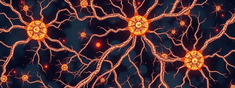
Neurons
Neurons
Individual cells in the nervous system; receives, integrates, and transmits information.
Cell body (soma)
Cell body (soma)
Region of the neuron defined by the presence of the cell nucleus.
Mitochondrion
Mitochondrion
Cellular organelle providing metabolic energy for the cell's processes.
Golgi apparatus
Golgi apparatus
Signup and view all the flashcards
Ribosomes
Ribosomes
Signup and view all the flashcards
Input zone
Input zone
Signup and view all the flashcards
Dendrite
Dendrite
Signup and view all the flashcards
Integration zone
Integration zone
Signup and view all the flashcards
Conduction zone (axon)
Conduction zone (axon)
Signup and view all the flashcards
Output zone
Output zone
Signup and view all the flashcards
Motor neurons
Motor neurons
Signup and view all the flashcards
Sensory neurons
Sensory neurons
Signup and view all the flashcards
Interneurons
Interneurons
Signup and view all the flashcards
Multipolar neurons
Multipolar neurons
Signup and view all the flashcards
Bipolar neurons
Bipolar neurons
Signup and view all the flashcards
Unipolar neurons
Unipolar neurons
Signup and view all the flashcards
Dendritic arborization
Dendritic arborization
Signup and view all the flashcards
Neural plasticity
Neural plasticity
Signup and view all the flashcards
Dendritic spines
Dendritic spines
Signup and view all the flashcards
Neuron doctrine
Neuron doctrine
Signup and view all the flashcards
Synapses
Synapses
Signup and view all the flashcards
Presynaptic neuron
Presynaptic neuron
Signup and view all the flashcards
Neurotransmitter
Neurotransmitter
Signup and view all the flashcards
Postsynaptic neuron
Postsynaptic neuron
Signup and view all the flashcards
Synaptic cleft
Synaptic cleft
Signup and view all the flashcards
Axons
Axons
Signup and view all the flashcards
Anterograde transport
Anterograde transport
Signup and view all the flashcards
Retrograde transport
Retrograde transport
Signup and view all the flashcards
Glia
Glia
Signup and view all the flashcards
Astrocyte
Astrocyte
Signup and view all the flashcards
Microglia cell
Microglia cell
Signup and view all the flashcards
Oligodendrocytes
Oligodendrocytes
Signup and view all the flashcards
Schwann cells
Schwann cells
Signup and view all the flashcards
Myelin
Myelin
Signup and view all the flashcards
Myelination
Myelination
Signup and view all the flashcards
Internal carotid artery
Internal carotid artery
Signup and view all the flashcards
Anterior cerebral arteries
Anterior cerebral arteries
Signup and view all the flashcards
Middle cerebral arteries
Middle cerebral arteries
Signup and view all the flashcards
Vertebral arteries
Vertebral arteries
Signup and view all the flashcards
Basilar artery
Basilar artery
Signup and view all the flashcards
Posterior cerebral arteries
Posterior cerebral arteries
Signup and view all the flashcards
Study Notes
Neurons
- Neurons are individual cells responsible for receiving, integrating, and transmitting information within the nervous system, which contains 80-90 billion neurons.
Neuron Divisions
- Neuron divisions consist of:
- Cell body (soma): contains the cell nucleus
- Mitochondria: provides metabolic energy
- Golgi apparatus: packages cellular materials into vesicles for transport
- Ribosomes: translates genetic information to produce protein
Neuron Zones
- Functional zones include:
- Input zone: collects information from other neurons or sensory structures
- Dendrites are extensions of the cell body that receive synaptic inputs
- Integration zone: initiates neural electrical activity, located at the cell body
- Conduction zone: the axon transmits electrical signals away from the cell body, with axon collaterals forming branches
- Output zone: axon terminals or synaptic boutons transmit the neuron's activity across synapses to other neurons
Neuron Categories
- Neurons classified by function:
- Motor neurons: trigger movements via long axons synapsing on muscles
- Sensory neurons: carry messages from peripheral tissue to the spinal cord
- Interneurons: receive input from and send input to other neurons
Neuron Shapes
- Neuron categories include:
- Multipolar neurons: have one axon and multiple dendrites
- Bipolar neurons: have one axon and one dendrite
- Unipolar/monopolar neurons: have a single branch extending in two directions from the cell body, one side being the input and the other the output zone
Dendrites
- Dendritic arborization determines the complexity of a neuron's information processing
- Neural plasticity: the configuration of synapses on dendrites and the cell body changes with experience
- Dendritic spines increase the surface area for synapses and can rapidly change
Neuron Doctrine
- Neuron doctrine states that the brain is composed of cells that are distinct structurally, metabolically, and functionally, and it is composed of independent cells.
- Synapses are tiny gaps between neurons where information passes from one to another
- Presynaptic neuron (output): the region of a synapse that releases neurotransmitters
- Neurotransmitters: chemicals released from the presynaptic axon terminal for communication between neurons
- Postsynaptic neuron (input): the region of a synapse that receives and responds to neurotransmitters -Synaptic cleft: separates the pre- and postsynaptic membranes
Axons
- Axons are single nerve fibers that carry action potentials from the cell body
- Axonal transport:
- Anterograde transport moves material toward the axon terminals; retrograde transport moves materials back to the cell body using motor proteins
- Axons function in:
- Rapid transmission of electrical signals
- Slower transportation of substances
Glial Cells
- Glia provides neurons with information and affects neuronal function
- Glial cell composition:
- 75% oligodendrocytes
- 17% astrocytes
- 7% microglia
- Astrocytes swell in response to injury and cause edema, which damages neurons
Types of Glial Cells
- Astrocyte: regulates local blood flow, maintains the blood-brain barrier, attaches to the neuron's soma, and monitors neuronal synapses
- It may participate in transmitting information between neurons, contribute to new synapse formation and the shortening of surplus synapses, and influence local brain chemistry
- Microglia: changes shape/size, forms a containment zone when there is damage
- It removes debris and is involved in pain perception and neuronal remodeling
- Oligodendrocytes: create myelination in the brain and spinal cord
- Nodes of Ranvier are small uninsulated patches between the beads.
Schwann Cells
- Schwann cells are glial cells that form myelin in the PNS
- Myelin: fatty insulation around an axon that increases the speed of nerve impulses
- Myelination: ensheathing axons in myelin, and increases the speed of electrical signals that jump from node to node.
Vascular System
- The internal carotid artery supplies 80% of the brain's blood and supplies blood to the anterior and middle cerebral arteries
- Anterior cerebral arteries supply the medial frontal and parietal lobes and are interconnected by the anterior communicating artery
- Middle cerebral arteries supply most of the lateral surface of the cerebral hemispheres
- Vertebral arteries supply remaining 20% that join to form the basilar artery, supplying blood to the brainstem and posterior cerebral arteries
- Posterior cerebral arteries arise from the basilar artery and supply blood to the posterior aspects of the cerebral hemispheres, cerebellum, and brainstem
- Circle of Willis: a vascular structure at the base of the brain formed by communicating arteries
Blood Brain Barrier
- The blood-brain barrier makes the movement of substances from blood vessels into brain cells more difficult
Ventricular System
- Cerebrospinal fluid acts as a shock absorber, a medium for material exchange, and removes toxins
- Ventricular system: fluid-filled cavities that produce and distribute cerebrospinal fluid(CSF)
- Lateral ventricle: lateral portion of the ventricular system within each hemisphere
- Choroid plexus: vascular portion of the ventricles lining that secretes cerebrospinal fluid
- Third ventricle: conducts cerebrospinal fluid from the lateral ventricle to the fourth ventricle
- Fourth ventricle: receives cerebrospinal fluid from the third ventricle and surrounds cerebellum and brainstem
Glymphatic System
- Glymphatic system: a lymphatic system in the brain that removes wastes, and nutrients and signaling compounds
Early Development
- Epigenetics: factors affecting gene expression
- Ontogeny: process when individual grows
- Mature brain: Over 80 billion neurons and quadrillion synapses
- Zygote: fertilized egg
- Embryo: earliest developmental stage (10 weeks)
- Fetus: developing stage after embryo
- Genotype: all inherited genetic information
- Phenotype: physical traits change
- Gene expression: cells transcribe particular genes to make proteins
- Cell differentiation: cells acquire characteristics Cell-cell interactions: cells development affect neighboring ones
Radial Glial Cells
- Radial glial cells: guide migration in development in the emerging cerebral hemispheres.
- CAM: cell adhesion molecule found on the surface of cells for migration and pathfinding.
Neural Development - Ectoderm
- Zygote divides after 12 hours after fertilisation.
- Neural crest: forms the PNS
- Layers of the embryo after 18 days thicken to form the neural plate which folds inward, forming the neural groove
- Neural tube becomes ventricles (fluid filled)
- Head of the neural tube forms:
- Prosencephalon (forebrain): which then forms cerebrum (telencephalon and diencephalon)
- Mesencephalon: midbrain
- Rhombencephalon (hindbrain: metencephalon (pons and cerebellum) & myelencephalon (medulla) → cerebellum and brainstem
- Spinal cord: transmits signals between Brian and rest of body
Nervous System Development - Stages
-
- Neurogenesis: non-neural cells divide.
-
- Cell migration: Cells move to establish distinct nerve cell populations and radial glial cells guide migrating neurons
-
- Differentiation: transformation of precursors
-
- Synaptogeniesis: forming Synaptic connections
-
- Neuronal cell deals: Many nerve cells die
-
- Synapse rearrangement: Some synapses are lost, so development to refine synapses.
- Synaptic pruning is due to experience - for Flexibility in the brain.
Terms
- Neurogenesis: production/growth of nerve cells
- Ventricular zone: region lining cerebral ventricles. displays mitosis = glials and neurons
- Adult neurogenesis: production of new neurons in the adult brain
- Apoptosis: "surplus" cells die
- Sensitive period: permanently altered during development due to experience/treatment
- Epigenetics: effects of gene expressions - no nucleotide sequence change
- Cell migration: cells move from origin to final spot
- Synaptogenesis: establish Synaptic connections = growth
Degeneration & Factors
- Retrograde degeneration: destruction of the nerve
- Anterograde degeneration/Wallerian: Loss of part of axon from injury
- Neurotrophic factor: Target-derived chemical that "feeds" to help survive. Nerve growth (NGF) A substance affects growth of neurons
- Brain-derived: that keep some classes of neurons alive.
The Role of Experience
- Experience-expectant development: input for development to unfold + time windows
- Caregiving: newborns require support and caregiving, which assists the developing immature infant. Is predictable, and sensitive
- Perceptual Discrimination: Fx. specific discrimination tuned to environment
- Neural plasticity: permits developmental perturbations that produce vulnerability
- Experience-dependent: Unique to individuals. Eg. a language
Prenatal Abilities & Newborn Abilities
- 3 Months: Basic physical reflex
- 4 Months: Reacts to light and sounds
- 5 Months: Movements being more controlled
- 6 Months: Learning can be observed
- 7 Months: Electrical activity was detected
- The last trimester: gyrification takes Place.
- Newborn preferable - prefers response to maternal stimuli.
- Infancy – dramatic brain changes, the elimination of unused Synapses. They Are born with Small brains, that reached half size of adult after the first year
- Myelination: Early growth = white matter growth.
Subcortical Mechanisms
-
- Fetal/newborn cognition is helped by mechanisms Face detection mechanisms are low sensitive = development to bias
- Central Nervous System - Brian and Spinal cord
- Meninges - DAP
Cerebrum
- Cerebrum: largest part with high cognitive function. cortex = divided into 2 hemispheres and the 4 lobes
- Pyramidal cell is the most prominent cell
- White matter has axons with myelin/ Grey matter: cell bodies (dendrites) with/out myelin
- Corpus callosum: band of axons links 2 hemispheres.
Frontal Lobe
- Frontal Lobe - Anterior portion and responsible for: complex cognitive functions / plan / behaviour , motor.
- Precentral gyrus (motor cortex): motor functions
- Brocas Area – language left
- Prefrontal cortex: and impulse control
- orbitofrontal cortex-centre: and motivation/reward, regulating Here
Parietal Lobe
- Parietal Lobe - Cortex between Frontal and Occipital lobes on Each Hemisphere: register yd re input like pressure and Muscle
- Postcentral gyrus Supratnarginal gyrus + Angle gyrus Area
- Temporal lobe – learning / auditory
- Olfactory bulb (smell)
Occipital Lobe and Major Features
- Occipital Lobe - Cortex on posterior
- (eye functions) : visual information + impulses
- Postcentral Gyrus and front the cortex on central with sensory body, and sulcus,
- precentral = Front for the Movernent.
- Sylvian Sulcus is fold divides Tempora and is Frontal
- Central Sulcus divides/ Frontal. Telencephalon frontal Subdivision = hemispheres
Nucleus, Putamen and Globus Pallidus
- Caudate Nucleus within the voluntary
- Putamen: structure in basal ganglia coordinates the movement
- Globus Pallidus: for decision making functions + motivation
Diencephalon
- Diencephalon: in the forebrain: information + functions thalamus consists = Nuclei relay sensory information except
Hypothalamic functions + is part of brainstem:
- nuclei (regulation , mm)
- regulates autonomous (hormonal)
Mesencephalon
- brainstem Midbrain is sensory and motor _ Tectum: part of system Process both audity visual . _ Tegmentur main body – motor is
Metaencephalon Cerebellum
- Responsible For movement and pons motor
Medullo Oblongata is part of brain stem
- cardiovasvular/ respiratory functions and reflexes
- Subcortical- for the cortex. Basal Ganglia: of structures in movements Receives information Thalamus cortex to
Limbric System
- The Limbric system – for emotion: located above brainstem
- hypothalamus - for endrocine system
- Stria terminalis a band connects others
- Olfactory bulb for smell
- The septum (regulating-emotion) nucleus: structures in that Are part of
Peripheral Nervous System
- Peripheral nervous system (Outside of Skull - Column) for transmits information + sensory, motor nerves
- Somatics nervous known as voulntarly
_ 12 Cranial nerves for Muscle and neck
_ Spinal Nerves group from Spinal cord for
- Cervical: necks , shoulder
- Thoracic: / Upper chest
- Lumbar : in legs
- the sacral at base -genitals
- ventral side: Motor and front
- Dorsal side ; sensory
Nervous System
- Autonomic : internal and involuntary
- Autonomic Ganglia : outside the CNS
- Sympathetic: Actions to PREPARE and Flight.
Other Terms
- Parasympathetic: to digest/ rest
- Enteric: to help gut controlled BY CNS
- NEUROLINGO brain
- Three = of 3 VIEWS brain. to see
The 3 Planes
- divisions
- Horizontal Plane : lower
- : Vertical halves
- _ Coronal Plane: + backwards
Neurotransmission
- Neurotransmission- transformation. Neurotransmitters Are The - basis . Criterion Is: -axon The contains Enzymes. _ That receptor The Application / activity
Terms
- K+(Potassium)Na sodium CI-Chloride. Ca+ ( Calcium): -Receptor
- Molecules transmitter. the, agonist Binds = antagonist. the.
Non-Competitive
_Modulator, that - without ligand's. = At the molecule cell. Is - In/Exogenous = study = An Gated ion channel - channel. channel The Channel . Membrane /Exocytosis. to: To AID that into
Neurotransmitter (Systems) and Brain Stem
- Brain –Acetylcholine with , attention+ Memory for (skeletal ) alzheimer
- Main from raphe nuclei. have - for Alertness from 1,2 Beta serotonin ( 5HT) _ Memory
- Multiple DA (dopamine VTA) -reward : cognition. is for movement. -the ( and is has(↓), (
- Inhibitory: GLUTAMATA with nervous Excitatory, Plasticity GABA _ In Anxiety Dendrites Synthesis.
-
Electrical by+ more - A"s
Effects
- Pre -Drug: Effects - synthesis/Blockade of. Release _Channel/Release.
Physiologic
Post- synaptic processes :Receptors/ Modulation ,4, . (Neurophysiology – function)
Charged Paticles
- Particles 1 by K++Sodium _ C1+ ( Calcium); An of gain -A A +ve –(cation, charge
- fluid. membrane/ Electrical
Soduim Pump
- _ Pump/ the/ + the is: It + 2 K++/Cycle:
- to: At Resting potentials; membrane
- (balance) In is. more.
Signals & Release
-
- Transmillter unidireclinal, Common . Sumatiom an. in: It release - flow is"
The Action & Propagation
-: Increase of In + - Until40 mV + to
- -increase+ K+ The, - to and is ~150. km is
EPSP /IPSP
- Excitatory: Glutamate, Noradrenaline, Dopamine results in:
- De-polarization of neurons (+):
- ↑ neural transmission and produces
- Alertness, focus, etc
- Inhibitory: GABA, Serotonin, Taurine results in:
- Hyper-polarisation of neurons (-):
- ↓ neural transmission and produces
- Relaxation, sleep, etc
Neuro-Imaging
- information for: Fx: basis + brain/Visual cell/ Golgi
- -isotope /label for
- immunes /antibodies. tissue -: nucleic _ tracer inject Brain + study
Structural Imaging & Tomography
- : to see , Monitor
- Scanning: an an overall stroke / lesion
- Resonance and MRI, waves
- By- . . + For Model
DTI STRUCTURAL IMAGING
to =>Measures fibre in of there
Functional imaging Dynamic
-
the different, like : MEC _ non electrical are -to over
-
signal for close surface sleep PET -gamma scanner and -detailed Exposure is/
-
Hormones :Messengers AND Produced
Hormones are chemical messengers and systems.
Peptides & Action
They Are for , (ACH/ are action Hormone :with for = Portal : with system. Hormone and ( insulin).
Gland: The -
The + A:Feedback to, - with or is + balance with and stress
hormones the and
Studying That Suits You
Use AI to generate personalized quizzes and flashcards to suit your learning preferences.


