Podcast
Questions and Answers
What primary role do glial cells play in neuronal function?
What primary role do glial cells play in neuronal function?
- Generating action potentials for faster communication.
- Providing structural support and synthesizing myelin.
- Transmitting electrical signals directly between neurons.
- Providing neurons with raw materials, chemical signals, and structural elements. (correct)
Which of the following is the MOST accurate description of the neuron doctrine?
Which of the following is the MOST accurate description of the neuron doctrine?
- The brain is composed of interconnected cells that share metabolic functions.
- The brain and spinal cord operate independently of each other.
- The brain functions as a single, continuous network without individual cells.
- The brain is composed of cells that are distinct structurally, metabolically, and functionally. (correct)
Which functional zone of a neuron is primarily responsible for integrating incoming information to determine whether to produce a neural signal?
Which functional zone of a neuron is primarily responsible for integrating incoming information to determine whether to produce a neural signal?
- Input zone
- Output zone
- Integration zone (correct)
- Conduction zone
What is the primary function of the Golgi apparatus in a neuron?
What is the primary function of the Golgi apparatus in a neuron?
Which type of neuron is MOST commonly found in the nervous system and characterized by a single axon and multiple dendrites?
Which type of neuron is MOST commonly found in the nervous system and characterized by a single axon and multiple dendrites?
Which of the following describes the function of the nodes of Ranvier?
Which of the following describes the function of the nodes of Ranvier?
In which direction does anterograde axonal transport move materials?
In which direction does anterograde axonal transport move materials?
What is the primary role of astrocytes in the brain?
What is the primary role of astrocytes in the brain?
What is the significance of dendritic arborization in neurons?
What is the significance of dendritic arborization in neurons?
During neural development, what role do radial glial cells play?
During neural development, what role do radial glial cells play?
Which arteries primarily supply blood to the medial frontal and parietal lobes of the brain?
Which arteries primarily supply blood to the medial frontal and parietal lobes of the brain?
What is the function of the Circle of Willis?
What is the function of the Circle of Willis?
What is the role of the choroid plexus in the ventricular system?
What is the role of the choroid plexus in the ventricular system?
Which of the following is a key function of the glymphatic system?
Which of the following is a key function of the glymphatic system?
Which of the following structures is MOST directly involved in motor functions, decision-making, reward processing and action selection?
Which of the following structures is MOST directly involved in motor functions, decision-making, reward processing and action selection?
What is the role of cell adhesion molecules (CAMs) during neural development?
What is the role of cell adhesion molecules (CAMs) during neural development?
What process is defined as the developmental stage in which cells acquire distinctive characteristics, such as those of neurons, as a result of expressing particular genes?
What process is defined as the developmental stage in which cells acquire distinctive characteristics, such as those of neurons, as a result of expressing particular genes?
During brain development, what is the main event occurring during synaptogenesis?
During brain development, what is the main event occurring during synaptogenesis?
What is the term for the programmed cell death that occurs during development, in which surplus cells are eliminated??
What is the term for the programmed cell death that occurs during development, in which surplus cells are eliminated??
What is the primary function of the somatic nervous system?
What is the primary function of the somatic nervous system?
Which division of the autonomic nervous system is responsible for the 'fight or flight' response?
Which division of the autonomic nervous system is responsible for the 'fight or flight' response?
What is a key function of the enteric nervous system?
What is a key function of the enteric nervous system?
Which structure serves as a relay station for sensory information (except smell) going to the cerebral cortex?
Which structure serves as a relay station for sensory information (except smell) going to the cerebral cortex?
What is the function of the cerebellum?
What is the function of the cerebellum?
Which of the following best describes the function of the hypothalamus?
Which of the following best describes the function of the hypothalamus?
Flashcards
Neurons
Neurons
Individual cells in the nervous system that receive, integrate, and transmit information.
Cell Body (Soma)
Cell Body (Soma)
The region of the neuron defined by the presence of the cell nucleus.
Mitochondrion
Mitochondrion
A cellular organelle providing metabolic energy for the cell's processes.
Golgi Apparatus
Golgi Apparatus
Signup and view all the flashcards
Ribosomes
Ribosomes
Signup and view all the flashcards
Input Zone
Input Zone
Signup and view all the flashcards
Dendrite
Dendrite
Signup and view all the flashcards
Integration Zone
Integration Zone
Signup and view all the flashcards
Conduction Zone (Axon)
Conduction Zone (Axon)
Signup and view all the flashcards
Output Zone (Axon Terminals)
Output Zone (Axon Terminals)
Signup and view all the flashcards
Motor Neurons
Motor Neurons
Signup and view all the flashcards
Sensory Neurons
Sensory Neurons
Signup and view all the flashcards
Interneurons
Interneurons
Signup and view all the flashcards
Multipolar Neurons
Multipolar Neurons
Signup and view all the flashcards
Bipolar Neurons
Bipolar Neurons
Signup and view all the flashcards
Unipolar/Monopolar Neurons
Unipolar/Monopolar Neurons
Signup and view all the flashcards
Neural Plasticity
Neural Plasticity
Signup and view all the flashcards
Neuron Doctrine
Neuron Doctrine
Signup and view all the flashcards
Synapses
Synapses
Signup and view all the flashcards
Presynaptic Neuron (Output)
Presynaptic Neuron (Output)
Signup and view all the flashcards
Neurotransmitter
Neurotransmitter
Signup and view all the flashcards
Postsynaptic Neuron (Input)
Postsynaptic Neuron (Input)
Signup and view all the flashcards
Synaptic Cleft
Synaptic Cleft
Signup and view all the flashcards
Axons
Axons
Signup and view all the flashcards
Anterograde Transport
Anterograde Transport
Signup and view all the flashcards
Study Notes
Neurons
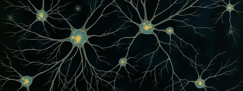
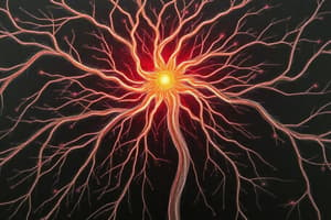
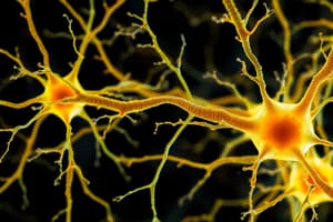
- Neurons are the key cells of nervous system, receiving, integrating, and transmitting information
- There are about 80-90 billion neurons in the human nervous system
Neuron Divisions
- Cell body (soma) contains the cell nucleus
- Mitochondria provide metabolic energy for cellular processes
- The Golgi apparatus, found in eukaryotic cells, packages cellular materials into vesicles for transport
- Ribosomes in the cell body translate genetic information into protein
Functional Zones
- Input zone receives information from other neurons or sensory structures
- Dendrites are extensions of the cell body that receive synaptic inputs
- Integration zone initiates neural electrical activity, located at the cell body
- Conduction zone's axon conducts electrical signals away from the cell body
- Axon collaterals are multiple branches that the axon splits into
- Output zone's axon terminals or synaptic boutons are specialized swellings at the axon's end where neuron activity transmits across synapses
Neuron Categories by Function
- Motor neurons trigger movements via long axons synapsing on muscles
- Sensory neurons carry messages from peripheral tissue to the spinal cord
- Interneurons receive input from other neurons and send input to other neurons
Neuron Categories by Shape
- Multipolar neurons have a single axon and multiple dendrites, the most common type in the nervous system
- Bipolar neurons have one axon and one dendrite, common in sensory systems like vision
- Unipolar/monopolar neurons have a single branch from the cell body extending in two directions: one input zone with dendritic branches, and one output zone with axon terminals
Dendritic Arborization
- The degree of dendrite branching reflects a neuron's information processing complexity
Neural Plasticity
- Synapse configuration and layout on dendrites and the cell body constantly change based on experience and environment
- Dendritic spines increase the surface area for synapses and can be quickly altered to remodel neural connections continually
Neuron Doctrine
- The neuron doctrine states the brain is composed of structurally, metabolically, and functionally distinct cells
- The brain is composed of independent cells
- Synapses are tiny gaps between neurons where information passes
- The presynaptic neuron (output) region releases neurotransmitter into the synapse
- Neurotransmitters are chemicals released from the presynaptic axon terminal that enable communication between neurons
- The postsynaptic neuron (input) region receives and responds to neurotransmitters
- The synaptic cleft is the gap separating the pre- and postsynaptic membranes
Axons
- Axons are nerve fibers and single extensions of a nerve cell, carrying action potentials from the cell body to other neurons
Axonal Transport
- Anterograde transport moves material toward the axon terminal using motor proteins to move transport vesicles
- Retrograde transport moves materials back to the cell body from the axon terminal and synaptic cleft using motor proteins.
- Axons have two functions: rapid electrical signal transmission on the outside, and slower substance transportation inside
Glial Cells
- Glia affects neuronal function by providing information like raw materials, chemical signals, and structural elements
- Glial cells are almost as numerous as neurons
- 75% of glial cells are oligodendrocytes, 17% are astrocytes, and 7% are microglia
- Astrocytes can swell in response to brain injury, causing edema
Types of Glial Cells: Astrocytes
- Astrocytes are star-shaped glial cells that regulate local blood flow to supply active neurons
- Astrocytes maintain the blood-brain barrier, attach to the soma/cell body of neurons, receive synapses, monitor neuronal synapses, and may participate in information transmission
- Astrocytes contribute to forming/shortening synapses and influence local brain chemistry, potentially contributing to diseases like epilepsy
Types of Glial Cells: Microglia
- Microglia are small glial cells that change shape and size, acting as the brain's cleanup crew
- Microglia are active, extending/withdrawing processes to form containment zones around injuries, remove debris, and are potentially involved in pain perception and neuronal remodeling
Types of Glial Cells: Oligodendrocytes
- Oligodendrocytes are glial cells that create myelination in the brain and spinal cord
- Oligodendrocytes attach to axons and wrap them in layers of myelin, creating an appearance of slender beads
- Nodes of Ranvier are small uninsulated patches between the myelin beads where ions can escape the membrane
- Oligodendrocytes provide chemical signals that enhance the structural integrity of axons
Types of Glial Cells: Schwann Cells
- Schwann cells are glial cells that form myelin in the peripheral nervous system (PNS)
- Myelin is fatty insulation around an axon formed by glial cells that boosts nerve impulse speed
- Myelin cells are separated by nodes of Ranvier
- Myelination is the process of ensheathing axons in myelin, continuing for 10-15 years after birth and increasing electrical signal speed as signals jump from node to node
- Some axons lack myelin but are still surrounded by Schwann or oligodendrocyte cells.
Vascular System
- The internal carotid artery supplies the brain with approximately 80% of its blood
- These arteries ascend on the left and right sides of the neck, supplying the anterior and middle cerebral arteries
- Anterior cerebral arteries supply the medial frontal and parietal lobes and connect via the anterior communicating artery
- Middle cerebral arteries supply most of the lateral surface of the cerebral hemispheres
- Vertebral arteries supply remaining 20% of blood.
- These arteries ascend the vertebrae, enter the base of the skull, and join to form the basilar artery
- A basilar artery is formed by the fusion of the vertebral arteries, supplying blood to the brainstem and posterior cerebral arteries
- Posterior cerebral arteries arise from the basilar artery, providing blood to posterior aspects of the cerebral hemispheres, cerebellum, and brainstem
- The circle of Willis is a vascular structure at the base of the brain that is formed by communicating arteries, interconnecting the major cerebral arteries
Blood-Brain Barrier
- The blood-brain barrier makes the movement of substances from blood vessels into brain cells more difficult
Ventricular System
- The cerebrospinal fluid (CSF) acts as a shock absorber, facilitates material exchange between blood vessels and brain tissue, and removes toxins
- The ventricular system comprises fluid-filled cavities within the brain, producing, and distributing CSF
- Lateral ventricles are complexly shaped portions of the ventricular system within each brain hemisphere
- Choroid plexus is a vascular portion of the lining of the ventricles that secretes CSF and is composed of ependymal cells
- The third ventricle is the midline ventricle that conducts cerebrospinal fluid from the lateral ventricle to the fourth ventricle
- The fourth ventricle is the passageway within the pons that receives cerebrospinal fluid from the third ventricle and releases it to surround the brain and spinal cord, located between the cerebellum and the brainstem
GLYMPHATIC SYSTEM
- The glymphatic system is a lymphatic system in the brain involved in waste removal, nutrient transport, and signaling compound movement
Early Development
- Epigenetics is the study of factors that affect gene expression without changing the genes
- Ontogeny is the process by which an individual changes during its lifetime, growing and aging
- The mature brain has over 80 billion neurons and a quadrillion synapses
- Zygote: a fertilized egg
- Embryo: earlist stage of development (first 10 weeks)
- Fetus: developing stage after embryo
- Genotype: all inherited genetic information
- Phenotype: physical characteristics of an individual that changes constantly
- Gene expression: The process by which a cell transcribes a particular gene and makes the protein it encodes
- Cell differentiation is the developmental stage in which cells acquire distinctive characteristics by expressing particular genes
- Cell-cell interactions are the general process during development in which one cell affects the differentiation of other cells, usually neighboring cells
- Radial glial cells are glial cells that form early in development, span the width of the emerging cerebral hemispheres, and guide migrating neurons from inner to outer surfaces of the emerging nervous system
- Cell adhesion molecule (CAM): A protein guides cell migration and/or axonal pathfinding on the cell surface.
Neural Development
-
Ectoderm forms from the Zygote (fertilized egg) approximately starts dividing 12 hours after fertilization
-
Neural crest forms the peripheral nervous system (PNS)
-
One of three cell layers in the embryo from 18 days, it thickens, forming the neural plate, which folds inward to create the neural groove
-
The neural folds fuse, thus forming the neural tube
-
The inside of the neural tube becomes fluid filled ventricles
-
The head of the neural tube forms the
- Prosencephalon (forebrain)
- Mesencephalon (midbrain)
- Rhombencephalon (hindbrain)
-
Prosencephalon (forebrain) forms the cerebrum, telencephalon and diencephalon
-
Mesencephalon is the midbrain
-
Rhombencephalon (hindbrain) forms the metencephalon (pons and cerebellum) and myelencephalon (medulla), as well as the cerebellum and brainstem
-
The spinal cord transmits neural signals between the brain and the rest of the body
Development of the Nervous System
- Cells are produced in the ventricular zone (mitosis)
- Neurogenesis is the mitotic division of non-neural cells to produce neurons
- Cell migrations (brain nuclei and layers of the cerebral cortex) are movements of cells to establish distinct nerve cell populations. Radial glial cells guide the migrations
- Differentiation is the transformation of precursor cells into distinctive neuron and glial cell types
- Synaptogenesis creates synaptic connections as axons and dendrites grow and doubles brain size in the first year
- Neuronal cell death is the selective death of many nerve cells
- Synapse rearrangement involves the loss of certain synapses and the development of others to refine synaptic connections
- Synaptic pruning, the loss of gray matter, is affected by experience, and is essential for brain flexibility
- Neurogenesis: the production of nerve cells
- The ventricular zone, also called the ependymal layer, lines the cerebral ventricles that displays mitosis, providing neurons early in development as well as glial cells
- Adult neurogenesis: the production of new neurons in the adult brain
- Apoptosis: a developmental process where “surplus” cells die
- Gene expression: the process by which a cell transcribes a particular gene and makes the protein it encodes
- Cell differentiation: developmental stage in which cells acquire distinctive characteristics
Further terms regarding development
- Sensitive period is the period during development when the organism can be permanently altered by a particular experience or treatment
- Epigenetics studies factors affecting gene expression without changing the nucleotide sequence of the genes
- Cell migration: movement of cells from site of origin to final location
- Cell differentiation: developmental stage where cells acquire distinctive characteristics
- Synaptogenesis: establishment of synaptic connections as axons and dendrites grow
Degeneration
- Retrograde degeneration is the destruction of the nerve cell body following injury to its Axon
- Anterograde degeneration, also called Wallerian degeneration, is the loss of the distal portion of an axon resulting from injury.
Neurotrophic Factors
- Neurotrophic factor (trophic factor) is a target-derived chemical to help certain neurons survive
- Nerve growth factor (NGF) is a substance that markedly affects the growth of neurons in spinal ganglia and in the ganglia of the sympathetic nervous system
- Brain-derived neurotrophic factor is a protein purified from animal brains that can keep some classes of neurons alive
The Role of Experience
- Experience-expectant development: brain requires a particular input for development to unfold typically and experience exerts time windows
Caregiving: Newborns and Infants
- Newborns require care and support from adult caregivers
- Parenting provides regulation for the immature infant
- Caregivers provide essential input
- Children develop secure attachment through predictable, responsible, and sensitive caregiving
Brain Development Facts
- Neural plasticity permits developmental perturbations that produce vulnerability
- Experience-dependent development is unique to individuals and for example, specific language, or face type of ones community
- Perceptual discrimination becomes tuned to environmentally relevant distinctions by 9-12 months of life
- Prenatal abilities are Basic physical reflexes begin at 3 months, react to lights and sounds at 4 months, movements become more controlled at 5 months, Learning is observed at 6 months, and electrical activity can be detected at 7 months
- Complex gyrification takes place as well final migration of neurons during the last trimester of gestation
- Newborns preferentially respond to maternal voice, speech and native language and prefer to look at face-like stimuli
- Infancy has dramatic change in structural and functional connectivity. The explosion of synaptic connections and elimination of unused synapses.
Facts About Babies Brains
- babies are born with small brains and there are various explanations
- obstetric dilemma hypothesis: the human pelvis constraints childbirth
- metabolism hypothesis: infants are born when
- after the first year, the brain has reached half the size of the adult brain.
- Myelination: White matter integrity has All major white matter bundles are visible but they are weakly myelinated. Early development is characterized by rapid increase in myelination and white matter.
- Fetal and newborn cognitive abilities are thought to be supported initially by subcortical mechanisms
- Face detection mechanism sensitive to low spatial frequency information
- Subcortical mechanisms serve to bias visual input to developing cortical circuit
Central Nervous System
- Central nervous system (CNS): the brain and spinal cord
- Meninges: DAP - Dura mater, arachnoid and pia mater (layers of protection)
Cerebrum
- Cerebrum is the largest part of the brain which is responsible for higher cognitive functions, sensory processing, voluntary movement, and complex behaviors and is divided into two hemispheres and four lobes: -Cerebral Cortex has six distinct layers -Pyramidal cell is the most prominent cell with a large pyramid shaped cell body.
Brain Structure
- White matter consists mostly of axons with white myelin sheaths
- Gray matter contains more cell bodies and dendrites which lack myelin
- Corpus callosum is the main band of axons that connects the two cerebral hemispheres.
Frontal Lobe
- The frontal lobe is the most anterior portion of the cerebral cortex
- Responsible for complex cognitive functions such as decision-making, planning, problem-solving, emotional control, and personality
- It is also responsible for motor control with specific areas:
- Precentral gyrus (motor cortex) is the center for motor functions
- Broca's area is responsible for language production and is located at left
- Prefrontal cortex is responsible for cognition and behavior controls such as attention, impulse regulation, and emotional regulation
- Orbitofrontal cortex contains reward and motivation centers, evaluates rewards, and regulates behavior accordingly
Parietal Lobe
- A large region of the cortex lying between the frontal and occipital lobes of each cerebral hemisphere
- Primarily responsible for registering external/outer sensory inputs such as touch, pain, weight, and temperature as well as internal sensitive impressions such as movement of muscles
- Functions also include:
- Recognition/identification of objects
- Spatial orientation and attention
- Recognition and understanding of written text and speech
- Specific areas:
- Postcentral gyrus/somatosensory cortex which receives somatosensory information to receive bodily movement, touch and an impression for "short" over the body
- Association cortex is the identification of objects
- Supramarginal gyrus which is for position and posture, both yours and others
- Angular gyrus which is for word choice, reading etc
- Wernicks area is for recognition, understanding and interpretation of written text and speech
Temporal Lobe and Major Sulci
- Temporal Lobe: A large lateral cortical region of each cerebral hemisphere, the temporal lobe is continuous with the parietal lobe posteriorly and is separated from the frontal lobe by the Sylvian fissure
- Important parts:
- learning and memory.
- processing auditory signals.
- Olfactory bulb, which is responsible for our sense of smell
- Occipital Lobe is a large region of the cortex covering much of the posterior part of each cerebral hemisphere which is mainly involved in sight
- Specific areas process visual impulses and to helps understand and recognize what we see
- Major Gyri and Sulci
- Postcentral gyrus: is the strip of parietal cortex, just behind the central sulcus, that receives somatosensory information from the entire body
- Precentral gyrus: is the strip of frontal cortex, just in front of the central sulcus, that is crucial for motor control
- Sylvian sulcus/fissure: is a deep fold in the brain, that separates the temporal lobe from the frontal lobe and parietal lobe
- Central sulcus/fissure: separates the frontal lobe from the parietal lobe. Located laterally and stretches from top to bottom
Brain Terms: Telen, Dien, Thalamus, Hypothalamus
- Telencephalon: is the frontal subdivision of the forebrain that includes the cerebral hemispheres when fully developed
- Caudate Nucleus: within the basal ganglia it plays a role in controlling voluntary movements and cognitive functions
- Putamen: Also within the basal ganglia it coordinates movements and regulating motor control
- Globus Pallidus: is within the basal ganglia and is involved in motor functions and plays a role in decision-making, reward processing and action selection
- Diencephalon: It is the posterior part of the forebrain, including the thalamus and hypothalamus. It directs sensory information and controls basic physiological functions.
- Thalamus: it consists of nuclei that relay sensory information to the cortex, except for smell (olfactory bulb). It has roles in regulating consciousness, sleep, and alertness.
- It serves as a relay station for sensory information to the cerebral cortex and plays a role in regulating consciousness,sleep, and alertness
Brain Terms: Hypothalamus and Other Brain Parts
- Hypothalamus: (hunger, thirst, temperature regulation, mm) it controls the pituitary gland thus controlling the endocrine, hormonal system and regulates autonomic nervous system
- Hypothalamus controls Hormones and Homeostasis
- Mesencephalon: is a part of the brainstem with sensory and motor components
- the midbrains sensory system is the tectum which has 2 regions processing visual (superior colliculi) and auditory (inferior colliculi) infor
- the main body of the midbrain is the tegmentum which has important motor centers
- the midbrains motor centers are the substantia nigra (basal ganglia in the midbrain) which has neurons releasing dopamine and red nucleus communications with motor neurons in the spinal cord.
- Substantia Nigra: within the basal ganglia, it helps control movement
- Reticular formation is a group in sleep, arousal, temperature , motor control extends from midbrain to medulla
Studying That Suits You
Use AI to generate personalized quizzes and flashcards to suit your learning preferences.


