Podcast
Questions and Answers
Neurons always fire simultaneously to ensure efficient brain function.
Neurons always fire simultaneously to ensure efficient brain function.
False (B)
Mirror neurons fire only when an individual performs an action, but not while observing the same action performed by others.
Mirror neurons fire only when an individual performs an action, but not while observing the same action performed by others.
False (B)
Astrocytes are responsible for producing myelin sheaths around axons in the central nervous system.
Astrocytes are responsible for producing myelin sheaths around axons in the central nervous system.
False (B)
The ventral horns of the spinal cord primarily process sensory information from the body.
The ventral horns of the spinal cord primarily process sensory information from the body.
Microglia are the primary signaling cells in the central nervous system.
Microglia are the primary signaling cells in the central nervous system.
Electrical synapses involve the release of neurotransmitters into a synaptic cleft.
Electrical synapses involve the release of neurotransmitters into a synaptic cleft.
According to Cajal’s neuron theory, the brain functions as a continuous network without distinct areas of signaling interest.
According to Cajal’s neuron theory, the brain functions as a continuous network without distinct areas of signaling interest.
Action potentials can be transmitted directly between smooth muscle fibers through chemical synapses.
Action potentials can be transmitted directly between smooth muscle fibers through chemical synapses.
Connexin 43 is a key connexin found in neurons, facilitating communication between pre- and post-synaptic terminals.
Connexin 43 is a key connexin found in neurons, facilitating communication between pre- and post-synaptic terminals.
Effective synaptic transmission requires neurotransmitter depletion in the presynaptic terminal.
Effective synaptic transmission requires neurotransmitter depletion in the presynaptic terminal.
Nitric oxide (NO) is stored in vesicles in the presynaptic terminal before being released.
Nitric oxide (NO) is stored in vesicles in the presynaptic terminal before being released.
Glutamate primarily acts as an inhibitory neurotransmitter in the brain.
Glutamate primarily acts as an inhibitory neurotransmitter in the brain.
Ionotropic receptors are G-protein coupled receptors that influence various cellular functions over a longer term.
Ionotropic receptors are G-protein coupled receptors that influence various cellular functions over a longer term.
GABAb receptors increase chloride ion conductance, leading to inhibition.
GABAb receptors increase chloride ion conductance, leading to inhibition.
5-HT3 receptors are metabotropic receptors that mediate slow synaptic transmission.
5-HT3 receptors are metabotropic receptors that mediate slow synaptic transmission.
Muscarinic receptors regulate only heart rate and exocrine gland secretion, but not visceral motility.
Muscarinic receptors regulate only heart rate and exocrine gland secretion, but not visceral motility.
Benzodiazepines directly mimic GABA to enhance GABAa receptor activity.
Benzodiazepines directly mimic GABA to enhance GABAa receptor activity.
Pain is sensed through mechanoreceptors and thermoreceptors.
Pain is sensed through mechanoreceptors and thermoreceptors.
The labeled line principle suggests that specific nerve pathways correspond to multiple types of sensations.
The labeled line principle suggests that specific nerve pathways correspond to multiple types of sensations.
Rapidly adapting receptors continue to respond over time.
Rapidly adapting receptors continue to respond over time.
Meissner’s corpuscles detect vibration and pressure changes, and are located deep within the skin's dermal layers.
Meissner’s corpuscles detect vibration and pressure changes, and are located deep within the skin's dermal layers.
Larger receptive fields are associated with higher sensitivity.
Larger receptive fields are associated with higher sensitivity.
In spatial summation, the perceived stimulus gets stronger as stimulation frequency increases over time.
In spatial summation, the perceived stimulus gets stronger as stimulation frequency increases over time.
Lateral inhibition decreases the ability to discriminate between closely spaced stimuli.
Lateral inhibition decreases the ability to discriminate between closely spaced stimuli.
All sensory information is relayed through the medulla oblongata before reaching the cortex.
All sensory information is relayed through the medulla oblongata before reaching the cortex.
The primary somatosensory area II provides highly precise localization of body parts.
The primary somatosensory area II provides highly precise localization of body parts.
The face is represented in the posterior part of the somatosensory cortex.
The face is represented in the posterior part of the somatosensory cortex.
The cortex is organized into horizontal layers, each serving specific sensory modalities.
The cortex is organized into horizontal layers, each serving specific sensory modalities.
The anterolateral pathway transmits fine touch and proprioceptive information.
The anterolateral pathway transmits fine touch and proprioceptive information.
Allodynia refers to decreased sensitivity to pain.
Allodynia refers to decreased sensitivity to pain.
A slower injury will elicit a stronger pain response than a rapid one.
A slower injury will elicit a stronger pain response than a rapid one.
The refractive index of air is significantly greater than 1.
The refractive index of air is significantly greater than 1.
A concave lens converges light rays to a focal point.
A concave lens converges light rays to a focal point.
The vitreous humor is located in front of the lens.
The vitreous humor is located in front of the lens.
Cones are most effective at night and in the dark.
Cones are most effective at night and in the dark.
Metarhodopsin II increases cGMP levels in photoreceptor cells.
Metarhodopsin II increases cGMP levels in photoreceptor cells.
Ganglion cells spread a message by depolarizing to signal to the brain.
Ganglion cells spread a message by depolarizing to signal to the brain.
Rod photoreceptors are more responsive to the red-yellow wavelength.
Rod photoreceptors are more responsive to the red-yellow wavelength.
Acute trauma temporarily masks pain through parasympathetic nervous system activation.
Acute trauma temporarily masks pain through parasympathetic nervous system activation.
Magnocellular ganglion cells process color and fine detail for detailed visual acuity.
Magnocellular ganglion cells process color and fine detail for detailed visual acuity.
Flashcards
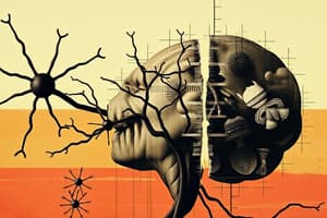
Neurons
Neurons
Primary cells in the CNS with structures like dendritic arborization and axons ending in synaptic terminals. They have common structures like the Golgi apparatus, ER, mitochondria, and nucleus.
Neuron Functionality
Neuron Functionality
Neurons modify their membrane potential via ion channels, crucial for generating action potentials for neuronal communication.
Neuronal Activity
Neuronal Activity
Neurons firing asynchronously in groups which allows synchronous firing is essential for complex brain functions.
Mirror Neurons
Mirror Neurons
Signup and view all the flashcards
Neuronal Count
Neuronal Count
Signup and view all the flashcards
Neural Stem Cells
Neural Stem Cells
Signup and view all the flashcards
Oligodendrocytes
Oligodendrocytes
Signup and view all the flashcards
Astrocytes
Astrocytes
Signup and view all the flashcards
Spinal Cord Matter
Spinal Cord Matter
Signup and view all the flashcards
Microglia
Microglia
Signup and view all the flashcards
Electrical Synapses
Electrical Synapses
Signup and view all the flashcards
Chemical Synapses
Chemical Synapses
Signup and view all the flashcards
Spinal Cord Level
Spinal Cord Level
Signup and view all the flashcards
Lower Brain
Lower Brain
Signup and view all the flashcards
Higher Brain
Higher Brain
Signup and view all the flashcards
Reticular Theory
Reticular Theory
Signup and view all the flashcards
Neuron Theory
Neuron Theory
Signup and view all the flashcards
Electrical Synapses
Electrical Synapses
Signup and view all the flashcards
Chemical Synapses
Chemical Synapses
Signup and view all the flashcards
Reticular Theory
Reticular Theory
Signup and view all the flashcards
Neuron Theory
Neuron Theory
Signup and view all the flashcards
Glial Cells
Glial Cells
Signup and view all the flashcards
Gap Junctions
Gap Junctions
Signup and view all the flashcards
Presynaptic Terminal
Presynaptic Terminal
Signup and view all the flashcards
Postsynaptic Terminal
Postsynaptic Terminal
Signup and view all the flashcards
Neurotransmitter Availability
Neurotransmitter Availability
Signup and view all the flashcards
Synaptic Fatigue
Synaptic Fatigue
Signup and view all the flashcards
Acidosis and Alkalosis
Acidosis and Alkalosis
Signup and view all the flashcards
Nitric Oxide (NO)
Nitric Oxide (NO)
Signup and view all the flashcards
Acetylcholine (ACh)
Acetylcholine (ACh)
Signup and view all the flashcards
Catecholamines
Catecholamines
Signup and view all the flashcards
Dopamine
Dopamine
Signup and view all the flashcards
Serotonin
Serotonin
Signup and view all the flashcards
GABA
GABA
Signup and view all the flashcards
Glutamate
Glutamate
Signup and view all the flashcards
Ionotropic Receptors
Ionotropic Receptors
Signup and view all the flashcards
Metabotropic Receptors
Metabotropic Receptors
Signup and view all the flashcards
Nicotinic Receptors
Nicotinic Receptors
Signup and view all the flashcards
GABAa Receptors
GABAa Receptors
Signup and view all the flashcards
GABAb Receptors
GABAb Receptors
Signup and view all the flashcards
Study Notes
Neurons: The Key Cells of the Central Nervous System
- Neurons are the main cells in the central nervous system (CNS) and have special parts like dendritic arborization and axons that end in synaptic terminals.
- They also have common cell parts like the Golgi apparatus, endoplasmic reticulum (ER), mitochondria, and nucleus.
- Neurons can change their membrane potential and ion concentrations inside and outside the cell using ion channels.
- This ability is important for creating action potentials, which neurons use to communicate.
- Action potentials can be recorded to identify specific functions and brain activity.
- Neurons do not fire at the same time; they work in groups where some are active and others are resting.
- This asynchronous firing is important for complex brain functions.
Recording Neuronal Activity
- Researchers can record neuron activity by placing electrodes in the brain.
- When a neuron fires, it makes a distinct sound, while inactive areas produce a steady noise.
- Mirror neurons fire both when an individual performs an action and when they see someone else perform the same action, which are important for understanding empathy and social behaviors.
Neuron Population in the Brain
- The human brain has about 100 billion neurons that create complex networks through synapses.
- Neurons receive signals from about 200,000 synaptic connections, integrating information before sending a single output signal.
Neural Stem Cells and Their Role
- Neural stem cells create all neural lineages in the CNS and are marked by SOX2 and NEXIN.
- They are important for adult neurogenesis and tissue repair after injury or inflammation.
Cell Populations of the CNS
- Neurons are the main signaling cells in the CNS.
- Oligodendrocytes produce myelin sheaths for axons, which help speed up signal transmission.
- A single oligodendrocyte can myelinate 20 to 50 axons.
- Astrocytes support neurons and maintain the blood-brain barrier.
Spinal Cord Anatomy and Function
- The spinal cord is made up of grey matter (cell bodies) and white matter (myelinated axons).
- Damage to neurons can cause loss of function, such as in amyotrophic lateral sclerosis (ALS).
- The ventral horns of the spinal cord control motor functions, while the dorsal horns process sensory information.
Spinal Cord Injury and Repair
- Spinal cord injuries cause damage that leads to blood-brain barrier leakage and glial scar formation, which prevents neuron regeneration.
- Devices are being developed to bypass lesions in the spinal cord, offering hope for restoring mobility.
Microglia: The Immune Cells of the CNS
- Microglia act as the immune defense in the CNS, performing phagocytosis and synaptic pruning and are involved in neurodegenerative diseases like Alzheimer's and multiple sclerosis.
- Microglia eliminate unused synapses, which can relate to memory loss and cognitive decline.
Neurotransmission and Synapses
- Electrical synapses allow direct ion flow between cells.
- Chemical synapses involve neurotransmitter release across a synaptic cleft, which modulates neuron activity.
- Over 40 neurotransmitters have been identified, including acetylcholine, GABA, and serotonin, each with a unique signaling role.
Levels of CNS Function
- The spinal cord level controls reflexes and basic walking movements.
- The lower brain/subcortical level manages subconscious activities, like respiration.
- The higher brain/cortical level involves complex thought processes and precise responses.
Integrative Function of the Nervous System
- The brain integrates sensory information and generates appropriate responses, filtering out irrelevant stimuli.
- The nervous system uses electrical and chemical signals to communicate between neurons.
Historical Perspectives: Reticularists vs. Neuronists
- Golgi's reticular theory suggested a network of interconnected neurons.
- Cajal's neuron theory emphasized that neurons are discrete signaling units, and are the fundamental components of the brain.
Action Potentials and Synaptic Transmission
- Action potentials can be transmitted directly between smooth muscle fibers and cardiac muscle cells through electrical synapses, allowing for rapid communication.
- Chemical synapses operate on a one-way conduction principle, where neurotransmitters are released from the presynaptic terminal into the synaptic cleft and bind to receptors on the postsynaptic terminal.
Reticularists vs Neuronists
- Camillo Golgi's reticular theory suggested the brain functions as a continuous network (reticulum) with no distinct areas of interest.
- Ramon y Cajal countered this view, positing that neurons are individual units that communicate via axons and dendrites.
- Both shared the Nobel Prize in 1906 for their contributions to understanding the nervous system.
The Role of Neuroglia in Communication
- Glial cells, including astrocytes, oligodendrocytes, and microglia, regulate neuronal excitability and maintain homeostasis.
- These cells communicate with neurons and each other through gap junctions, facilitating metabolic exchanges and influencing neurodegenerative disorders.
Electric Synapses and Gap Junctions
- Gap junctions are formed by connexins and allow for direct communication between cells, enabling the exchange of ions and small molecules.
- Connexin 43 is a key connexin found in astrocytes, facilitating communication between pre- and post-synaptic terminals.
Chemical Synapses and Their Characteristics
- The presynaptic terminal contains neurotransmitters that are released into the synaptic cleft.
- The postsynaptic terminal contains receptors that respond to neurotransmitters, potentially triggering an action potential.
- Synaptic areas may involve additional cell types, such as astrocytes and microglia, which modulate synaptic activity.
- Chemical synapses can be excitatory or inhibitory, allowing for complex signaling.
- Glutamate is primarily excitatory, while GABA is inhibitory.
Synaptic Transmission Dynamics
- The presynaptic terminal must have sufficient neurotransmitters to release for effective transmission.
- Rapid stimulation can lead to neurotransmitter depletion and neuronal fatigue.
- Changes in pH can depress or enhance neuronal activity.
- Reduced oxygen levels (hypoxia) decrease neuronal excitability.
- Caffeine increases excitability, while anesthetics raise the threshold for action potentials.
- The delay in neurotransmitter release and receptor activation is approximately 0.5 milliseconds.
Types of Neurotransmitters and Their Roles
- Nitric Oxide (NO) is a reactive gas synthesized as needed, involved in vasodilation and inflammation.
- Acetylcholine (ACh) is key for communication between the CNS and peripheral systems, influencing muscle contraction and autonomic functions.
- Catecholamines, including dopamine, norepinephrine, and epinephrine, are derived from tyrosine and involved in mood regulation and neurodegenerative disorders.
- Dopamine affects reward pathways and motor control, originating from the ventral tegmental area and substantia nigra.
- Norepinephrine affects attention and arousal, originating from the locus coeruleus.
- Serotonin regulates mood, sleep-wake cycles, and pain perception, originating in the raphe nuclei of the brainstem.
- GABA is the primary inhibitory neurotransmitter, reducing neuronal excitability.
- Glutamate is the main excitatory neurotransmitter, essential for synaptic plasticity but can cause excitotoxicity if overactive.
- Glycine acts as an inhibitory neurotransmitter in the spinal cord.
Receptors and Signal Transduction
- Ionotropic receptors are ion channels that open in response to neurotransmitter binding, facilitating rapid synaptic transmission.
- Metabotropic receptors, like G-Protein Coupled Receptors (GPCRs), are involved in longer-term signaling cascades, influencing various cellular functions.
- Nicotinic receptors respond to acetylcholine, facilitating muscle contraction and neurotransmitter release.
- Glutamate receptors (AMPA, kainate, and NMDA) mediate excitatory transmission, with NMDA receptors particularly important for synaptic plasticity.
- GABAa receptors are ionotropic receptors that increase chloride ion conductance, leading to inhibition.
- GABAb receptors are metabotropic receptors involved in slower inhibitory signaling.
GABA Receptors and Their Functions
- GABAb receptors are metabotropic receptors that are coupled with G-proteins.
- They play a role in inhibitory neurotransmission by modulating the release of neurotransmitters.
- GABAc receptors are found primarily in the retina.
- Also metabotropic but distinct from GABAb receptors.
- GABAa receptors are pentameric, forming a central pore that is permeable to chloride ions (Cl-) and bicarbonate ions.
- Benzodiazepines, barbiturates, and neurosteroids enhance the current through GABAa receptors.
- Picrotoxin blocks the channel, inhibiting its function.
- GABAa receptors play an inhibitory role at excitatory synapses by increasing the threshold potential required to trigger an action potential.
Ionotropic Serotonin Receptors
- 5-HT3 receptors are pentameric receptors that can be either homomeric or heteromeric.
- They conduct monovalent or divalent cations and mediate rapid excitatory transmission.
- Antagonists of 5-HT3 receptors are utilized as antiemetics, anxiolytics, and antipsychotics.
Metabotropic Receptors
- Metabotropic receptors induce changes in ionic conductance and trigger biochemical modifications and cellular metabolic processes.
- They mediate effects that are distinct from the receptor that binds the signaling molecule or neurotransmitter.
Muscarinic Receptors for Acetylcholine (ACh)
- Muscarinic receptors are found both peripherally and centrally.
- M1, M3, and M5 receptors are coupled to G-proteins that activate phospholipase C (PLC), leading to the production of inositol trisphosphate (IP3) and diacylglycerol (DAG).
- M2 and M4 receptors inhibit adenylate cyclase, reducing intracellular levels of cyclic AMP (cAMP).
- Muscarinic receptors regulate heart rate, visceral motility, and exocrine gland secretion.
- Antagonists include atropine (causes pharmacological mydriasis) and scopolamine (treats motor disturbances).
Dopamine Receptors (D1 and D2)
- D1 receptors increase cAMP and are primarily postsynaptic.
- D2 receptors decrease cAMP and are found both postsynaptically and presynaptically, modulating dopamine release.
- D1 and D2 receptors are present in the cortex (prefrontal and frontal areas), limbic system, and nigrostriatal dopaminergic system (involved in Parkinson's disease).
Catecholamine Receptors (Adrenaline and Noradrenaline)
- α1 receptors activate PLC and close potassium (K+) channels, leading to depolarization.
- α2 receptors inhibit adenylate cyclase and activate K+ channels, causing hyperpolarization.
- β1, β2, and β3 receptors activate adenylate cyclase, increasing cAMP.
- These receptors are crucial for regulating the autonomic nervous system, heart rate, smooth muscle contraction, and metabolism.
Metabotropic Serotonin Receptors
- Serotonin acts on one ionotropic receptor (5-HT3) and at least 10 metabotropic receptors.
- 5-HT2 receptors stimulate PLC, leading to calcium release, are present in smooth muscle, stimulating contraction and inducing platelet aggregation.
- 5-HT2 receptors are involved in attention, feeding, circadian rhythms, and the onset of depressive and anxious states.
Metabotropic Glutamate Receptors (mGluRs)
- mGluRs are widespread in the brain and can be excitatory or inhibitory.
- mGluR1 and mGluR5 activate PLC, leading to excitation by closing K+ channels and opening calcium (Ca++) channels.
- mGluR2-3, mGluR4, 6-8 exhibit inhibitory activity, reducing calcium currents.
Metabotropic GABA Receptors (GABAb)
- GABAb receptors inhibit adenylate cyclase and open K+ channels, leading to slow depolarization.
- Presynaptically, GABAb receptors increase the threshold potential required for neurotransmitter release.
Potentiation of Endogenous Neurotransmitters
- Cocaine does not act as a neurotransmitter but increases the duration dopamine spends at the terminal, highlighting the concept of potentiation in neurotransmitter activity.
Understanding Sensory Receptors
- Sensory Selectivity:
- Mechanoreceptors respond to mechanical stimuli.
- Thermoreceptors detect temperature changes.
- Nociceptors respond to physical or chemical damage, signaling pain.
- Electromagnetic receptors respond to light.
- Chemoreceptors are involved in taste, smell, and monitoring gas levels.
- Pain Perception: Mediated by nociceptors, which respond to chemical, thermal, or physical stimuli.
Modality Of Sensation And Labeled Line Principle
- Modality of Sensation: Nerve fibers transmit impulses related to specific types of sensations.
- Labeled Line Principle: The area of the CNS a nerve connects to determines the sensation felt.
- Receptor Potential: Sensory receptors change their membrane potential to generate a receptor potential, which is a readable message for the CNS.
Action Potentials And Stimulus Intensity
- Weak stimulus may not generate an action potential (sub-threshold).
- As stimulus intensity increases, action potentials are fired in a generally linear relationship until a maximum firing rate is reached.
- Rapidly Adapting Receptors: Respond quickly but stop firing after a short duration.
- Slowly Adapting Receptors: Continue to respond over time.
Sensory Receptor Types And Their Functions
- Free Nerve Endings: Detect pain and temperature.
- Meissner’s Corpuscles: Sensitive to light touch, located in fingertips and lips.
- Merkel’s Discs: Detect sustained touch, found in fingertips.
- Pacinian Corpuscles: Sensitive to vibration and pressure changes.
- Ruffini’s Endings: Respond to heavy touch and pressure, adapt slowly.
Receptive Fields And Sensitivity
- Receptive Field: The area of skin innervated by a single receptor.
- Larger receptive fields correspond to lower sensitivity, while smaller fields correspond to higher sensitivity.
- Discrimination of Stimuli: Ability to distinguish between stimuli depends on receptive field size and receptor density.
Summation Mechanisms in Sensory Processing
- Spatial Summation: Recruitment of multiple fibers when a strong stimulus is applied.
- Temporal Summation: Increase in stimulation frequency over time to enhance perception.
Neuronal Transmission and Facilitation
- Neuronal Pools: Groups of neurons that process sensory information.
- Excited Zone: Terminals stimulate a single neuron.
- Facilitated Zone: Terminals facilitate adjacent neurons.
- Lateral Inhibition: Ability to discriminate between stimuli by inhibiting adjacent neurons.
Pathways of Sensory Information
- Convergence: Multiple neurons synapse onto a single neuron, integrating information.
- Divergence: A single neuron activates multiple pathways, amplifying the signal.
- Reverberatory Circuits: Allow prolonged activation of neurons through positive feedback loops, important for memory.
Sensory Information Processing in the CNS
- Thalamic Relay: All sensory information is relayed through the thalamus, maintaining spatial orientation.
- Somatosensory Cortex: Located posterior to the central sulcus, responsible for processing somatic sensory information.
Cortical Maps and Sensory Areas
- Brodmann Areas: The cortex is divided into areas for different sensory functions.
- Primary Somatosensory Area I: High localization of body parts.
- Primary Somatosensory Area II: Poor localization, with body parts represented in order.
- Cortical Homunculus: Representation of the body in the brain, showing areas dedicated to different body parts based on sensitivity.
E. Kandel’s Insights on Perception
- Our perception of the world is limited to what we can sense.
Classification of Somatic Senses
- Mechanoreceptive Somatic Senses: Touch, pressure, vibration, and position senses.
- Thermoreceptive Senses: Detect heat and cold.
- Pain Senses: Related to nociception.
Tactile Receptors and Their Functions
- Free Nerve Endings: Detect pain and temperature.
- Meissner’s Corpuscles: Sensitive to light touch.
- Pacinian Corpuscles: Detect vibration and pressure changes.
- Type Aβ Fibers: Fast fibers responsible for precise touch.
- Type C Fibers: Slow fibers involved in poorly localized touch and tickle sensations.
Pathways to Conscious Sensation
- Dorsal Column-Medial Lemniscus System: Fast pathway for touch and pressure sensations.
- Anterolateral System: Slower pathway for pain and temperature sensations.
Important Concepts in Sensory Processing
- Cortical Representation: The brain's cortex is organized such that different body parts are represented in specific areas, like the face is represented anteriorly, arms centrally, and legs posteriorly.
- Primary somatosensory area is crucial for precise localization of sensations.
- Sensory Homunculus: Representation showing how different body parts are allocated space in the sensory cortex.
- Areas with more sensory receptors, like the lips, occupy larger cortical regions.
Structure of the Cortex
- The cortex is convoluted to accommodate a large number of neurons and is organized into vertical columns, each serving specific sensory modalities.
- Cortex Layer Organization:
- Layer I: Few cells, interconnections between cortical areas.
- Layers II & III: Send axons to the opposite cortex via the corpus callosum.
- Layer IV: Main layer for sensory input.
- Layer V: Projects to basal ganglia, brain stem, and spinal cord.
- Layer VI: Returns signals to the thalamus.
- Dorsal Column-Medial Lemniscus Pathway: Responsible for transmitting fine touch and proprioceptive information and shows divergence at each synaptic stage, enhancing the ability to discriminate stimuli.
- Anterolateral Pathway: Transmits pain and thermal sensations and crosses to the opposite side of the spinal cord immediately upon entry, which is different from the dorsal column pathway.
Pain and Sensation Characteristics
- Definition of Pain: An unpleasant sensory or emotional experience associated with actual or potential tissue damage.
- Types of Pain:
- Fast Pain: Sharp, acute pain felt quickly after a stimulus, transmitted by Aδ fibers.
- Slow Pain: Dull, aching pain that develops more slowly, transmitted by C fibers.
Pain Receptors and Nociception
- Nociceptors: Free nerve endings that respond to mechanical, thermal, and chemical stimuli, widespread in the skin and certain internal tissues.
- Hyperalgesia: Increased sensitivity to pain.
- Allodynia: Pain due to a stimulus that does not normally provoke pain.
- Pain perception is closely linked to the rate of tissue damage rather than the total amount of damage
Clinical Implications of Sensory Processing
- Damage to theses Brodmann’s Areas can lead to conditions like amorphosynthesis, where individuals lose the ability to recognize complex objects.
- Neurosurgeons may stimulate specific areas of the cortex in awake patients to identify critical regions before performing surgeries, particularly in cases of tumors.
Pain Response to Temperature
- As temperature increases, a small portion of the population begins to feel pain at 44 degrees Celsius, while most people experience pain at around 45 degrees Celsius.
- Certain individuals, like professional cooks or chefs, may have a higher tolerance and require greater pressure to feel pain.
Blood Pressure Measurement Techniques
- Riva Rocci used the sphygmomanometer for measuring arterial pressure.
- Steps for Measuring Systolic Pressure:
- Position the cuff over the cubital fossa of the forearm.
- Inflate the cuff until the radial artery is occluded, stopping the pulse.
- Slowly decrease the pressure until the radial pulse reappears, which indicates the systolic pressure on the manometer.
- Korotkov discovered that blood flow produces sound when it transitions from laminar to turbulent flow as pressure is applied to a blood vessel.
- Korotkov sounds are critical for determining blood pressure.
- Process of Measuring Diastolic Pressure:
- Place the cuff on the arm to block blood flow, eliminating the radial pulse.
- As pressure decreases, Korotkov sounds are heard, indicating systolic pressure.
- Once the pressure drops sufficiently, the blood flow returns to laminar, and the sounds cease, indicating diastolic pressure.
- Muscle pain can occur quickly during exercise, while it may take longer to develop in a resting state.
- Pain is often triggered by stimuli affecting the body surface, whether chemical or mechanical.
- Ischemia is triggered by reduced blood flow and oxygen to tissues. Ischemia can cause significant pain due to the release of mediators like chemokines and cytokines, which activate nociceptors.
Studying That Suits You
Use AI to generate personalized quizzes and flashcards to suit your learning preferences.