Podcast
Questions and Answers
What is the study of the nervous system?
What is the study of the nervous system?
Neurobiology
Which two organ systems are dedicated to maintaining internal coordination?
Which two organ systems are dedicated to maintaining internal coordination?
- The endocrine and nervous systems. (correct)
- The digestive and endocrine systems.
- The cardiovascular and endocrine systems.
- The respiratory and nervous systems.
Name one of the anatomical subdivisions of the nervous system.
Name one of the anatomical subdivisions of the nervous system.
Central nervous system or peripheral nervous system
The central nervous system (CNS) consists of the brain and spinal cord.
The central nervous system (CNS) consists of the brain and spinal cord.
What is a bundle of nerve fibers wrapped in connective tissue called?
What is a bundle of nerve fibers wrapped in connective tissue called?
What is a knotlike swelling in a nerve where the cell bodies of neurons are concentrated?
What is a knotlike swelling in a nerve where the cell bodies of neurons are concentrated?
What is the sensory division of the PNS also known as?
What is the sensory division of the PNS also known as?
What type of motor division (efferent) carries signals to glands, cardiac muscle, and smooth muscle over which we have no voluntary control?
What type of motor division (efferent) carries signals to glands, cardiac muscle, and smooth muscle over which we have no voluntary control?
Which division of the autonomic nervous system tends to arouse the body for action?
Which division of the autonomic nervous system tends to arouse the body for action?
Which division of the autonomic nervous system tends to have a calming effect?
Which division of the autonomic nervous system tends to have a calming effect?
All cells are excitable and respond to stimuli.
All cells are excitable and respond to stimuli.
What do neurons secrete when an electrical signal reaches the end of a nerve fiber to stimulate the next cell?
What do neurons secrete when an electrical signal reaches the end of a nerve fiber to stimulate the next cell?
What is another name for sensory neurons?
What is another name for sensory neurons?
What is another name for association neurons?
What is another name for association neurons?
What is cytoplasm called in an axon?
What is cytoplasm called in an axon?
The axon (nerve fiber) originates on a mound on the soma called the _____ hillock.
The axon (nerve fiber) originates on a mound on the soma called the _____ hillock.
A neuron can have multiple axons.
A neuron can have multiple axons.
What is the name of the location where each branch ends in the axon that create a junction with the next cell?
What is the name of the location where each branch ends in the axon that create a junction with the next cell?
What type of neurons are those with one axon and multiple dendrites, and considered to be the most common type?
What type of neurons are those with one axon and multiple dendrites, and considered to be the most common type?
Which type of neurons have one axon and one dendrite?
Which type of neurons have one axon and one dendrite?
What type of neurons carry sensory signals to the spinal cord?
What type of neurons carry sensory signals to the spinal cord?
Movement from the soma down the axon is called _____ transport.
Movement from the soma down the axon is called _____ transport.
Movement up the axon toward the soma is called _____ transport.
Movement up the axon toward the soma is called _____ transport.
What is the motor protein for anterograde transport?
What is the motor protein for anterograde transport?
What is another name for supportive cells?
What is another name for supportive cells?
Name one of the type of neuroglia that only occur in the CNS.
Name one of the type of neuroglia that only occur in the CNS.
Which supportive cells somewhat resemble an octopus with as many as 15 armlike processes?
Which supportive cells somewhat resemble an octopus with as many as 15 armlike processes?
Which supportive cells produce cerebrospinal fluid (CSF)?
Which supportive cells produce cerebrospinal fluid (CSF)?
Which supportive cells are small macrophages that develop from white blood cells?
Which supportive cells are small macrophages that develop from white blood cells?
Which supportive cells are the most abundant glia and constitute over 90% of the tissue in some brain areas?
Which supportive cells are the most abundant glia and constitute over 90% of the tissue in some brain areas?
Name one of the supportive cells located in the PNS.
Name one of the supportive cells located in the PNS.
Production of the myelin sheath is called _____.
Production of the myelin sheath is called _____.
Nerve fibers of the CNS have no neurilemma or endoneurium.
Nerve fibers of the CNS have no neurilemma or endoneurium.
What are the gaps between the segments in the myelin sheath called?
What are the gaps between the segments in the myelin sheath called?
What is the short section of nerve fiber between the axon hillock and the first glial cell called?
What is the short section of nerve fiber between the axon hillock and the first glial cell called?
Signal conduction occurs along the axoplasm of a fiber.
Signal conduction occurs along the axoplasm of a fiber.
What part of the neuron is vulnerable to trauma, but may regenerate of the soma is intact and at least some neurilemma?
What part of the neuron is vulnerable to trauma, but may regenerate of the soma is intact and at least some neurilemma?
Muscle fibers deprived of nerve supply shrink, a process called _____ atrophy.
Muscle fibers deprived of nerve supply shrink, a process called _____ atrophy.
What did Santiago Ramon y Cajal demonstrate in the late 1800s?
What did Santiago Ramon y Cajal demonstrate in the late 1800s?
What is a difference in the concentration of charged particles between one point and another?
What is a difference in the concentration of charged particles between one point and another?
What is a flow of charged particles from one point to another?
What is a flow of charged particles from one point to another?
What is a charge difference called across the plasma membrane?
What is a charge difference called across the plasma membrane?
For every 1 ATP consumed by the Na+-K+ pump, how many Na+ are pumped out of the cell, and K+ are brought in?
For every 1 ATP consumed by the Na+-K+ pump, how many Na+ are pumped out of the cell, and K+ are brought in?
What is any change that shifts the membrane voltage to a less negative value called?
What is any change that shifts the membrane voltage to a less negative value called?
The result of inhibitory local potentials is _____.
The result of inhibitory local potentials is _____.
Action potentials do get weaker with distance.
Action potentials do get weaker with distance.
What is it called during an action potential and for a few milliseconds after, the neuron cannot be stimulated to fire again?
What is it called during an action potential and for a few milliseconds after, the neuron cannot be stimulated to fire again?
What is it called when an action potential is generated only from node to node?
What is it called when an action potential is generated only from node to node?
In synapses between two neurons, which is the first neuron(releases neurotransmitter)?
In synapses between two neurons, which is the first neuron(releases neurotransmitter)?
Who discovered the first neurotransmitter, acetylcholine?
Who discovered the first neurotransmitter, acetylcholine?
What are hormones, neuropeptides, and other messengers that modify synaptic transmission called?
What are hormones, neuropeptides, and other messengers that modify synaptic transmission called?
What is the ability of neurons to process information, store and recall it, and make decisions?
What is the ability of neurons to process information, store and recall it, and make decisions?
Any voltage change that raises the membrane potential closer to the threshold is termed and _____ postsynaptic potential (EPSP).
Any voltage change that raises the membrane potential closer to the threshold is termed and _____ postsynaptic potential (EPSP).
Any voltage change that hyperpolarizes the membrane and makes it more negative than the RMP is termed an _____ postsynaptic potential (IPSP).
Any voltage change that hyperpolarizes the membrane and makes it more negative than the RMP is termed an _____ postsynaptic potential (IPSP).
What is the process of adding up ESPSs and ISPSs and responding to their net effect called?
What is the process of adding up ESPSs and ISPSs and responding to their net effect called?
What is it called when one neuron enhances the effect of another?
What is it called when one neuron enhances the effect of another?
What is a pathway through the brain called that is the physical basis of memory?
What is a pathway through the brain called that is the physical basis of memory?
What is the ability to hold something in mind for just a few seconds?
What is the ability to hold something in mind for just a few seconds?
What are the 4 categories of neurotransmitters?
What are the 4 categories of neurotransmitters?
What is the most important mechanism in the labeled line code?
What is the most important mechanism in the labeled line code?
What amino acids are part of the GABA?
What amino acids are part of the GABA?
Neurons function in ensembles, what are they called?
Neurons function in ensembles, what are they called?
What are the different modes of action if neurotransmitters?
What are the different modes of action if neurotransmitters?
What ways does neurotransmitters function?
What ways does neurotransmitters function?
What are catecholamines?
What are catecholamines?
What are part of monoamines that are not involved in subclass catecholamines?
What are part of monoamines that are not involved in subclass catecholamines?
What G protein binds with an enzyme?
What G protein binds with an enzyme?
What is the result of binding G protein that converts ATP to____?
What is the result of binding G protein that converts ATP to____?
An interval in synaptic events
An interval in synaptic events
_________ occurs when neurotransmitter stops being released and when neurotransmitter already present in the synaptic cleft is removed.
_________ occurs when neurotransmitter stops being released and when neurotransmitter already present in the synaptic cleft is removed.
The receptor of adrenergic synapse
The receptor of adrenergic synapse
The 3 ways of neurtransmitter removal
The 3 ways of neurtransmitter removal
What absorbs the neurotransmitter and return it to neurons in the CNS?
What absorbs the neurotransmitter and return it to neurons in the CNS?
It is produced by neurotransmitters
It is produced by neurotransmitters
It inhibit spinal interneurons from transmitting pain
It inhibit spinal interneurons from transmitting pain
Stimulates the presynaptic neuron to release more neurotransmitter
Stimulates the presynaptic neuron to release more neurotransmitter
What amino acids produxes EPSP?
What amino acids produxes EPSP?
What amino acids produces IPSP?
What amino acids produces IPSP?
Amino acids that produces both IPSP and EPSP
Amino acids that produces both IPSP and EPSP
What influences neural integration?
What influences neural integration?
Types of refractory period
Types of refractory period
Types of summation
Types of summation
______ about intensity of a stimulus
______ about intensity of a stimulus
Ways of encoding qualtitative information
Ways of encoding qualtitative information
Ability of synapse to be added, removed or modified is ____
Ability of synapse to be added, removed or modified is ____
Synapses ina certain pathway become modified so that signal travel more easily
Synapses ina certain pathway become modified so that signal travel more easily
4 neural circuits
4 neural circuits
Determines the function of neural pool
Determines the function of neural pool
Involved in longer-lasting memories
Involved in longer-lasting memories
Memories lasting for a few hours may involve ____
Memories lasting for a few hours may involve ____
2 forms of long-term memory
2 forms of long-term memory
Where are the receptors of somatic found?
Where are the receptors of somatic found?
Cells and organs that responds
Cells and organs that responds
Nerve emerge from the CNS through ___
Nerve emerge from the CNS through ___
Cluster if cell bodies in the white matter
Cluster if cell bodies in the white matter
White matter are myelinated
White matter are myelinated
Gray matters are unmyelinated
Gray matters are unmyelinated
Characteristic of reflex
Characteristic of reflex
Dorect route from sensory, interneuron to an effector
Dorect route from sensory, interneuron to an effector
Physiological characteristics of neurons/nerve cells
Physiological characteristics of neurons/nerve cells
Involuntary contractions
Involuntary contractions
The golden-brown pigment and wear-and-tear granules
The golden-brown pigment and wear-and-tear granules
Help in identifying tissues and unique to neurons
Help in identifying tissues and unique to neurons
Anaxonic neurons are found in ____
Anaxonic neurons are found in ____
Soma range
Soma range
Axon range
Axon range
External to neurilemma
External to neurilemma
A thin sleeve of fibrous connective tissue
A thin sleeve of fibrous connective tissue
The thickoutermost coil of schwann cell
The thickoutermost coil of schwann cell
Formation of hardened scar tissue when neurons are damaged
Formation of hardened scar tissue when neurons are damaged
Composition of myelin sheath
Composition of myelin sheath
Speed at which nerve signals are conducted along a nerve fiber depends on ____
Speed at which nerve signals are conducted along a nerve fiber depends on ____
Near the injury, schwann, the basal lamina, and the neurilemma form a ____
Near the injury, schwann, the basal lamina, and the neurilemma form a ____
Nerve injury is followed by some degree of ____
Nerve injury is followed by some degree of ____
RMP Factors
RMP Factors
Characteristics that distinguish local potentials from action potential
Characteristics that distinguish local potentials from action potential
Nervous system consumes so much of these because it works continually
Nervous system consumes so much of these because it works continually
Distinguishing characteristics in action potential
Distinguishing characteristics in action potential
The biggest part of the brain
The biggest part of the brain
The part of the brain that is between the cerebrum and brain stem
The part of the brain that is between the cerebrum and brain stem
Connects the spinal cord
Connects the spinal cord
Gyri:____ ; Sulci:____
Gyri:____ ; Sulci:____
Flashcards
Neurobiology
Neurobiology
The study of the nervous system.
Endocrine System
Endocrine System
Maintains internal coordination through hormones.
Nervous System Function
Nervous System Function
Coordinates body tasks by receiving, processing, and issuing commands.
Central Nervous System (CNS)
Central Nervous System (CNS)
Signup and view all the flashcards
Peripheral Nervous System (PNS)
Peripheral Nervous System (PNS)
Signup and view all the flashcards
Nerve
Nerve
Signup and view all the flashcards
Ganglion
Ganglion
Signup and view all the flashcards
Sensory (Afferent) Division
Sensory (Afferent) Division
Signup and view all the flashcards
Motor (Efferent) Division
Motor (Efferent) Division
Signup and view all the flashcards
Effectors
Effectors
Signup and view all the flashcards
Somatic Motor Division
Somatic Motor Division
Signup and view all the flashcards
Visceral Motor Division
Visceral Motor Division
Signup and view all the flashcards
Sympathetic Division
Sympathetic Division
Signup and view all the flashcards
Parasympathetic Division
Parasympathetic Division
Signup and view all the flashcards
Excitability
Excitability
Signup and view all the flashcards
Conductivity
Conductivity
Signup and view all the flashcards
Secretion (Neurons)
Secretion (Neurons)
Signup and view all the flashcards
Sensory (Afferent) Neurons
Sensory (Afferent) Neurons
Signup and view all the flashcards
Interneurons
Interneurons
Signup and view all the flashcards
Motor (Efferent) Neurons
Motor (Efferent) Neurons
Signup and view all the flashcards
Soma (Neuron)
Soma (Neuron)
Signup and view all the flashcards
Dendrites
Dendrites
Signup and view all the flashcards
Axon
Axon
Signup and view all the flashcards
Synaptic Knob
Synaptic Knob
Signup and view all the flashcards
Multipolar Neurons
Multipolar Neurons
Signup and view all the flashcards
Study Notes
- Neurobiology studies the nervous system.
- The endocrine and nervous systems maintain internal coordination.
- The nervous system coordinates in three steps:
- Receiving information via sense organs and sensory nerve endings, transmitting messages to the spinal cord and brain.
- Processing the information, relating it to past experiences, and determining a response.
- Issuing commands to muscle and gland cells for response.
Anatomical Subdivisions
- The central nervous system (CNS) and peripheral nervous system (PNS) are the two main subdivisions.
- CNS includes the brain and spinal cord, protected by the cranium and vertebral column.
- Nerves emerge from the CNS through foramina in the skull and vertebral column.
- PNS comprises all nervous system components outside the brain and spinal cord, including nerves and ganglia.
- A nerve consists of nerve fibers (axons) wrapped in connective tissue.
- A ganglion is a knot-like swelling in a nerve where neuron cell bodies concentrate.
Functional Divisions of the PNS
- Sensory (afferent) division carries sensory signals from receptors to the CNS, informing it of stimuli. -- Somatic sensory division carries signals from skin, muscles, bones, and joints. -- Visceral sensory division carries signals from thoracic and abdominal viscera.
- Motor (efferent) division carries signals from the CNS to gland and muscle cells (effectors), which carry out the body's responses. -- Somatic motor division carries signals to skeletal muscles, producing voluntary and involuntary (somatic reflexes) contractions. -- Visceral motor division (autonomic nervous system) carries signals to glands, cardiac muscle, and smooth muscle, resulting in visceral reflexes, which are not under voluntary control
Autonomic Nervous System Divisions
- Sympathetic division arouses the body for action, such as accelerating heartbeat, increasing respiratory airflow, and inhibiting digestion.
- Parasympathetic division has a calming effect, such as slowing down heartbeat and stimulating digestion.
Properties of Neurons
- Neurons, or nerve cells, have three key physiological properties for communication:
- Excitability (irritability): Responding to stimuli.
- Conductivity: Producing electrical signals in response to stimuli.
- Secretion: Secreting neurotransmitters to stimulate the next cell when an electrical signal reaches the nerve fiber end.
Functional Classes of Neurons
- Sensory (afferent) neurons: Detect stimuli and transmit information to the CNS. -- Begin in body organs and end in the CNS. -- Some receptors are neurons (e.g., for pain and smell), while others are separate cells communicating with sensory neurons (e.g., for taste and hearing).
- Interneurons (association neurons): Located entirely within the CNS. -- Receive signals from other neurons, perform integrative functions, and "make decisions" about responses. -- Account for about 90% of neurons in the human body. -- Interconnect sensory and motor pathways in the CNS.
- Motor (efferent) neurons: Send signals predominantly to muscle and gland cells, carrying signals away from the CNS.
Neuron Structure
- Soma (neurosoma, cell body, or perikaryon) is the control center containing a single, centrally located nucleus with a large nucleolus. -- Cytoplasm includes mitochondria, lysosomes, a Golgi complex, inclusions, extensive rough ER, and a cytoskeleton. -- Cytoskeleton consists of microtubules and neurofibrils (bundles of actin filaments), compartmentalizing rough ER into Nissl bodies. -- Nissl bodies are unique to neurons and aid in tissue identification. -- Mature neurons do not divide, but CNS stem cells can. -- Cytoplasmic inclusions include glycogen granules, lipid droplets, melanin, and lipofuscin (wear-and-tear granules from lysosome digestion that accumulates with age).
- Dendrites are thick processes branching from the soma, serving as the primary site for receiving signals from other neurons. -- Number varies from one to thousands, providing precise pathways.
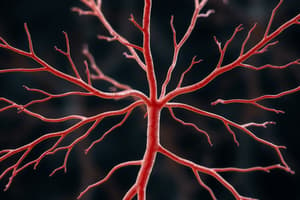
- Axon (nerve fiber) originates from the axon hillock on the soma. -- Cylindrical and relatively unbranched, but may have axon collaterals. -- Specialized for rapid nerve signal conduction to remote points. -- Cytoplasm is the axoplasm, and its membrane is the axolemma. -- A neuron has no more than one axon. -- Schwann cells and a myelin sheath associate with the axon. -- Somas range from 5–135 µm in diameter, while axons range from 1-20 µm and from millimeters to over a meter in length. -- The distal end has a terminal arborization of fine branches, each ending in a synaptic knob (terminal button) forming a junction (synapse) with the next cell. -- Synapse contains synaptic vesicles filled with neurotransmitters.
Neuron Structure Classification
- Multipolar neurons have one axon and multiple dendrites, most common neuron type.
- Bipolar neurons have one axon and one dendrite, including olfactory cells, retina neurons, and inner ear sensory neurons.
- Unipolar neurons have a single process leading away from the soma, carrying sensory signals to the spinal cord. -- Also called pseudounipolar – start as bipolar but fuse processes. -- The process branches like a T, with a peripheral fiber from a sensation source and a central fiber into the spinal cord. -- Dendrites are branching receptive endings, while the rest of the fiber is considered the axon due to myelin and action potential production.
- Anaxonic neurons have multiple dendrites but no axon, communicate through dendrites without action potentials, found in the brain, retina, and adrenal medulla.
Axonal Transport
- Axonal transport moves proteins, organelles, and materials along the axon to and from the cell soma.
- Anterograde transport moves from the soma down the axon.
- Retrograde transport moves up the axon toward the soma.
- Materials travel along microtubules of the cytoskeleton using kinesin (anterograde) or dynein (retrograde) motor proteins.
- Two types of axonal transport exist: fast and slow. -- Fast axonal transport occurs at 20 to 400 mm per day, and can be anterograde or retrograde. -- Fast anterograde transports mitochondria, synaptic vesicles, organelles, axolemma components, calcium ions, enzymes to the axon's distal end. -- Fast retrograde returns used synaptic vesicles and informs the soma of conditions at the axon terminals. -- Some pathogens use fast retrograde transport to invade the nervous system, entering the axon tips and being transported to the soma. -- Slow axonal transport (axoplasmic flow) occurs at 0.5 to 10 mm per day and is always anterograde. -- Slow axonal transport moves enzymes and cytoskeletal components, renews axoplasm in mature neurons, and supplies axoplasm for developing or regenerating neurons. -- Damaged nerve fibers regenerate at a speed governed by slow axonal transport.
Supportive Cells (Neuroglia)
- Neurons are outnumbered 50 to 1 by neuroglia, or glial cells, which bind neurons and form a supportive framework.
- Six types of neuroglia exist, each with a unique function; four are only in the CNS:
- Oligodendrocytes: CNS cells resembling an octopus with processes that spiral around nerve fibers, forming an insulating myelin sheath for faster nerve conduction.
- Ependymal cells: CNS cells resembling cuboidal epithelium lining brain and spinal cord cavities with no basement membrane and root-like processes. -- Produce and circulate cerebrospinal fluid (CSF) using cilia.
- Microglia: Small macrophages from white blood cells (monocytes) that wander through the CNS, phagocytizing dead tissue and microorganisms, clustering in damaged areas.
- Astrocytes: Abundant, star-shaped CNS cells with diverse functions. -- They form a supportive framework for nervous tissue. -- Perivascular feet extensions contact capillaries and stimulate the formation of a tight blood-brain barrier. -- Convert blood glucose to lactate for neurons. -- Secrete nerve growth factors that promote neuron growth and synapse formation. -- Communicate electrically with neurons and influence signaling. -- Regulate chemical composition of tissue fluid, absorbing neurotransmitters and potassium ions. -- Form hardened scar tissue when neurons are damaged (astrocytosis or sclerosis).
- Schwann cells: PNS cells that envelope nerve fibers, producing a myelin sheath. -- Assist in regeneration of damaged fibers.
- Satellite cells: PNS cells that surround neurosomas in ganglia, providing electrical insulation and regulating the chemical environment.
The Myelin Sheath
- The myelin sheath is an insulating layer around a nerve fiber, composed of 20% protein and 80% mixed lipids.
- Myelination begins in the 14th fetal week, rapidly proceeds in infancy, and completes in late adolescence. -- Children under 2 years should not be on low-fat diets.
- In the PNS, a Schwann cell spirals around a single nerve fiber, forming layers of membrane with almost no cytoplasm. -- The outermost coil is the neurilemma, containing the Schwann cell body and nucleus. -- External to the neurilemma are the basal lamina and endoneurium (fibrous connective tissue).
- In the CNS, each oligodendrocyte myelinates multiple nerve fibers in its vicinity. -- Cannot migrate around fibers, pushing new myelin layers under older ones. -- Nerve fibers of the CNS have no neurilemma or endoneurium.
- Schwann cells or oligodendrocytes are needed to cover a single nerve fiber. -- The myelin sheath is segmented, with gaps called nodes of Ranvier. -- Myelin-covered segments are called internodes (0.2 to 1.0 mm long). -- The initial segment is the nerve fiber section between the axon hillock and the first glial cell, and the axon hillock plus initial segment form the trigger zone.
Unmyelinated Fibers
- Many CNS and PNS nerve fibers are unmyelinated, but PNS fibers are enveloped by Schwann cells.
- One Schwann cell harbors 1 to 12 unmyelinated fibers in grooves, folding its plasma membrane around the fiber. -- This wrapping is the neurilemma (mesaxon). -- Nerve fibers have individual channels, but small fibers may bundle. -- A basal lamina surrounds the Schwann cell and fibers.
Nerve Signal Conduction
- The speed of nerve signals depends on fiber diameter and the presence/absence of myelin.
- Signal conduction occurs on the fiber surface, not in the axoplasm.
- Large fibers have more surface area and conduct more rapidly than small fibers.
- Nerve signals travel at 0.5 to 2.0 m/sec in small unmyelinated fibers and 3 to 15 m/sec in myelinated fibers of the same size.
- In large myelinated fibers, nerve signals travel as fast as 120 m/sec.
- Unmyelinated fibers are sufficient for many processes; fast myelinated fibers are vital for motor commands to skeletal muscles.
Vulnerability and Regeneration
- PNS nerve fibers are vulnerable to trauma but can regenerate if the soma is intact and there is some neurilemma.
- Somatic motor neuron regeneration involves the following steps: -- Distal fiber to the injury cannot survive without synthesizing protein; Schwann cells degenerate, and macrophages clean up tissue debris. -- The soma swells, its ER breaks up, the nucleus moves off-center; some neurons may die. -- The axon stump may sprout growth processes while the distal end degenerates. -- Muscle fibers shrink due to denervation atrophy. -- Schwann cells, basal lamina, and neurilemma form a regeneration tube, producing cell-adhesion molecules and nerve growth factors. -- One axon growth process finds the tube, growing rapidly (3-5 mm/day). -- The tube guides the sprout back to target cells, reestablishing synaptic contact. -- The soma shrinks and muscle fibers regrow.
- Regeneration is imperfect; some fibers connect to the wrong muscle fibers, or die, resulting in functional deficit.
- Damaged CNS nerve fibers cannot regenerate due to bone enclosure.
Electrophysiology of Neurons
- Santiago Ramon y Cajal showed that the nervous pathway is a series of cells separated by synapses, known as the neuron doctrine.
- Neurophysiology addresses how neurons generate electrical signals and transmit meaningful messages.
- An electrical potential is a concentration difference of charged particles between two points; an electrical current is a flow of charged particles. -- When an electrical potential exists, they are in a polarized state.
- Living cells are polarized due to the resting membrane potential (RMP) across the plasma membrane. -- In a resting neuron, the membrane potential is –70 mV. -- Electrical currents in the body are created by ion flow (Na+ and K+) through gated channels.
- The resting membrane potential exists because electrolytes are unequally distributed between ECF and ICF, determined by: -- Ion diffusion down concentration gradients. -- Selective membrane permeability to ions. -- Electrical attraction of cations and anions.
Potassium Ions Influence on RMP
- Potassium ions (K+) have significant influence on RMP since the plasma is most permeable to K+. -- Cytoplasmic anions cannot escape the cell due to size/charge, K+ diffusion out leaves a negative charge in ICF. -- Negative ICF attracts K+ back, reaching equilibrium between diffusion and electrical attraction, stopping net K+ diffusion, when K+ is 40 times as concentrated in the ICF. -- If K+ were the only ion involved, RMP would be about -90 mV.
Sodium Ions Influence on RMP
- Sodium ions (Na+) - Na+ is 12 times more concentrated in the ECF. -- Some Na+ diffuses down the concentration gradient into the cell and is attracted by the anions in the ICF.
- The Na+-K+ pump compensates for Na+ and K+ leakage.
- For every 1 ATP, 3 Na+ are pumped out and 2 K+ are brought in.
- The Na+-K+ pump is 70% of the nervous system's ATP usage. Net effect of K+ diffusion outward, Na+ diffusion inward is -70 mV RMP.
Local Potential
- Changes in local potential are triggered by the stimulus in a neuron.
- The response begins at the dendrite, spreads though the soma, travels to the axon, and ends at the synapse.
- Stimulation at the dendrites open the Na+ gates so that Na+ flows into the cell neutralizing the negative charges.
- Depolarization is any change that makes the membrane voltage less negative.
- Incoming Na+ ions diffuse for a short distance and produce a current from the stimulus to the trigger zone. Four characteristics distinguish local potentials from action potentials
Local vs Action Potentials
- Local -- Graded; vary depending on stimulus -- They weaken the farther they travel from the stimulus -- They are reversible -- Either inhibitory or excitatory
Triggering an Action Potential
- Action potentials have a rapid shift in the membrane voltage produced by voltage-regulated ion gates in the plasma membrane. -- Only if enough voltage-regulated gates are there will action potentials occur -- The trigger zone contains 350-500 gates compared to the 50 to 75 in a soma.
- If a local potential spreads into the trigger zone it may be able to generate an action potential if it opens up the gates.
- The sequence of an action potential is: -- the Na+ arrives at the axon hillock for a local potential -- The local potential rises high enough to open the voltage-regulated gates. -- The voltage-regulated Na+ gates open rapidly and the gates slowly depolarize further, causing a positive feedback loop. -- The Na+ gates inactivate and begin closing. -- The K+ gates are fully open so K+ exits and repolarizes the membrane. -- K+ gates are kept open longer so that more K+ leaves the cell than Na+ entered, hyperpolarizing the membrane. -- Na+ diffusion into the cell and the removal of K+ gradually restore the RMP.
- The action potential only affects a small thin layer of ions near the membrane.
Action Potential Characteristics
- Action Potentials follow an all-or-nothing law and are not graded (if it doesn't reach the threshold, it is not generated)
- Action potentials are nondecremental, they do not weaken
- Action potentials are irreversible and after initiated, they will go to completion.
Refractory Period
- The neuron cannot be stimulated during the action potential and shortly after
- The refractory period has two phases: -- The absolute refractory period: no stimulus will cause a new action potential, this happens when it returns to RMP, or as long as the Na+ gates are open. -- The relative refractory period: an unusually strong stimulate will trigger a new action potential.
- The refractory period only happens when an action potential has already occurred.
- Signal conduction is the opposite in unmyelinated and myelinated fibers.
Electrical Signals in Unmyelinated Fibers
- Unmyelinated fibers have voltage-regulated ions along the entire length.
- An action potential occurs when Na+ enters the axon and depolarizes, exciting the voltage-regulated gates.
- Chain reaction continues as the gates travel
- Signal does not travel along the axon, instead produces a new potential ahead of it. -Action potentials do not travel backward because the membrane signal is in its refractory period. Electrical signals in the neuron are not a current in a wire.
Electrical Signals in Myelinated Fibers
- Very few gates in the myelinating internodes, in ranvier there are an abundance of them. -- Signals travel through the internode and Na+ travels down the axolemma, which is not fast, but decremental.
- These gates are voltage regulated and every ranvier has an abundance of gates.
- Saltatory Conductance is when diffusing ions reach a node and creates a new signal
Synapses
- A Synapse happens at the end of the axon between two neurons, the first is the presynaptic and the second is the postsynaptic.
- Pre synaptic can form an axodentritic, axosomatic, or axoaxonic synapse.
- A motor neuron may have 10,000 synaptic knobs that connect to a spinal motor neuron from other motor neurons.
- In the cerebellum their could be as many as 100,000.
Neurotransmitters
- Acetylcholine was the first to be tested in 1921 by Otto Loewi
- At first electrical signals were not considered possible because of the synaptic cleft, but new research shows it is possible in some excited cells like cardiac muscle.
- The structure of a chemical synapsis occurs on the synaptic knob, a component of the presynaptic neuron containing synaptic vesicles.
- The vesicles in the neuron are docked at the membrane near the site, and others constitute a reserve pool further back.
- The postsynaptic neuron doesn't release vesicles like the pre-synaptic neuron
- the postsynaptic instead contains receptor proteins and ligand-regulated ion gates.
- More than 100 likely neurotransmitters have been identified.
- There are four categories: -- Acetylcholine -- Amino Acids -- Monoamines -- Neuro peptides: Some of them function like hormones and can be produced by the digestive system.
- Small organic compounds that function with the synapse in the following ways. -- Produced by presynaptic neuron -- Triggered by stimulation -- They bind to receptors -- Alter the physiology
Actions of a Neurotransmitter
- 3 Actions are illustrated from an excitatory Cholinergic synapse.
- The arrival of a nerve signal opens Voltage regulated calcium gates
- Ca2+ will enter triggering exocytosis where it releases ACh
- The empty vesicles drop into the cytoplasm where they are refilled: vesicles in the pool move to active sites:
- ACh diffuses across and binds to ligand gated gates to enter the cell while K+ leaves.
Synapses
- An adrenergic synapse employs norepinephrine as a neurotransmitter which its receptors are a transmembrane protein.
- Binding of NE to receptors causes the G protein to dissociate from it, binding with cyclase to change ATP into cyclic AMP. -- These adrenergic snapses amplify, that is each NE causes tons of formation.
- Synaptic events need 0.5 msec to occur
- Cessation of a signal occurs when neurotransmitter stops releasing
The Synaptic Cleft
- Removal is accomplished: -- Through diffusion, which is later absorbed by astrocytes. Also reuptake where it reabsorbs and breakdowns by Monoamin oxidase. There's also the process of degradation where enzymes breakdown.
- Neuromodulators are messengers that change synaptic transmissions
- A family of neuro modulators which have small peptides inhibits spinal interneurons from sending pain signals
- Nitric Oxide releases and triggers a postsynaptic neuron.
Neural Integration
- The ability of neurons to process information, store it
- neural integration is based on neurotransmitters as well.
- Any voltages made higher reach the threshold closer or from -30/-55 is termed an ESPS.
- Any voltage change that makes the membrane more negative it's termed an IPSP Because of ion leakage, all Neurons fire at the same kind of Rate.
- There are chemicals that can produce:
- The primary chemicals are glutamate asparatate ePSP, glycine gaba is IPSP. acH is both.
- Stimulation, facilitation, and inhibition influence neural actions.
Action of Neural Circuits
- Stimulation - Allows to make all decisions
- A reaction doesn't produce enough stimulus to produce more, rather It takes 30 EPSPs to become something and can be added in 2 ways -- Temporarily which means it can cause EPSPs that are quickly generated to become cumulative.
- There is spatial summation when a spatial epsp is from several locations that add up. the Neurons routinely working in groups to move. -- Facilitation Enhances and helps with example from two of them to indcue. -- pre-synaptic inhibition reduces the work.
- The way nervous system makes and understand the code.
The Labeled Line Code
- The most effective is the line code, which carries specific stimuli. A Quanatative Information on the intensity of sstimuls
- Has the recruitment.
- This has an upperlimit to what neurons are.
Neural Pools
- Neurons function and can contain millions functions are determined by circuits, info arrives at the one one input. the neuro ncan make the cell fire. the neuro stimulates them.
Parallel Circuits
- Consists of what passes through the variety and principle which contains:
- divergin- one neerve fiber synapses that sends it through some pathways. the nerve fiber channels to one that helps evalue different systems. In reverbation it is stimulates some have back to the main one. parallel different change, and at different times. may continue til stim stops. no feedback , output stope
Memory
- physical bass - brain pathway is calles trace itss ablility to sync add be modified. is a certain pathway , called potentiuation, holds it for a few.
- it is called sync potentials.
Short Term Memory
- stm second and hours the memory is hold for a certain amount of hours.
Limbic System
- involved because of its involvement with emotional. It's important in reading because of its reverberatory. longer in short term more synaptic can happen. rapid causes a build of calcium this gets stronger. In long term the calc levels stay elevated jog earlier memorie
Studying That Suits You
Use AI to generate personalized quizzes and flashcards to suit your learning preferences.