Podcast
Questions and Answers
Damage to which lobe would most likely result in difficulties with understanding spoken language?
Damage to which lobe would most likely result in difficulties with understanding spoken language?
- Occipital Lobe
- Frontal Lobe
- Parietal Lobe
- Temporal Lobe (correct)
Which of the following best describes the composition and location of gray matter within the central nervous system (CNS)?
Which of the following best describes the composition and location of gray matter within the central nervous system (CNS)?
- Primarily composed of myelinated axons, found in the deep nuclei only.
- Primarily composed of nerve cell bodies, found in the cortical layer and deep nuclei. (correct)
- Primarily composed of glial cells, evenly distributed throughout the CNS.
- Primarily composed of fiber tracts, connecting different parts of the CNS.
The anterior spinal artery supplies which portion of the spinal cord and originates from which arteries?
The anterior spinal artery supplies which portion of the spinal cord and originates from which arteries?
- The lateral columns; originating from the posterior inferior cerebellar arteries.
- The posterior one-third; originating from the internal carotid arteries.
- The anterior two-thirds; originating from the vertebral arteries. (correct)
- The entire spinal cord; originating from the basilar artery.
What is the primary function of the choroid plexus, and in which structures is it located within the brain?
What is the primary function of the choroid plexus, and in which structures is it located within the brain?
Which of the following accurately describes the flow of cerebrospinal fluid (CSF) from its production site to its reabsorption point?
Which of the following accurately describes the flow of cerebrospinal fluid (CSF) from its production site to its reabsorption point?
In a patient experiencing increased intracranial pressure due to impaired CSF reabsorption, which of the following structures is most likely to be malfunctioning?
In a patient experiencing increased intracranial pressure due to impaired CSF reabsorption, which of the following structures is most likely to be malfunctioning?
A patient presents with a lesion affecting the posterior inferior cerebellar artery (PICA). Which of the following neurological deficits is most likely to be observed, considering the PICA's area of supply?
A patient presents with a lesion affecting the posterior inferior cerebellar artery (PICA). Which of the following neurological deficits is most likely to be observed, considering the PICA's area of supply?
Which statement accurately describes the role of cerebrospinal fluid (CSF) in both normal and pathological conditions within the central nervous system?
Which statement accurately describes the role of cerebrospinal fluid (CSF) in both normal and pathological conditions within the central nervous system?
A patient presents with loss of pain and temperature sensation on the left side of their body. Assuming a single lesion, where is the most likely location of this lesion in the spinal cord?
A patient presents with loss of pain and temperature sensation on the left side of their body. Assuming a single lesion, where is the most likely location of this lesion in the spinal cord?
A patient has difficulty with fine motor movements and exhibits increased muscle tone on the right side of their body following a spinal cord injury. Which spinal tract is most likely affected?
A patient has difficulty with fine motor movements and exhibits increased muscle tone on the right side of their body following a spinal cord injury. Which spinal tract is most likely affected?
Damage to the anterior spinal artery can lead to anterior cord syndrome. Which set of symptoms would most likely be observed in a patient with this syndrome?
Damage to the anterior spinal artery can lead to anterior cord syndrome. Which set of symptoms would most likely be observed in a patient with this syndrome?
A patient presents with loss of discriminative touch and proprioception on the right side of their body. Where is the most likely location of the lesion?
A patient presents with loss of discriminative touch and proprioception on the right side of their body. Where is the most likely location of the lesion?
A patient exhibits ipsilateral loss of motor function, ipsilateral loss of discriminative touch and proprioception, and contralateral loss of pain and temperature sensation. Which condition is most likely?
A patient exhibits ipsilateral loss of motor function, ipsilateral loss of discriminative touch and proprioception, and contralateral loss of pain and temperature sensation. Which condition is most likely?
Following a traumatic injury, a patient develops a syrinx in the central canal of the spinal cord. Which of the following is the mechanism of this condition?
Following a traumatic injury, a patient develops a syrinx in the central canal of the spinal cord. Which of the following is the mechanism of this condition?
A clinician assesses a patient with a spinal cord injury and notes spasticity and brisk deep tendon reflexes below the level of the lesion. What does this indicate about the affected corticospinal tract?
A clinician assesses a patient with a spinal cord injury and notes spasticity and brisk deep tendon reflexes below the level of the lesion. What does this indicate about the affected corticospinal tract?
Which component of the nervous system connects the brain and spinal cord to the periphery and visceral tissues?
Which component of the nervous system connects the brain and spinal cord to the periphery and visceral tissues?
During the relative refractory period of a neuron, what is the primary factor contributing to the higher threshold required to initiate an action potential?
During the relative refractory period of a neuron, what is the primary factor contributing to the higher threshold required to initiate an action potential?
Which of the following comparisons accurately describes the difference between continuous and saltatory conduction?
Which of the following comparisons accurately describes the difference between continuous and saltatory conduction?
Which of the following is a key difference between electrical and chemical synapses?
Which of the following is a key difference between electrical and chemical synapses?
If a drug selectively blocked metabotropic glutamate receptors (mGluRs) in the CNS, what would be the most likely direct effect?
If a drug selectively blocked metabotropic glutamate receptors (mGluRs) in the CNS, what would be the most likely direct effect?
How do GABAA receptors mediate their inhibitory effects in the CNS?
How do GABAA receptors mediate their inhibitory effects in the CNS?
What is the primary mechanism by which acetylcholinesterase (AChE) terminates signaling at cholinergic synapses?
What is the primary mechanism by which acetylcholinesterase (AChE) terminates signaling at cholinergic synapses?
Which of the following describes a primary role of astrocytes in neuronal function?
Which of the following describes a primary role of astrocytes in neuronal function?
Which of the following best describes the concept of temporospatial summation in synaptic transmission?
Which of the following best describes the concept of temporospatial summation in synaptic transmission?
What directly triggers the release of acetylcholine (ACh) into the synaptic cleft at the neuromuscular junction?
What directly triggers the release of acetylcholine (ACh) into the synaptic cleft at the neuromuscular junction?
Which component of the neuromuscular junction is responsible for propagating the action potential to the muscle fiber?
Which component of the neuromuscular junction is responsible for propagating the action potential to the muscle fiber?
What is the immediate effect of acetylcholine (ACh) binding to its receptors on the skeletal muscle membrane?
What is the immediate effect of acetylcholine (ACh) binding to its receptors on the skeletal muscle membrane?
Which of the following best describes the role of voltage-gated calcium channels at the neuromuscular junction?
Which of the following best describes the role of voltage-gated calcium channels at the neuromuscular junction?
What is the endplate potential (EPP) and how is it generated?
What is the endplate potential (EPP) and how is it generated?
Which of the following describes the sequence of events at the neuromuscular junction (NMJ) that leads to muscle contraction?
Which of the following describes the sequence of events at the neuromuscular junction (NMJ) that leads to muscle contraction?
Which of the following does NOT directly participate in signal transduction at the neuromuscular junction (NMJ)?
Which of the following does NOT directly participate in signal transduction at the neuromuscular junction (NMJ)?
What is the primary function of the neuromuscular junction (NMJ)?
What is the primary function of the neuromuscular junction (NMJ)?
A patient presents with a lesion affecting the anterior white commissure of the spinal cord. Which sensory deficit would you most likely observe?
A patient presents with a lesion affecting the anterior white commissure of the spinal cord. Which sensory deficit would you most likely observe?
A stroke involving the caudal medulla affects the medial lemniscal pathway. What sensory deficits would result from this?
A stroke involving the caudal medulla affects the medial lemniscal pathway. What sensory deficits would result from this?
Which spinal cord tract plays a crucial role in coordinating movement, particularly by facilitating flexor muscle activity in the upper limbs and originating from the red nucleus?
Which spinal cord tract plays a crucial role in coordinating movement, particularly by facilitating flexor muscle activity in the upper limbs and originating from the red nucleus?
A patient exhibits loss of fine touch and proprioception in the left leg. Imaging reveals a spinal cord lesion. Which specific region of the spinal cord is most likely affected?
A patient exhibits loss of fine touch and proprioception in the left leg. Imaging reveals a spinal cord lesion. Which specific region of the spinal cord is most likely affected?
Damage to the anterior corticospinal tract in the spinal cord would most likely result in:
Damage to the anterior corticospinal tract in the spinal cord would most likely result in:
A patient has a lesion in the spinal cord that results in the loss of pain and temperature sensation a few segments below the level of the lesion on the opposite side of the body. Which of the following explains this phenomenon?
A patient has a lesion in the spinal cord that results in the loss of pain and temperature sensation a few segments below the level of the lesion on the opposite side of the body. Which of the following explains this phenomenon?
Compared to the spinothalamic tract, what is a key difference in the origin and termination of the Dorsal Column-Medial Lemniscal Pathway?
Compared to the spinothalamic tract, what is a key difference in the origin and termination of the Dorsal Column-Medial Lemniscal Pathway?
How does the location of the fasciculus gracilis and fasciculus cuneatus within the posterior columns relate to the body region they serve?
How does the location of the fasciculus gracilis and fasciculus cuneatus within the posterior columns relate to the body region they serve?
Which of the following accurately describes the path of the rubrospinal tract?
Which of the following accurately describes the path of the rubrospinal tract?
What is the primary role of the alpha motor neuron (α-motor neuron)?
What is the primary role of the alpha motor neuron (α-motor neuron)?
During the myotatic reflex, which type of sensory receptor is primarily responsible for detecting changes in muscle length?
During the myotatic reflex, which type of sensory receptor is primarily responsible for detecting changes in muscle length?
What is the functional significance of reciprocal innervation in the myotatic reflex?
What is the functional significance of reciprocal innervation in the myotatic reflex?
Why is the myotatic reflex classified as a monosynaptic reflex?
Why is the myotatic reflex classified as a monosynaptic reflex?
An individual with damage to their Ia interneurons would likely exhibit which of the following during a patellar tendon tap?
An individual with damage to their Ia interneurons would likely exhibit which of the following during a patellar tendon tap?
Where are Golgi tendon organs (GTOs) located, and what do they primarily monitor?
Where are Golgi tendon organs (GTOs) located, and what do they primarily monitor?
How does the inverse myotatic reflex (Golgi tendon reflex) protect muscles from injury?
How does the inverse myotatic reflex (Golgi tendon reflex) protect muscles from injury?
Flashcards
Temporal Lobe
Temporal Lobe
Processes auditory information, language comprehension, memory formation, and facial/object recognition.
Occipital Lobe
Occipital Lobe
The primary visual cortex responsible for processing visual information.
Limbic Lobe
Limbic Lobe
Involved in emotional regulation.
Gray Matter
Gray Matter
Signup and view all the flashcards
White Matter
White Matter
Signup and view all the flashcards
Anterior Spinal Artery
Anterior Spinal Artery
Signup and view all the flashcards
Choroid Plexus
Choroid Plexus
Signup and view all the flashcards
Role of CSF
Role of CSF
Signup and view all the flashcards
Relative Refractory Period
Relative Refractory Period
Signup and view all the flashcards
Saltatory Conduction
Saltatory Conduction
Signup and view all the flashcards
Synapse
Synapse
Signup and view all the flashcards
Neurotransmitter
Neurotransmitter
Signup and view all the flashcards
Glutamate
Glutamate
Signup and view all the flashcards
GABA and Glycine
GABA and Glycine
Signup and view all the flashcards
Acetylcholine
Acetylcholine
Signup and view all the flashcards
Biogenic Amines
Biogenic Amines
Signup and view all the flashcards
Lateral Corticospinal Tract
Lateral Corticospinal Tract
Signup and view all the flashcards
Spinothalamic Tract
Spinothalamic Tract
Signup and view all the flashcards
Posterior Column
Posterior Column
Signup and view all the flashcards
Neuromuscular Junction (NMJ)
Neuromuscular Junction (NMJ)
Signup and view all the flashcards
Anterior Spinal Cord Syndrome
Anterior Spinal Cord Syndrome
Signup and view all the flashcards
NMJ Components
NMJ Components
Signup and view all the flashcards
Syringomyelia
Syringomyelia
Signup and view all the flashcards
ACh Release
ACh Release
Signup and view all the flashcards
Brown-Séquard Syndrome
Brown-Séquard Syndrome
Signup and view all the flashcards
Endplate Potential
Endplate Potential
Signup and view all the flashcards
Ipsilateral Motor Loss
Ipsilateral Motor Loss
Signup and view all the flashcards
Peripheral Nervous System (PNS)
Peripheral Nervous System (PNS)
Signup and view all the flashcards
Ca2+ and Contraction
Ca2+ and Contraction
Signup and view all the flashcards
Calcium Influx
Calcium Influx
Signup and view all the flashcards
Neurotransmitter Release
Neurotransmitter Release
Signup and view all the flashcards
Receptor Binding
Receptor Binding
Signup and view all the flashcards
Anterior Corticospinal Tract function
Anterior Corticospinal Tract function
Signup and view all the flashcards
Spinothalamic Tract function
Spinothalamic Tract function
Signup and view all the flashcards
Spinothalamic Tract lesion effect
Spinothalamic Tract lesion effect
Signup and view all the flashcards
DC-Medial Lemniscal Pathway function
DC-Medial Lemniscal Pathway function
Signup and view all the flashcards
DC-Medial Lemniscal Pathway lesion effect
DC-Medial Lemniscal Pathway lesion effect
Signup and view all the flashcards
Rubrospinal Tract function
Rubrospinal Tract function
Signup and view all the flashcards
Rubrospinal Tract specifics
Rubrospinal Tract specifics
Signup and view all the flashcards
Dorsal Column-Medial Lemniscal Pathway neuron count
Dorsal Column-Medial Lemniscal Pathway neuron count
Signup and view all the flashcards
Rubrospinal Tract
Rubrospinal Tract
Signup and view all the flashcards
Alpha Motor Neuron
Alpha Motor Neuron
Signup and view all the flashcards
Myotatic Reflex
Myotatic Reflex
Signup and view all the flashcards
Muscle Spindles
Muscle Spindles
Signup and view all the flashcards
Ia Nerve Afferents
Ia Nerve Afferents
Signup and view all the flashcards
Reciprocal Innervation
Reciprocal Innervation
Signup and view all the flashcards
Monosynaptic
Monosynaptic
Signup and view all the flashcards
Inverse Myotatic Reflex
Inverse Myotatic Reflex
Signup and view all the flashcards
Study Notes
Cellular Components of the Nervous System
- Neurons are essential to the nervous system.
- They feature a functional organization with the soma, dendrites, and axon.
- The axon may have a myelin sheath.
- Neurons come in multipolar, pseudounipolar, and bipolar types.
- Synapses can be axodendritic, axosomatic, or axoaxonic.
- Axodendritic synapses link axons to dendrites, enabling temporal-spatial summation.
- Glial cells include: astroglia, oligodendroglia, Schwann cells, microglia, polydendrocytes, and ependymal cells.
- Astroglia includes astrocytes and Müller cells and is part of the blood-brain barrier and tripartite synapses.
- Astrocytes are fibrous (in white matter) or protoplasmic (in gray matter).
- Oligodendroglia is myelinating cells in the central nervous system.
- Schwann cells are myelinating cells in the peripheral nervous system.
- Microglia removes debris and presents antigens as immune cells.
- Polydendrocytes are stem cells generating glia and neurons, implicated in demyelinating disorders and network cross-talk.
- Ependymal cells are epithelial cells lining ventricles that separate cerebrospinal fluid (CSF) from the neuropil; also, they help produce CSF via the choroid plexus.
- The components of the blood-brain barrier include endothelial cells with tight junctions and astrocyte processes to maintain homeostasis, and it facilitates substance transport via diffusion or active transport.
Basic Neurophysiology
- Neurophysiology studies ion movements across membranes for signal transduction, action potentials, and neurotransmitter action.
- Ion movement across membranes plays a crucial role in neurophysiology.
- An action potential is depolarization moving down the axon.
- It starts when membrane voltage drops to a threshold, opening voltage-activated ion channels for Na+ inflow to allow flow into the neuron.
- Action potentials show an all-or-none response and self-propagate.
- Repolarization follows behind the action potential.
- Axons enter an absolute refractory period that prevents additional firing.
- After sufficient Na+ resets, there's a relative refractory period that needs a higher threshold.
- Action potential speed relies on axon diameter and myelination.
- Continuous conduction involves the total axon membrane (unmyelinated neurons).
- Saltatory conduction (myelinated neurons) makes depolarization jump between nodes of Ranvier, accelerating it.
- Synaptic transmission can be electrical or chemical.
- A synapse is a functioning connection between two neurons.
- Electrical synapses use gap junctions.
- Chemical synapses use vesicles.
- Synaptic signal transduction uses inhibitory (IPSP) and excitatory postsynaptic potentials (EPSP).
- Temporospatial summation effectively evokes potential.
- Receptors are either ionotropic or metabotropic.
Main Neurotransmitters
- Neurotransmitters include glutamate, GABA, glycine, acetylcholine, biogenic amines, ATP, and neuropeptides.
- Glutamate is the main excitatory neurotransmitter in the CNS, working on NMDA, AMPA, kainate, and mGluRs receptors.
- Its precursor is glutamine, which is supplied by astrocytes.
- GABA and glycine serve as chief inhibitory neurotransmitters in the CNS.
- GABA links to ionotropic GABAA and GABAC receptors, causing Cl- influx and hyperpolarization.
- The metabotropic GABAB receptor activates K+ channels and blocks Ca2+ channels, thereby hyperpolarizing.
- Acetylcholine is within the peripheral nervous system (PNS), CNS, and neuromuscular junctions.
- Nicotinic acetylcholine receptors are ionotropic, while muscarinic acetylcholine receptors are metabotropic.
- Acetylcholinesterase deactivates acetylcholine.
- Biogenic amines: This class includes catecholamines (dopamine, norepinephrine, epinephrine), histamine, and serotonin.
- ATP acts as an energy carrier, cotransmitter, and neuromodulator.
- Neuropeptides: These encompass substance P, metenkephalin, opioids, and corticotropin-releasing hormone.
General Organization and Major Anatomical Structures of the CNS
- The brain has the forebrain, brainstem, and hindbrain.
- The forebrain includes the telencephalon (cerebrum and basal ganglia) and the diencephalon (thalamus, hypothalamus, and subthalamus), and the thalamus is a gateway to the cerebral cortex.
- The hypothalamus manages autonomic, endocrine functions, plus impacts behavior, whereas the subthalamus changes voluntary motor activity.
- The brainstem contains the midbrain.
- Hindbrain has the medulla, pons, and cerebellum.
- The spinal cord is divided into cervical, thoracic, lumbar, sacral, and coccygeal areas.
- Meninges cover both the brain and spinal cord and have three layers including: dura mater, arachnoid mater, and pia mater.
Lobes of the Brain and Important Landmarks
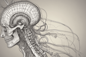
- The primary lobes of the brain include frontal, parietal, temporal, occipital, and limbic lobes.
- The longitudinal fissure separates the cerebral hemispheres.
- The central sulcus divides the frontal from parietal lobes.
- The lateral fissure (Sylvius) separates the frontal and parietal lobes from the temporal lobe.
- The parieto-occipital sulcus sits on the medial surface, splitting the occipital from parietal and temporal lobes.
- The calcarine fissure is on the medial surface of the occipital lobe
- Gyrus: This is a ridge on the cerebral cortex.
- Sulcus: A valley between gyri on the cerebrl cortex.
- Deep sulcus: AKA as a fissure.
- Precentral gyrus: It is located anterior to the central sulcus, and happens to be the primary motor area.
- Postcentral gyrus: It is located posterior to the central sulcus, and happens to be the primary somatosensory area.
- Superior temporal gyrus: It is involved with higher-level auditory processing, including speech and sound localization.
- Parahippocampal gyrus: Critical for memory formation, spatial orientation, and scene recognition.
- Fusiform gyrus: It is strongly involved in facial recognition.
- Cingulate gyrus: It relates to motivation, spotting mistakes, and making decisions.
Functions of Different Lobes
- Frontal lobe: This lobe controls voluntary movement, plans and coordinates complex actions, enables language production/speech, supports high-level cognitive functions/emotional processes, and promotes decision-making/social behavior.
- Parietal lobe: It interprets sensory data relating to touch, pressure, warmth, and discomfort, in addition to involving with language and reading.
- Temporal lobe: It processes auditory data, and supports language comprehension/memory, plus facial/object recognition.
- Occipital lobe: It forms the primary visual cortex.
- Limbic lobe: It is essential for emotional regulation.
Arrangements of Gray and White Matter
- Gray matter contains nerve cell bodies and occurs in the cortical layer and certain deep nuclei.
- White matter has fiber tracts (axons) joining distinct portions of the CNS, most of which is myelinated.
Structure of the Blood-Brain Barrier (BBB)
- The blood-brain barrier (BBB) is a selective barrier that protects the brain from harmful substances while allowing essential nutrients to enter.
- It's built from tight junctions between endothelial cells, astrocyte end-feet surrounding blood vessels, and a thick basement membrane.
- The BBB stops large molecules, pathogens, and toxins from getting through, so neuronal function stays optimal.
Vessels that Supply the CNS
- Internal Carotid Artery (ICA): Features cervical, petrous, cavernous, and cerebral segments.
- Vertebral-Basilar Arterial system: The spinal artery forms through branches stemming off vertebral arteries, giving a blood supply to the spinal cord's anterior two-thirds.
- Posterior spinal arteries are sourced from the vertebral arteries or else from posterior inferior cerebellar arteries (PICAs), so delivering a supplement of blood for that posterior one-third on spinal cord.
Anatomy and function of the Choroid Plexus
- The choroid plexus produces cerebrospinal fluid (CSF) across all four ventricles; the choroid epithelium is made of ependymal cells that morph into cuboidal epithelium, along with capillaries.
Circulation of Cerebral Spinal Fluid (CSF)
- CSF secreted from choroid plexus travels out ventricles, starting from the lateral and moving in sequence through that third, then on into the ventricle, and finally gets in CSF filling inside that narrow central canal through getting exited from this ventricle past foramina( Luschka/Magendie).
- CSF passes though the subarachnoid area right to arachnoid granulations allowing itself drained back into the bloodstream through this passage or villi, which goes through where the superior sagittal sinus lies within itself.
Somatotopy and Blood Supply Occlusions
- Somatotopy: Each part on something you touch links back onto some matching spot located inside your central system.
- Impaired blood supply with blockages could further bring about focal deficits based precisely for what brain location ends up impacted plus their subsequent topographical layout mapping.
- Primary motor cortex lesions: If precentral cortex areas get broken, contralateral muscle malfunction or weakness happen most commonly as outcome scenarios.
- Primary sensory lesions: If post central gyrus get broken, contralateral muscle malfunction or weakness happen most commonly as outcome scenarios.
Circle of Willis
- The Circle of Willis is an arterial anastomosis that connects the internal carotid and vertebral-basilar systems.
- It consists of anterior communicating artery that connects the two ACAs, while the posterior communicating arteries connects ICAs to PCAs to ensure good blood supply to the brain.
Spinal Cord Functional Anatomy
- Spinal Meninges have three layers like what is happening with someone's human counterparts: the dura mater situated furthest from where injury could take place right on through protective barrier and where arachnoid follows next which further houses liquid called ‘cerebral’, then there is one layer positioned down quite close by, which shields with that too.
- White Matter has axons arranged into groups otherwise called "tracts" mostly, and it occupies area outside around those regions filled solely via grey matter,
- Gray Matter occurs towards cord's center , encompassing neurons inside all throughout central structure
- Rexed's Laminae consist mostly grey sections grouped alongside.
- Lamina II manages a level including sense around what some might refer in order as being ‘painful,’.
- Blood Supply: Anterior spinal artery giving blood on to this lower surface about 66%, while posterior ones assist about 33% to keep on well maintained health.
- Adamkiewicz blood carrier: If this particular artery sustains injuries this makes paraplegia.
Motor Components of the Spinal Cord
- Motor information leaves the spinal cord by way of anterior roots.
- Lower motor neurons (LMNs) exist from the spinal cord at each level.
- Autonomic fibers have cell bodies in the horn too.
- The anterior corticospinal tract transmits from the cerebral cortex over toward to the muscles around that central zone.
- The lateral corticospinal tract is sourced from the cerebrum but even crosses inside that brainstem, so reaching LMNs with any spinal location.
- Motor neurons of the spinal cord use acetylcholine (ACh) as neurotransmitter within the muscle junction.
- When an action potential hits that nerve's end, Ca+ reaches that release neurotransmitters at opening and opening of synapse.
Spinal Reflexes
- The reflex arc is a basic neuronal circuit where the sensory stimulus initiates a motor response instantly.
- The sensory signals come from Ia nerve afferents from the spinal cord, that synapse with a-motor parts in those same muscles.
- Ia interneurons inhibit a-motor neurons towards antagonist parts to the body.
- Withdrawing one particular point by the sensory stimulus, typically something causing painful stimulus.
- Activation upon entering it prevents homonymous to all parts associated muscle activity as primary stimulus or pain generator starts sending high input signals on spinal connections.
Dorsal Column-Medial Lemniscus System (DC-ML)
- It relays awareness regarding fine touch sensitivity, vibratory perceptions, sense about where your parts happen at within space along touch, discrimination pressure etc, while DC–ML pathway then makes contact down with the medulla.
- Ascending in ipsilateral posterior columns along the top location around what there happens here for its relay spot,
- The information stemming for where that part is within relation when compared up higher gets routed down one direction, medially via its path along up inside those brain components.
Anterolateral System (Spinothalamic Tract)
The anterolateral component relays sensing touch, vibration, position and proprioception, though touch information about temp and pain starts one spinal area but eventually finds transmission to higher level destinations located up inside cortex
Descending and Rubrospinal Tract Characteristics
- descending tracks regulate function originating at Brodmann's location 4 on the sides with the main pathway
- Anterolateral column transmits motor knowledge on parts, therefore improving contralateral area's postural.
Clinical Significance of the NMJ
- NMJ function is affected from diseases along lines on what can manifest through gravis, but Guillain barre symptoms has a contribution aspect to where demyelination causes slow to block conduction between regions, so causing both sensory as along with motor impairment.
- Myasthenia gravis: This is an autoimmune illness, and it affects the acetylcholine receptors.
- Guillon Barre syndrome: In this autoimmune version associated in PNS, the schwann cells take a hit.
Types of nerve fiber arrangement
- peripheral cables exhibit cables arranged within called fasciculi, so enveloped in what’s named as ‘connective cables’.
- Perineurium: These give security covering all individual nerve part.
- Epineurium forms what’s thought with each layer found outermost so enveloping all about the components with fibers contained inner areas
Studying That Suits You
Use AI to generate personalized quizzes and flashcards to suit your learning preferences.