Podcast
Questions and Answers
The upper motor neurons are located in which of the following?
The upper motor neurons are located in which of the following?
- pre central gyrus (correct)
- auditory cortex
- post central gyrus
- visual cortex
Regarding the white matter of the cerebral hemisphere, all of the following are examples of association type of fibers except:
Regarding the white matter of the cerebral hemisphere, all of the following are examples of association type of fibers except:
- optic radiation (correct)
- superior longitudinal fasciculus
- fronto-occipital fasciculus
- uncinated fasciculus
Lateral ventricle is the cavity of:
Lateral ventricle is the cavity of:
- Metencephalon
- Diencephalon
- Telencephalon (correct)
- Mesencephalon
Which cerebral lobe is located immediately posterior to central sulcus & superior to lateral sulcus?
Which cerebral lobe is located immediately posterior to central sulcus & superior to lateral sulcus?
Which of the following is a branch of internal carotid artery?
Which of the following is a branch of internal carotid artery?
Meckel's cartilage of 1st pharyngeal arch gives rise to all of the following except:
Meckel's cartilage of 1st pharyngeal arch gives rise to all of the following except:
Thyroid gland during its development descends in front of the following structures except
Thyroid gland during its development descends in front of the following structures except
The nasolacrimal duct is formed along the line of fusion of lateral nasal process & maxillary process by invagination of:
The nasolacrimal duct is formed along the line of fusion of lateral nasal process & maxillary process by invagination of:
In skeletal muscles, which of the following is correct?
In skeletal muscles, which of the following is correct?
Which type of epithelium lines the bronchi of lungs?
Which type of epithelium lines the bronchi of lungs?
Which of the following co... is responsible for anaphylactic shock?
Which of the following co... is responsible for anaphylactic shock?
Regarding transverse section of spinal cord which of the following is true:
Regarding transverse section of spinal cord which of the following is true:
Pituitary basophils are characteristically PAS-positive because of the following structural characteristics?
Pituitary basophils are characteristically PAS-positive because of the following structural characteristics?
Section of sympathetic ganglion can be identified by the presence of which of the following
Section of sympathetic ganglion can be identified by the presence of which of the following
Which bone forms part of the roof of the orbit?
Which bone forms part of the roof of the orbit?
Which nerve provides motor innervation to the superior oblique muscle?
Which nerve provides motor innervation to the superior oblique muscle?
The ciliary ganglion is located between which two structures?
The ciliary ganglion is located between which two structures?
Which of the following is a derivative of the sympathetic root of the ciliary ganglion?
Which of the following is a derivative of the sympathetic root of the ciliary ganglion?
What is the primary arterial supply to the orbit?
What is the primary arterial supply to the orbit?
Which bone contributes to both the lateral wall and floor of the orbit?
Which bone contributes to both the lateral wall and floor of the orbit?
A patient presents with a restriction in upward gaze after a blow to the orbit. Which structure is most likely trapped in a floor fracture?
A patient presents with a restriction in upward gaze after a blow to the orbit. Which structure is most likely trapped in a floor fracture?
Which clinical condition is characterized by infection of the lacrimal sac?
Which clinical condition is characterized by infection of the lacrimal sac?
Which of the following statements about lacrimation is FALSE?
Which of the following statements about lacrimation is FALSE?
A patient reports diplopia and inability to look laterally in the affected eye. Which nerve is most likely damaged?
A patient reports diplopia and inability to look laterally in the affected eye. Which nerve is most likely damaged?
A 45-year-old patient with chronic sinusitis develops a medial wall orbital fracture. Which sinus is most likely involved?
A 45-year-old patient with chronic sinusitis develops a medial wall orbital fracture. Which sinus is most likely involved?
A patient has a dilated pupil unresponsive to light but reacts to accommodation. Dysfunction in which structure is most likely?
A patient has a dilated pupil unresponsive to light but reacts to accommodation. Dysfunction in which structure is most likely?
A patient presents with excessive tearing and a swelling at the medial aspect of the lower eyelid. Which condition should be suspected?
A patient presents with excessive tearing and a swelling at the medial aspect of the lower eyelid. Which condition should be suspected?
A lesion in the Edinger-Westphal nucleus affects which function?
A lesion in the Edinger-Westphal nucleus affects which function?
Which of the following is part of the outer fibrous coat of the eyeball?
Which of the following is part of the outer fibrous coat of the eyeball?
What is the primary function of the cornea?
What is the primary function of the cornea?
Which muscle is responsible for elevating the upper eyelid?
Which muscle is responsible for elevating the upper eyelid?
Which nerve supplies the lateral rectus muscle?
Which nerve supplies the lateral rectus muscle?
The superior oblique muscle is responsible for which primary action?
The superior oblique muscle is responsible for which primary action?
Which artery is the main blood supply to the eyeball and orbit?
Which artery is the main blood supply to the eyeball and orbit?
A lesion affecting the oculomotor nerve (CN III) would result in all of the following EXCEPT:
A lesion affecting the oculomotor nerve (CN III) would result in all of the following EXCEPT:
A patient presents with diplopia and difficulty looking downward while reading. Which muscle is likely paralyzed?
A patient presents with diplopia and difficulty looking downward while reading. Which muscle is likely paralyzed?
Which vein drains venous blood from the orbit into the cavernous sinus?
Which vein drains venous blood from the orbit into the cavernous sinus?
A patient exhibits drooping of the upper eyelid and difficulty elevating the eye. Which nerve is most likely affected?
A patient exhibits drooping of the upper eyelid and difficulty elevating the eye. Which nerve is most likely affected?
A child presents with misaligned eyes due to an imbalance in extraocular muscle action. This condition is called:
A child presents with misaligned eyes due to an imbalance in extraocular muscle action. This condition is called:
A patient with a cavernous sinus thrombosis may exhibit all of the following EXCEPT:
A patient with a cavernous sinus thrombosis may exhibit all of the following EXCEPT:
A 60-year-old patient has double vision and cannot move their left eye laterally. Which cranial nerve is most likely affected?
A 60-year-old patient has double vision and cannot move their left eye laterally. Which cranial nerve is most likely affected?
A lesion in the sympathetic fibers supplying the levator palpebrae superioris muscle would result in:
A lesion in the sympathetic fibers supplying the levator palpebrae superioris muscle would result in:
Which structure primarily converts light into electrical signals for transmission to the brain?
Which structure primarily converts light into electrical signals for transmission to the brain?
Flashcards
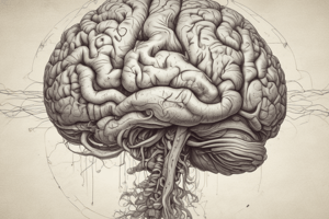
Where are upper motor neurons located?
Where are upper motor neurons located?
Upper motor neurons are the nerve cells that originate in the cerebral cortex and control voluntary movement.
What type of fibers are NOT association fibers?
What type of fibers are NOT association fibers?
Association fibers connect different areas within the same cerebral hemisphere.
What is the cavity of the Telencephalon?
What is the cavity of the Telencephalon?
The lateral ventricle is the largest fluid-filled cavity within the brain, located within the telencephalon.
Which lobe sits behind the central sulcus and above the lateral sulcus?
Which lobe sits behind the central sulcus and above the lateral sulcus?
Signup and view all the flashcards
Which artery is a branch of the internal carotid?
Which artery is a branch of the internal carotid?
Signup and view all the flashcards
What does Meckel's cartilage not give rise to?
What does Meckel's cartilage not give rise to?
Signup and view all the flashcards
Which structure does the thyroid gland NOT descent in front of?
Which structure does the thyroid gland NOT descent in front of?
Signup and view all the flashcards
What forms the nasolacrimal duct?
What forms the nasolacrimal duct?
Signup and view all the flashcards
What is found at the active site in skeletal muscle?
What is found at the active site in skeletal muscle?
Signup and view all the flashcards
What type of epithelium lines the bronchi?
What type of epithelium lines the bronchi?
Signup and view all the flashcards
Which component is responsible for anaphylactic shock?
Which component is responsible for anaphylactic shock?
Signup and view all the flashcards
What is TRUE about a tranverse section of the spinal cord?
What is TRUE about a tranverse section of the spinal cord?
Signup and view all the flashcards
Why are pituitary basophils PAS-positive?
Why are pituitary basophils PAS-positive?
Signup and view all the flashcards
What is the defining feature of a sympathetic ganglion?
What is the defining feature of a sympathetic ganglion?
Signup and view all the flashcards
Which bone forms part of the roof of the orbit?
Which bone forms part of the roof of the orbit?
Signup and view all the flashcards
Which nerve supplies the superior oblique muscle?
Which nerve supplies the superior oblique muscle?
Signup and view all the flashcards
Where is the ciliary ganglion located?
Where is the ciliary ganglion located?
Signup and view all the flashcards
Which muscle is a derivative of the sympathetic root of the ciliary ganglion?
Which muscle is a derivative of the sympathetic root of the ciliary ganglion?
Signup and view all the flashcards
What is the primary arterial supply to the orbit?
What is the primary arterial supply to the orbit?
Signup and view all the flashcards
Which bone contributes to both the lateral and floor of the orbit?
Which bone contributes to both the lateral and floor of the orbit?
Signup and view all the flashcards
Which muscle is most likely trapped in an orbital floor fracture?
Which muscle is most likely trapped in an orbital floor fracture?
Signup and view all the flashcards
Which condition is caused by an infection of the lacrimal sac?
Which condition is caused by an infection of the lacrimal sac?
Signup and view all the flashcards
Which statement about Lacrimation is FALSE?
Which statement about Lacrimation is FALSE?
Signup and view all the flashcards
Damage to which nerve causes difficulty looking laterally?
Damage to which nerve causes difficulty looking laterally?
Signup and view all the flashcards
Which sinus is most likely involved in a medial wall orbital fracture?
Which sinus is most likely involved in a medial wall orbital fracture?
Signup and view all the flashcards
What structure is most likely affected by a dilated pupil that doesn't react to light?
What structure is most likely affected by a dilated pupil that doesn't react to light?
Signup and view all the flashcards
What condition is suspected with excessive tearing and swelling at the medial aspect of the lower eyelid?
What condition is suspected with excessive tearing and swelling at the medial aspect of the lower eyelid?
Signup and view all the flashcards
What function is affected by a lesion in the Edinger-Westphal nucleus?
What function is affected by a lesion in the Edinger-Westphal nucleus?
Signup and view all the flashcards
Which structure is part of the outer fibrous coat of the eyeball?
Which structure is part of the outer fibrous coat of the eyeball?
Signup and view all the flashcards
What is the primary function of the cornea?
What is the primary function of the cornea?
Signup and view all the flashcards
Which muscle is responsible for lifting the upper eyelid?
Which muscle is responsible for lifting the upper eyelid?
Signup and view all the flashcards
Which nerve supplies the lateral rectus muscle?
Which nerve supplies the lateral rectus muscle?
Signup and view all the flashcards
What is the superior oblique muscle responsible for?
What is the superior oblique muscle responsible for?
Signup and view all the flashcards
Which artery is the main blood supply to the eyeball and orbit?
Which artery is the main blood supply to the eyeball and orbit?
Signup and view all the flashcards
What would a lesion affecting the oculomotor nerve NOT result in?
What would a lesion affecting the oculomotor nerve NOT result in?
Signup and view all the flashcards
Which muscle is likely paralyzed if the patient is having difficulty looking downwards?
Which muscle is likely paralyzed if the patient is having difficulty looking downwards?
Signup and view all the flashcards
Which vein drains venous blood from the orbit into the cavernous sinus?
Which vein drains venous blood from the orbit into the cavernous sinus?
Signup and view all the flashcards
Which nerve is most likely affected if a patient has drooping eyelids and difficulty elevating the eye?
Which nerve is most likely affected if a patient has drooping eyelids and difficulty elevating the eye?
Signup and view all the flashcards
What is the condition caused by misaligned eyes due to an imbalance in extraocular muscle action?
What is the condition caused by misaligned eyes due to an imbalance in extraocular muscle action?
Signup and view all the flashcards
What would a patient with a cavernous sinus thrombosis NOT exhibit?
What would a patient with a cavernous sinus thrombosis NOT exhibit?
Signup and view all the flashcards
Which cranial nerve is most likely affected in a patient with double vision and inability to move the eye laterally?
Which cranial nerve is most likely affected in a patient with double vision and inability to move the eye laterally?
Signup and view all the flashcards
What would a lesion in sympathetic fibers supplying the levator palpebrae superioris result in?
What would a lesion in sympathetic fibers supplying the levator palpebrae superioris result in?
Signup and view all the flashcards
Which structure primarily converts light into electrical signals?
Which structure primarily converts light into electrical signals?
Signup and view all the flashcards
Study Notes
Upper Motor Neurons Location
- Upper motor neurons are located in the pre-central gyrus.
Cerebral Hemisphere White Matter
- The uncinated fasciculus, superior longitudinal fasciculus, and fronto-occipital fasciculus are all examples of association fibers in the cerebral hemisphere's white matter.
- The optic radiation is not an association fiber.
Lateral Ventricle Cavity
- The lateral ventricle is a cavity of the telencephalon.
Cerebral Lobe Location
- The parietal lobe is located immediately posterior to the central sulcus and superior to the lateral sulcus.
Branches of Internal Carotid Artery
- The internal carotid artery's branch is the ophthalmic artery, not the superior cerebellar artery, posterior communicating artery, or inferior cerebellar artery.
Meckel's Cartilage
- Meckel's cartilage of the first pharyngeal arch does not give rise to the incus.
Thyroid Gland Development
- The thyroid gland develops in front of the trachea but not the mandible.
Nasolacrimal Duct Formation
- The nasolacrimal duct forms from the fusion of the lateral nasal process and maxillary process by the invagination of endoderm and ectoderm.
Skeletal Muscles
- Skeletal muscle characteristics include multiple peripheral nuclei, a troponin active site, but the H-band is within the I band, not in between two bands.
Bronchi Epithelium
- The bronchi of the lungs are lined with pseudostratified columnar ciliated epithelium.
Anaphylactic Shock
- Mast cells are responsible for anaphylactic shock.
Spinal Cord Transverse Section
- The anterior horn in a spinal cord transverse section is more prominent, and the gray matter is centrally located.
Pituitary Basophils
- Pituitary basophils are PAS-positive due to granules containing glycoproteins.
Sympathetic Ganglion Section
- Sympathetic ganglia sections are identifiable by well-developed satellite cells.
Orbit Roof Bone
- The frontal bone forms part of the orbit's roof.
Orbit Motor Innervation Nerve
- The trochlear nerve (CN IV) provides motor innervation to the superior oblique muscle.
Ciliary Ganglion Location
- The ciliary ganglion is located between the lateral rectus and optic nerve.
Primary Arterial Supply to Orbit
- The ophthalmic artery supplies the orbit.
Orbit Bone Contribution
- The zygomatic bone contributes to both the lateral wall and floor of the orbit.
Floor Fracture Entrapment
- The inferior rectus muscle is most likely trapped in an orbital floor fracture.
Lacrimal Sac Infection
- Dacryocystitis is characterized by infection of the lacrimal sac.
Oculomotor Nerve Dysfunction
- Dysfunction of the oculomotor nerve will cause ptosis (drooping eyelid), impaired eye elevation and adduction. The eye won't have impaired abduction.
Eye Movement Muscle Dysfunction
- The superior oblique muscle is responsible for intorsion, not depression, elevation, or adduction.
- A lesion in the superior oblique muscle causes difficulty looking downward.
- A lesion in the abducens nerve causes difficulty looking laterally.
- A lesion in the oculomotor nerve causes eyelid drooping (ptosis), and difficulty moving the eye up, down, and inwards.
- A lesion in the trochlear nerve affects the superior oblique muscle, which enables the eye to look down and in.
Venous Blood Drainage from Orbit
- Venous blood from the orbit drains to the cavernous sinus via the inferior ophthalmic vein.
Studying That Suits You
Use AI to generate personalized quizzes and flashcards to suit your learning preferences.