Podcast
Questions and Answers
Which of the following is the primary functional characteristic used to classify different types of neurons?
Which of the following is the primary functional characteristic used to classify different types of neurons?
- Size and shape of the cell body
- Arrangement of neurofilaments and neurotubules within the cytoskeleton
- The number of their axons
- The direction in which they propagate electrical signals (correct)
In the central nervous system (CNS), which glial cell type is responsible for myelinating axons, allowing for faster action potential propagation?
In the central nervous system (CNS), which glial cell type is responsible for myelinating axons, allowing for faster action potential propagation?
- Astrocytes
- Ependymal cells
- Microglia
- Oligodendrocytes (correct)
Which structural component is unique to the cerebellum and is essential for its role in motor coordination and balance?
Which structural component is unique to the cerebellum and is essential for its role in motor coordination and balance?
- Molecular layer
- Purkinje cell layer (correct)
- Granule cell layer
- Pyramidal cell layer
How does white matter in the spinal cord contribute to the functioning of the central nervous system?
How does white matter in the spinal cord contribute to the functioning of the central nervous system?
What primary role do satellite cells play in the peripheral nervous system (PNS)?
What primary role do satellite cells play in the peripheral nervous system (PNS)?
Which of the following is a key histological feature that differentiates the cerebral cortex from the cerebellar cortex?
Which of the following is a key histological feature that differentiates the cerebral cortex from the cerebellar cortex?
How do ependymal cells contribute to the health and function of the central nervous system?
How do ependymal cells contribute to the health and function of the central nervous system?
What is the primary functional consequence of myelination on nerve fibers?
What is the primary functional consequence of myelination on nerve fibers?
Which of the following best describes the functional relationship between the dura mater, arachnoid mater, and pia mater?
Which of the following best describes the functional relationship between the dura mater, arachnoid mater, and pia mater?
What is the significance of the blood-brain barrier formed in part by astrocytes in the CNS?
What is the significance of the blood-brain barrier formed in part by astrocytes in the CNS?
Which connective tissue layer directly surrounds individual nerve fibers in the peripheral nervous system (PNS)?
Which connective tissue layer directly surrounds individual nerve fibers in the peripheral nervous system (PNS)?
How does the arrangement of gray and white matter differ between the cerebrum and the spinal cord?
How does the arrangement of gray and white matter differ between the cerebrum and the spinal cord?
Which type of neuron is characterized by a single process that bifurcates into two longer processes, often found in sensory ganglia?
Which type of neuron is characterized by a single process that bifurcates into two longer processes, often found in sensory ganglia?
What distinguishes myelinated from unmyelinated nerve fibers at the microscopic level?
What distinguishes myelinated from unmyelinated nerve fibers at the microscopic level?
What is the role of microglia within nervous tissue?
What is the role of microglia within nervous tissue?
What is the primary function of the ganglionic layer in the cerebral cortex?
What is the primary function of the ganglionic layer in the cerebral cortex?
What is the role of a synapse, found in nervous tissue?
What is the role of a synapse, found in nervous tissue?
Which glial cell myelinates PNS axons?
Which glial cell myelinates PNS axons?
Where in the spinal cord are interneurons located?
Where in the spinal cord are interneurons located?
Which meningeal layer consists of flattened mesenchymal derived cells?
Which meningeal layer consists of flattened mesenchymal derived cells?
What cells line the brain ventricles and spinal cord's central canal?
What cells line the brain ventricles and spinal cord's central canal?
Which cells are known as the “phagocytes of nervous tissue”?
Which cells are known as the “phagocytes of nervous tissue”?
Which glial cell type allows for faster action potential propagation along axons?
Which glial cell type allows for faster action potential propagation along axons?
Which of the following cells are responsible for myelinating and insulating PNS axons?
Which of the following cells are responsible for myelinating and insulating PNS axons?
Which layer surrounds each bundle of nerve?
Which layer surrounds each bundle of nerve?
When observing a cross-section of the cerebellum, what layers would you expect to see inside-out?
When observing a cross-section of the cerebellum, what layers would you expect to see inside-out?
What would most likely be observed in an unmyelinated nerve fiber?
What would most likely be observed in an unmyelinated nerve fiber?
Which cell type protects CNS by engulfing agents and substances?
Which cell type protects CNS by engulfing agents and substances?
Which fibers conduct nerve impulses away from the cell body?
Which fibers conduct nerve impulses away from the cell body?
True or False: Gray Matter consist mostly of myelinated nerve fibers, some unmyelinated fibers, and glial cells.
True or False: Gray Matter consist mostly of myelinated nerve fibers, some unmyelinated fibers, and glial cells.
From outermost to innermost, what are the layers of the body?
From outermost to innermost, what are the layers of the body?
What are some of the components of the peripheral nervous system?
What are some of the components of the peripheral nervous system?
Where can pseudounipolar neurons mostly be found?
Where can pseudounipolar neurons mostly be found?
Which layer contains the basket cells?
Which layer contains the basket cells?
Is white matter located internally or externally inside the spinal cord?
Is white matter located internally or externally inside the spinal cord?
What is considered the germ layer from which nervous tissue is derived?
What is considered the germ layer from which nervous tissue is derived?
What is the main difference between the dorsal and ventral horns in the spinal cord?
What is the main difference between the dorsal and ventral horns in the spinal cord?
In the cerebral cortex, which layer is characterized by a high density of small, densely packed neurons and serves as a major recipient of input from other cortical areas?
In the cerebral cortex, which layer is characterized by a high density of small, densely packed neurons and serves as a major recipient of input from other cortical areas?
What structural characteristic differentiates the cerebellar cortex from the cerebral cortex?
What structural characteristic differentiates the cerebellar cortex from the cerebral cortex?
Which of the following best explains the arrangement of white matter in the spinal cord?
Which of the following best explains the arrangement of white matter in the spinal cord?
Which of the following accurately describes the primary function of astrocytes within the central nervous system (CNS)?
Which of the following accurately describes the primary function of astrocytes within the central nervous system (CNS)?
How do unmyelinated nerve fibers in the peripheral nervous system (PNS) differ structurally from myelinated nerve fibers?
How do unmyelinated nerve fibers in the peripheral nervous system (PNS) differ structurally from myelinated nerve fibers?
In the context of neuron morphology, what is the primary role of dendrites?
In the context of neuron morphology, what is the primary role of dendrites?
Which of the following is a key difference between a nerve and a tract?
Which of the following is a key difference between a nerve and a tract?
What functional role do satellite cells play in relation to neurons within ganglia of the peripheral nervous system?
What functional role do satellite cells play in relation to neurons within ganglia of the peripheral nervous system?
How do oligodendrocytes facilitate neural transmission in the central nervous system (CNS)?
How do oligodendrocytes facilitate neural transmission in the central nervous system (CNS)?
Which meningeal layer directly adheres to the surface of the brain and spinal cord, closely following their contours?
Which meningeal layer directly adheres to the surface of the brain and spinal cord, closely following their contours?
What is the functional significance of the arrangement of gray matter in the cerebral cortex?
What is the functional significance of the arrangement of gray matter in the cerebral cortex?
What role do ependymal cells play in maintaining the health and functionality of the central nervous system?
What role do ependymal cells play in maintaining the health and functionality of the central nervous system?
Where are pseudounipolar neurons typically found, and what is their primary functional role?
Where are pseudounipolar neurons typically found, and what is their primary functional role?
How does the endoneurium contribute to the function of a peripheral nerve?
How does the endoneurium contribute to the function of a peripheral nerve?
What is the primary function of microglia in the central nervous system (CNS)?
What is the primary function of microglia in the central nervous system (CNS)?
Which layer of the cerebellar cortex contains the cell bodies of Purkinje cells?
Which layer of the cerebellar cortex contains the cell bodies of Purkinje cells?
Which of the following distinguishes a spinal ganglion from an autonomic ganglion?
Which of the following distinguishes a spinal ganglion from an autonomic ganglion?
What would be the most likely consequence of damage to ependymal cells within the central nervous system?
What would be the most likely consequence of damage to ependymal cells within the central nervous system?
What structural feature is characteristic of myelinated nerve fibers in the peripheral nervous system, facilitating rapid nerve impulse conduction?
What structural feature is characteristic of myelinated nerve fibers in the peripheral nervous system, facilitating rapid nerve impulse conduction?
In a typical synapse, what role do synaptic vesicles play in neurotransmission?
In a typical synapse, what role do synaptic vesicles play in neurotransmission?
What is the primary difference between protoplasmic and fibrous astrocytes?
What is the primary difference between protoplasmic and fibrous astrocytes?
How do the functions of oligodendrocytes and Schwann cells differ in the nervous system?
How do the functions of oligodendrocytes and Schwann cells differ in the nervous system?
What is the significance of the blood-brain barrier (BBB) in central nervous system (CNS) physiology?
What is the significance of the blood-brain barrier (BBB) in central nervous system (CNS) physiology?
Which of the following accurately reflects the microscopic organization of the spinal cord?
Which of the following accurately reflects the microscopic organization of the spinal cord?
Which connective tissue layer(s) associate(s) with the Peripheral Nervous System?
Which connective tissue layer(s) associate(s) with the Peripheral Nervous System?
Flashcards
Neurons
Neurons
The basic functional units of the nervous system, specialized for rapid communication.
Dendrites
Dendrites
Extensions of a neuron that receive signals from other neurons.
Axon
Axon
A single, long extension of a neuron that transmits signals to other cells.
Axon hillock
Axon hillock
Signup and view all the flashcards
Ganglia
Ganglia
Signup and view all the flashcards
Multipolar neurons
Multipolar neurons
Signup and view all the flashcards
Astrocytes
Astrocytes
Signup and view all the flashcards
Oligodendrocytes
Oligodendrocytes
Signup and view all the flashcards
Microglia
Microglia
Signup and view all the flashcards
Ependymal cells
Ependymal cells
Signup and view all the flashcards
Dura mater
Dura mater
Signup and view all the flashcards
Arachnoid mater
Arachnoid mater
Signup and view all the flashcards
Pia mater
Pia mater
Signup and view all the flashcards
White matter
White matter
Signup and view all the flashcards
Gray matter
Gray matter
Signup and view all the flashcards
Cerebral cortex
Cerebral cortex
Signup and view all the flashcards
Cerebellar cortex
Cerebellar cortex
Signup and view all the flashcards
Granular layer
Granular layer
Signup and view all the flashcards
Purkinje cell layer
Purkinje cell layer
Signup and view all the flashcards
Dorsal horns
Dorsal horns
Signup and view all the flashcards
Ventral horns
Ventral horns
Signup and view all the flashcards
Ectoderm
Ectoderm
Signup and view all the flashcards
Nissl substance
Nissl substance
Signup and view all the flashcards
Myelin
Myelin
Signup and view all the flashcards
Nerves, ganglia, nerve endings
Nerves, ganglia, nerve endings
Signup and view all the flashcards
Schwann cells
Schwann cells
Signup and view all the flashcards
Satellite cells
Satellite cells
Signup and view all the flashcards
Epineurium
Epineurium
Signup and view all the flashcards
Perineurium
Perineurium
Signup and view all the flashcards
Endoneurium
Endoneurium
Signup and view all the flashcards
Internodal segments
Internodal segments
Signup and view all the flashcards
Nerve vs. Tract
Nerve vs. Tract
Signup and view all the flashcards
Spinal vs Autonomic Ganglia
Spinal vs Autonomic Ganglia
Signup and view all the flashcards
Study Notes
- Lord is the true source of light and wisdom.
- One can be granted a keen sense of understanding, retentive memory, and the ability to grasp things correctly through Him.
- Grace allows accuracy, skill, and thoroughness to express clearly.
- Guidance and progress are asked to be given at the start of work until completion.
- This petition is made through Christ.
- Amen ends the prayer.
Unit 4: Nervous Tissue
- Laboratory focus is on Human Histology (Laboratory) MT120225 for the second semester of A.Y. 2024-2025.
- The nervous system is split into 4 sections; overview of nervous tissue, the central nervous system, the peripheral nervous system, laboratory activities and a review.
Learning Outcomes
- The student should be able to:
- Differentiate neuron types based on structure.
- Identify neuroglial cells in the CNS and PNS.
- Distinguish brain and spinal cord regions by location and structure.
- Identify cerebellum's histologic layers.
- Differentiate myelinated and unmyelinated nerve fibers microscopically.
Nervous Tissue Overview
- Typical neuron morphology is discussed.
- Cell body, dendrites and axons are looked at and displayed through slides
Cells of the Nervous System
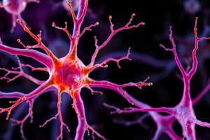
- Includes neurons and glial cells.
Neuron Classification
- Neurons can be classified as:
- Pseudounipolar
- Unipolar.
- Bipolar
- Multipolar
Synapse
- The synapse structure is shown, the point where neurons connect and communicate.
Central Nervous System
- The central nervous system is then looked at
- Glial cells, a type of Central Neuroglia, include:
- Astrocytes
- Oligodendrocytes
- Microglia
- Ependymal cells.
Central Neuroglia: Astrocytes
- Central neuroglia includes astrocytes.
- These astrocytes can be either:
- Protoplasmic;or
- Fibrous.
- Immunohistochemical staining of astrocytes in brain white matter using anti-GFAP Antibodies is another way to study astrocytes.
Central Neuroglia: Oligodendrocytes
- These are another type of central neuroglia
Central Neuroglia: Microglia
- These are another type of central neuroglia
Central Neuroglia: Ependymal Cells
- These are another type of central neuroglia
Connective Tissue of the CNS
- The CNS contains connective tissue, containing 3 meningeal layers:
- Dura Mater (outermost): This area includes dense irregular connective tissue, and connects to the periosteum of the skull.
- Arachnoid Mater (middle): This area has 2 components: a sheet of connective tissue in contact with dura mater, and loosely arranged trabeculae connecting to pia mater
- Pia Mater (innermost): This area is made of flattened mesenchymal derived cells.
CNS Structures
- White matter consists of mostly myelinated nerve fibers along with some unmyelinated and glial cells
- Gray matter consists of mostly neuronal cell bodies, unmyelinated fibers, and neuroglial cells
Cerebrum
- Is part of the Central Nervous System
Cerebral Cortex
- Cerebral cortex layers are:
- I Molecular layer.
- II External granular layer.
- III External pyramidal layer.
- IV Internal granular layer.
- V Internal pyramidal layer.
- VI Multiform layer.
- “Many Exes who Got Poisoned Inside GIT Promised to Move on” is a mnemonic for the layers.
Cerebellum
- Is part of the Central Nervous System
Spinal Cord
- Is part of the Central Nervous System
- Gray matter is H-shaped. It has:
- 2 dorsal horns containing interneurons that receive sensory fibers.
- 2 ventral horns containing multipolar motor neurons.
- White matter is located in the periphery and is made up of mostly myelinated ascending and descending fibers.
Lab Activities and Review
- Will cover:
- The germ layer where nervous tissue comes from.
- Outer brain/inner spinal cord region.
- Phagocytes of nervous tissue.
- Functional unit of nervous tissue.
- Rough endoplasmic reticulum equivalents.
- Cover neurons with single axons and multiple dendrites.
- Cover the lipoidal substance found in the nerve fibers of the CNS and PNS.
Peripheral Nervous System
- Components include: nerves, ganglia and nerve endings.
Glial Cell Types
- Cell types include:
- Astrocytes
- Oligodendrocytes
- Microglia
- Ependymal Cells
- Schwann Cells
- Satellite Cells.
Peripheral Neuroglia: Schwann Cells
- These are a type of peripheral neuroglia
Peripheral Neuroglia: Satellite Cells
- These are a type of peripheral neuroglia
Connective Tissue of the PNS
- Includes: Epineurium, Perineurium and Endoneurium
- Epineurium is the external coat of nerve
- Perineurium surrounds each nerve bundle
- Endoneurium surrounds each individual nerve fiber
Peripheral Nerve
- Shown through slides
Peripheral Ganglion
- Shown through slides
Myelinated Nerve Fibers
- The sections are enclosed by a myelin sheath.
- The sheath prevents the loss of nerve impulse.
- Circular constrictions are nodes of Ranvier.
- The internodal/schwann segments are also looked at
Peripheral Nerve
- Shown through another slide
Unmyelinated Nerve Fibers
- There are naked axons and no multiple wrapping to form the myelin sheath.
- Smaller diameter axons, schwann cells, and no nodes of Ranvier are noted.
Lab Activities and Review
- Will define the relationship between a nerve and a tract.
- Will define the relationship between the nucleus and the ganglion
- Will differentiate between a spinal ganglion and an autonomic ganglion.
Next session:
- Connective tissues will be looked at
Studying That Suits You
Use AI to generate personalized quizzes and flashcards to suit your learning preferences.


