Podcast
Questions and Answers
For faster action potential propagation within the CNS, axons are myelinated and insulated by ______.
For faster action potential propagation within the CNS, axons are myelinated and insulated by ______.
oligodendrocytes
In the CNS, the type of neuroglia that replicates to occupy the space of dying neurons is the ______.
In the CNS, the type of neuroglia that replicates to occupy the space of dying neurons is the ______.
astrocyte
The brain ventricles are lined by ______ cells that also contribute to the production of cerebrospinal fluid.
The brain ventricles are lined by ______ cells that also contribute to the production of cerebrospinal fluid.
ependymal
Within the brain and spinal cord, ______ act as phagocytic cells that remove waste and protect against harmful substances.
Within the brain and spinal cord, ______ act as phagocytic cells that remove waste and protect against harmful substances.
The ______ forms a part of the blood-brain barrier, limiting what substances can enter the CNS.
The ______ forms a part of the blood-brain barrier, limiting what substances can enter the CNS.
The outermost layer of the meninges, known as the ______, is composed of thick, dense irregular connective tissue.
The outermost layer of the meninges, known as the ______, is composed of thick, dense irregular connective tissue.
Located between the dura mater and the pia mater, the ______ features a system of loosely arranged trabeculae.
Located between the dura mater and the pia mater, the ______ features a system of loosely arranged trabeculae.
The ______, consisting of flattened mesenchymal-derived cells, is located innermost of the meningeal layers.
The ______, consisting of flattened mesenchymal-derived cells, is located innermost of the meningeal layers.
The cerebrum's outer region, known as ______, is mainly composed of neuronal cell bodies.
The cerebrum's outer region, known as ______, is mainly composed of neuronal cell bodies.
The cerebrum's inner tissue, known as ______, consists mostly of myelinated nerve fibers.
The cerebrum's inner tissue, known as ______, consists mostly of myelinated nerve fibers.
In the spinal cord, interneurons are found within the two ______.
In the spinal cord, interneurons are found within the two ______.
Located peripherally, the ______ of the spinal cord mainly contain ascending and descending myelinated fibers.
Located peripherally, the ______ of the spinal cord mainly contain ascending and descending myelinated fibers.
In the spinal cord, multipolar motor neurons are located in the two ______.
In the spinal cord, multipolar motor neurons are located in the two ______.
In the cerebral cortex, the ______ layer is characterized by a high density of small pyramidal cells.
In the cerebral cortex, the ______ layer is characterized by a high density of small pyramidal cells.
The ______ layer of the cerebral cortex is notable for containing medium-sized pyramidal cells.
The ______ layer of the cerebral cortex is notable for containing medium-sized pyramidal cells.
Deeper within the cerebral cortex, the ______ layer is characterized by its large pyramidal cells.
Deeper within the cerebral cortex, the ______ layer is characterized by its large pyramidal cells.
The innermost cortical layer, known as the ______ layer, consists of a diverse population of cells.
The innermost cortical layer, known as the ______ layer, consists of a diverse population of cells.
The ______ cell layer of the cerebellar cortex is situated between the molecular and granular layers.
The ______ cell layer of the cerebellar cortex is situated between the molecular and granular layers.
In the cerebellar cortex, densely packed ______ cells form the innermost layer.
In the cerebellar cortex, densely packed ______ cells form the innermost layer.
The outermost layer of the cerebellar cortex is the ______ layer.
The outermost layer of the cerebellar cortex is the ______ layer.
In the peripheral nervous system, ______ cells surround and insulate axons for faster action potential speed.
In the peripheral nervous system, ______ cells surround and insulate axons for faster action potential speed.
[Blank] cells electrically insulate PNS cell bodies and regulate nutrient and waste exchange in ganglia.
[Blank] cells electrically insulate PNS cell bodies and regulate nutrient and waste exchange in ganglia.
The external coat of a nerve, comprising dense connective tissue, is referred to as the ______.
The external coat of a nerve, comprising dense connective tissue, is referred to as the ______.
Bundles of nerve fibers are surrounded by ______, a protective connective tissue layer.
Bundles of nerve fibers are surrounded by ______, a protective connective tissue layer.
Each individual nerve fiber is surrounded by the ______, providing insulation and support.
Each individual nerve fiber is surrounded by the ______, providing insulation and support.
Nodes of Ranvier and internodal segments are characteristics of ______ nerve fibers.
Nodes of Ranvier and internodal segments are characteristics of ______ nerve fibers.
Unlike myelinated fibers, ______ nerve fibers feature smaller diameter axons and lack multiple wrappings; nodes of ranvier are not seen. They are still supported by Schwann cells.
Unlike myelinated fibers, ______ nerve fibers feature smaller diameter axons and lack multiple wrappings; nodes of ranvier are not seen. They are still supported by Schwann cells.
[Blank] are equivalent to the rough endoplasmic reticulum.
[Blank] are equivalent to the rough endoplasmic reticulum.
A ______ is a neuron with a single axon and multiple dendrites.
A ______ is a neuron with a single axon and multiple dendrites.
[Blank] is the lipoidal substance that covers the nerve fibers in the CNS and PNS.
[Blank] is the lipoidal substance that covers the nerve fibers in the CNS and PNS.
The ______ is the germ layer where the nervous tissue is derived.
The ______ is the germ layer where the nervous tissue is derived.
In the brain, gray matter is located in the ______ region and white matter is located in the ______ region; In the spinal cord, gray matter is located in the ______ region and the white matter is located in the ______ region.
In the brain, gray matter is located in the ______ region and white matter is located in the ______ region; In the spinal cord, gray matter is located in the ______ region and the white matter is located in the ______ region.
[Blank] is the phagocyte of the nervous tissue.
[Blank] is the phagocyte of the nervous tissue.
The ______ is the functional unit of a nervous tissue.
The ______ is the functional unit of a nervous tissue.
A ______ is a bundle of axons in the peripheral nervous system (PNS), while a ______ is a bundle of axons in the central nervous system (CNS).
A ______ is a bundle of axons in the peripheral nervous system (PNS), while a ______ is a bundle of axons in the central nervous system (CNS).
A ______ is a collection of neuron cell bodies outside the CNS, while a ______ is a collection of neuron cell bodies inside the CNS.
A ______ is a collection of neuron cell bodies outside the CNS, while a ______ is a collection of neuron cell bodies inside the CNS.
A ______ ganglion has neuron cell bodies in which processes extend to peripheral tissues while the ______ ganglion is only visceral motor neurons.
A ______ ganglion has neuron cell bodies in which processes extend to peripheral tissues while the ______ ganglion is only visceral motor neurons.
The types of neurons can be differentiated via the number of processes of their ______. Multipolar neurons have many; bipolar have two; unipolar have one.
The types of neurons can be differentiated via the number of processes of their ______. Multipolar neurons have many; bipolar have two; unipolar have one.
The differences between gray matter in the cerebrum versus the cerebellum is the number of relative ______ and ______.
The differences between gray matter in the cerebrum versus the cerebellum is the number of relative ______ and ______.
The three components of the peripheral nervous system consist of ______, ______, and ______.
The three components of the peripheral nervous system consist of ______, ______, and ______.
The part of the neuron that typically transmits signals away from the cell body is the ______.
The part of the neuron that typically transmits signals away from the cell body is the ______.
The type of neuron with one axon and multiple dendrites is classified as a ______ neuron.
The type of neuron with one axon and multiple dendrites is classified as a ______ neuron.
[Blank] cells in the central nervous system are responsible for myelinating axons, which speeds up signal transmission.
[Blank] cells in the central nervous system are responsible for myelinating axons, which speeds up signal transmission.
The brain and spinal cord are the main components of the ______ nervous system.
The brain and spinal cord are the main components of the ______ nervous system.
Within the cerebral cortex, the layer rich in small, densely packed neurons is known as the external ______ layer.
Within the cerebral cortex, the layer rich in small, densely packed neurons is known as the external ______ layer.
The ______ mater is the meningeal layer directly in contact with the brain and spinal cord.
The ______ mater is the meningeal layer directly in contact with the brain and spinal cord.
The outermost layer of the three meninges surrounding the brain and spinal cord is known as the ______ mater.
The outermost layer of the three meninges surrounding the brain and spinal cord is known as the ______ mater.
A key function of ______, a type of glial cell, is to phagocytize and remove cellular debris in the nervous tissue.
A key function of ______, a type of glial cell, is to phagocytize and remove cellular debris in the nervous tissue.
The cerebral cortex is organized into six layers, with layer I being identified as the ______ layer.
The cerebral cortex is organized into six layers, with layer I being identified as the ______ layer.
Unlike oligodendrocytes, ______ Cells myelinate nerve fibers in the peripheral nervous system, enabling faster nerve impulse conduction.
Unlike oligodendrocytes, ______ Cells myelinate nerve fibers in the peripheral nervous system, enabling faster nerve impulse conduction.
The dorsal and ventral ______ are key structures in the spinal cord's gray matter, with the dorsal horns receiving sensory information.
The dorsal and ventral ______ are key structures in the spinal cord's gray matter, with the dorsal horns receiving sensory information.
The abundant glial cells known as ______ have perivascular feet that contribute to the blood-brain barrier.
The abundant glial cells known as ______ have perivascular feet that contribute to the blood-brain barrier.
The area of the brain known as the ______ has a distinct histology, featuring a molecular layer, Purkinje cell layer, and granular layer.
The area of the brain known as the ______ has a distinct histology, featuring a molecular layer, Purkinje cell layer, and granular layer.
Areas of the central nervous system predominantly composed of myelinated axons are referred to as ______ matter.
Areas of the central nervous system predominantly composed of myelinated axons are referred to as ______ matter.
A ______ neuron is characterized by a single axon and a single dendrite extending from opposite sides of the cell body.
A ______ neuron is characterized by a single axon and a single dendrite extending from opposite sides of the cell body.
A ______ is a junction between two neurons where neurotransmitters are released to facilitate communication.
A ______ is a junction between two neurons where neurotransmitters are released to facilitate communication.
The ______ cells are ciliated and line the ventricles of the brain, aiding in the production and circulation of cerebrospinal fluid (CSF).
The ______ cells are ciliated and line the ventricles of the brain, aiding in the production and circulation of cerebrospinal fluid (CSF).
The covering of connective tissue that surrounds a nerve fascicle is called the ______.
The covering of connective tissue that surrounds a nerve fascicle is called the ______.
Groups of neuronal cell bodies located outside the central nervous system are called ______.
Groups of neuronal cell bodies located outside the central nervous system are called ______.
The nodes of ______ are gaps in the myelin sheath that expose the axon membrane to facilitate rapid nerve impulse conduction.
The nodes of ______ are gaps in the myelin sheath that expose the axon membrane to facilitate rapid nerve impulse conduction.
The germ layer from which nervous tissue is derived during embryonic development is the ______.
The germ layer from which nervous tissue is derived during embryonic development is the ______.
The functional unit of nervous tissue is the ______.
The functional unit of nervous tissue is the ______.
[Blank] are equivalent to the rough endoplasmic reticulum in neurons and are the sites of protein synthesis.
[Blank] are equivalent to the rough endoplasmic reticulum in neurons and are the sites of protein synthesis.
A neuron with a single axon and multiple dendrites is a ______ neuron.
A neuron with a single axon and multiple dendrites is a ______ neuron.
The lipoidal substance that covers nerve fibers in the CNS and PNS is called ______.
The lipoidal substance that covers nerve fibers in the CNS and PNS is called ______.
Flashcards
Neuron
Neuron
A nerve cell specialized for communication. It receives, processes, and transmits information to other cells.
Dendrites
Dendrites
Branch-like extensions of a neuron that receive signals from other neurons. They increase the surface area available for receiving these signals.
Axon hillock
Axon hillock
Area where the axon exits the neuron cell body.
Axon
Axon
Signup and view all the flashcards
Synapse
Synapse
Signup and view all the flashcards
Multipolar neuron
Multipolar neuron
Signup and view all the flashcards
Bipolar neuron
Bipolar neuron
Signup and view all the flashcards
Unipolar Neuron
Unipolar Neuron
Signup and view all the flashcards
Glial Cells
Glial Cells
Signup and view all the flashcards
Central Nervous System
Central Nervous System
Signup and view all the flashcards
Astrocytes
Astrocytes
Signup and view all the flashcards
Oligodendrocytes
Oligodendrocytes
Signup and view all the flashcards
Microglia
Microglia
Signup and view all the flashcards
Ependymal cells
Ependymal cells
Signup and view all the flashcards
Schwann cells
Schwann cells
Signup and view all the flashcards
Satellite cells
Satellite cells
Signup and view all the flashcards
Dura mater
Dura mater
Signup and view all the flashcards
Arachnoid mater
Arachnoid mater
Signup and view all the flashcards
Pia mater
Pia mater
Signup and view all the flashcards
White Matter
White Matter
Signup and view all the flashcards
Gray Matter
Gray Matter
Signup and view all the flashcards
Cerebrum
Cerebrum
Signup and view all the flashcards
Cerebellum
Cerebellum
Signup and view all the flashcards
Dorsal Horns
Dorsal Horns
Signup and view all the flashcards
Ventral Horns
Ventral Horns
Signup and view all the flashcards
Gray Matter
Gray Matter
Signup and view all the flashcards
Ectoderm
Ectoderm
Signup and view all the flashcards
Neuron
Neuron
Signup and view all the flashcards
Nissl Substance
Nissl Substance
Signup and view all the flashcards
Myelin
Myelin
Signup and view all the flashcards
Ganglion
Ganglion
Signup and view all the flashcards
Nerve (PNS)
Nerve (PNS)
Signup and view all the flashcards
Tract (CNS)
Tract (CNS)
Signup and view all the flashcards
Epineurium
Epineurium
Signup and view all the flashcards
Perineurium
Perineurium
Signup and view all the flashcards
Endoneurium
Endoneurium
Signup and view all the flashcards
Satellite cells
Satellite cells
Signup and view all the flashcards
Nodes of ranvier
Nodes of ranvier
Signup and view all the flashcards
Myelinated Nerve Fibers
Myelinated Nerve Fibers
Signup and view all the flashcards
Unmyelinated Nerve Fibers
Unmyelinated Nerve Fibers
Signup and view all the flashcards
Study Notes
- The topic is Unit 4: Nervous Tissue for Human Histology (Laboratory) MT120225
- The semester is Second Semester A.Y. 2024-2025
- The nervous system is being studied at UST General Santos - School of Health Sciences, Department of Medical Technology
Learning Outcomes
- Differentiate neuron types by structure.
- Identify neuroglial cells in the central nervous system, (CNS) and peripheral nervous system, (PNS).
- Distinguish brain and spinal cord regions by location and structural components.
- Identify cerebellum's histologic layers.
- Differentiate myelinated and unmyelinated nerve fibers microscopically.
Overview of Nervous Tissue
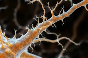
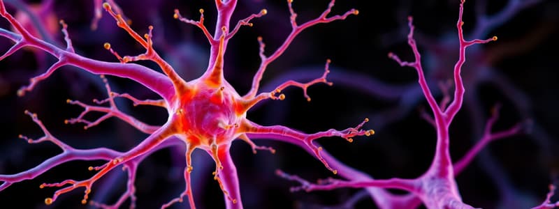
Morphology of a Typical Neuron
- Neurons have dendrites, multipolar neurons, Nissl substance, axon hillock, axons, arterioles, neuroglia, a nucleus, and a nucleolus.
- In the anterior horn of the human spinal cord one can observe neuroglial nuclei and Nissl bodies when stained with Toluidine Blue.
- Silver Impregnated Purkinje Neurons show dendrites in the Cerebellum.
Types of Neurons
- Neurons are classified into structural classes.
- Multipolar neurons have multiple dendrites and one axon.
- Bipolar neurons have one dendrite and one axon.
- Unipolar neurons arise from a single short process.
- Pseudounipolar neurons are a class of neurons
- Anaxonic neurons have many dendrites but no axon.
Classifications of Neurons
- Pseudounipolar neurons have satellite cells and cell bodies of sensory neurons.
- Bipolar neurons have axon hillocks, soma, neuroglia, and dendrites.
- Multipolar neurons have axons, axon hillocks, soma, dendrites, nuclei, and nucleolus.
Synapse
- Acetylcholine binds to receptor protein, causing ion gates to open
- Postsynaptic neurons have Sodium (Na+) ions
Central Nervous System
Glial Cells of the CNS
- Astrocytes form the blood-brain barrier and regulate interstitial fluid composition.
- Astrocytes provide structural support, aid neural development and occupy dying neuron space
- Oligodendrocytes myelinate CNS axons, enabling faster action potential propagation.
- Microglia are phagocytic cells, protecting the CNS by engulfing infectious agents.
- Ependymal cells line brain ventricles and the spinal cord's central canal.
- Ependymal cells produce and circulate cerebrospinal fluid (CSF).
- Protoplasmic and fibrous structures are central neuroglia, astrocytes.
- Anti-GFAP Antibodies are used in the immunohistochemical staining of astrocytes in brain white matter
Connective Tissue of the CNS / Meningeal Layers
- Dura Mater (outermost): Thick dense irregular connective tissue that is continuous with the periosteum of the skull.
- Arachnoid Mater (middle): Has two components being a sheet of connective tissue in contact with the dura mater and a system of loosely arranged trabeculae continuous with the pia mater.
- Pia Mater (innermost): Consists of flattened mesenchymal-derived cells.
- The meninges consist of the Dura mater, arachnoid mater and the pia mater.
Structures of the Central Nervous System
- White matter consists mostly of myelinated nerve fibers.
- White matter contains some unmyelinated fibers and glial cells.
- Gray matter consists mostly of neuronal cell bodies.
- Gray matter contains unmyelinated fibers and neuroglial cells.
Cerebrum
- The cerebrum is part of the central nervous system.
- The cerebrum has structures such as pia mater, gray matter, and white matter.
Cerebral Cortex
- The cerebral cortex has a molecular layer, external granular layer, external pyramidal layer, internal granular layer, internal pyramidal layer, and multiform layer
- Key components are Pia mater with blood vessels, neuroglial cells, small pyramidal cells, apical dendrites of pyramidal cells, medium-sized pyramidal cells, granule cells, dendrites of pyramidal cells, large pyramidal cells, and bundles of axons
Cerebellum
- The Cerebellum consists of the cerebellar cortex, the molecular layer granule cell layer, and white matter
- The Cerebellar Cortex consists of basket cells, dendrites of Purkinje cells, Purkinje cells with nucleus and nucleolus, Granular cell layer, white matter, Molecular cell layer, Purkinje cell layer, Golgi type II cells, Granule cells, Glomeruli and Axons
Spinal Cord
- Gray Matter (H shaped): 2 dorsal horns contain interneurons (receives sensory fibers from neurons in the spinal cord, 2 ventral horns contain multipolar motor neurons
- White Matter: Peripherally located and consists of ascending and descending fibers (mostly myelinated)
Peripheral Nervous System
- Nerve, ganglia, and nerve endings are components.
Glial Cells of the Peripheral Nervous System
- Schwann cells surround and insulate PNS axons.
- Schwann cells myelinate PNS axons, enabling faster action potential propagation.
- Satellite cells electrically insulate PNS cell bodies and regulate nutrient/waste exchange.
Peripheral Neuroglia
- Schwann Cells: Myelinated Nerve Fibers, Schwan cells, the Axon and the Myelin sheath
- Satellite Cells are located around neurons of ganglia in the PNS.
Connective Tissue of the PNS
- Epineurium is the external coat of the nerve.
- Perineurium surrounds each bundle of nerve.
- Endoneurium surrounds individual nerve fibers.
Peripheral Nerve
- Peripheral nerves have axons and myelin sheaths.
Ganglion
- Ganglion have blood vessels
Myelinated Nerve Fibers
- Myelinated nerve fibers are enclosed by a myelin sheath
- Myelinated nerve fibers prevent loss of nerve impulse
- Circular constrictions on myelinated nerve fibers are nodes of Ranvier
- Internodal segments or schwann segments are components of myelinated nerve fibers.
Unmyelinated Nerve Fibers
- Unmyelinated nerve fibers have naked axons.
- Have no multiple wrapping to form a myelin sheath
- Consist of smaller diameter axons
- Still consist of schwann cells
- Nodes of ranvier are not seen
Studying That Suits You
Use AI to generate personalized quizzes and flashcards to suit your learning preferences.