Podcast
Questions and Answers
What is the primary function of receptors in neurons?
What is the primary function of receptors in neurons?
- To interact with extracellular signals and trigger intracellular effects (correct)
- To synthesize neurotransmitter molecules
- To transmit electrical impulses to other neurons
- To maintain membrane polarization
Which type of channels in the axon membrane are responsible for membrane polarity reversal?
Which type of channels in the axon membrane are responsible for membrane polarity reversal?
- Voltage-gated Ca++ channels
- Potassium ion channels
- Calcium ion channels
- Voltage-gated Na+ channels (correct)
What distinguishes unipolar neurons from multipolar neurons?
What distinguishes unipolar neurons from multipolar neurons?
- Multipolar neurons are only found in the peripheral nervous system.
- Unipolar neurons have a single process emanating from the cell body. (correct)
- Multipolar neurons have a single axonal branch.
- Unipolar neurons have multiple dendrites.
What role do voltage-gated Ca++ channels play in neuron function?
What role do voltage-gated Ca++ channels play in neuron function?
Which type of neuron is most abundant in the central nervous system (CNS)?
Which type of neuron is most abundant in the central nervous system (CNS)?
What constitutes the basis of nervous system function according to the neural circuits?
What constitutes the basis of nervous system function according to the neural circuits?
What is the main mechanism through which most neurons communicate?
What is the main mechanism through which most neurons communicate?
What does the chromatophilic substance in neuronal cell bodies indicate?
What does the chromatophilic substance in neuronal cell bodies indicate?
What is the primary function of the cell body cytoplasm in neurons?
What is the primary function of the cell body cytoplasm in neurons?
Which type of proteins are synthesized in the cell body cytoplasm of neurons?
Which type of proteins are synthesized in the cell body cytoplasm of neurons?
Why are larger neurons more suited for conveying urgent information?
Why are larger neurons more suited for conveying urgent information?
What is the axon hillock's role in a neuron?
What is the axon hillock's role in a neuron?
Which of the following structures is absent in the axon of multipolar neurons?
Which of the following structures is absent in the axon of multipolar neurons?
What is the typical diameter range for cell bodies of neurons?
What is the typical diameter range for cell bodies of neurons?
What is the significance of the chromatophilic substance in neurons?
What is the significance of the chromatophilic substance in neurons?
In which area of a multipolar neuron is the cytoplasmic chromaticity relatively pale and diffuse?
In which area of a multipolar neuron is the cytoplasmic chromaticity relatively pale and diffuse?
What type of neuron is characterized by a single axon that bifurcates into central and peripheral branches?
What type of neuron is characterized by a single axon that bifurcates into central and peripheral branches?
Which type of synaptic vesicle is typically associated with inhibitory synapses and appears flattened under specific conditions?
Which type of synaptic vesicle is typically associated with inhibitory synapses and appears flattened under specific conditions?
What is the primary purpose of transporter proteins in the vesicle membrane?
What is the primary purpose of transporter proteins in the vesicle membrane?
What is the approximate diameter range of spherical synaptic vesicles?
What is the approximate diameter range of spherical synaptic vesicles?
Where are neuronal cell bodies for unipolar neurons typically found?
Where are neuronal cell bodies for unipolar neurons typically found?
Which neurotransmitters are typically contained in electron-dense synaptic vesicles?
Which neurotransmitters are typically contained in electron-dense synaptic vesicles?
What allows for neural plasticity, enabling learning and behavioral modification?
What allows for neural plasticity, enabling learning and behavioral modification?
What is a distinguishing feature of multipolar neurons compared to unipolar and bipolar neurons?
What is a distinguishing feature of multipolar neurons compared to unipolar and bipolar neurons?
What is the primary role of the postsynaptic density?
What is the primary role of the postsynaptic density?
Which factor primarily determines whether a synapse is excitatory or inhibitory?
Which factor primarily determines whether a synapse is excitatory or inhibitory?
What happens when calcium ions ($Ca^{++}$) influx into the presynaptic terminal?
What happens when calcium ions ($Ca^{++}$) influx into the presynaptic terminal?
Which neurotransmitter is associated with excitatory synapses?
Which neurotransmitter is associated with excitatory synapses?
What is the immediate effect of an action potential at the axon terminal?
What is the immediate effect of an action potential at the axon terminal?
How do synaptic vesicles release neurotransmitters into the synaptic cleft?
How do synaptic vesicles release neurotransmitters into the synaptic cleft?
What types of synapses are primarily formed between neurons?
What types of synapses are primarily formed between neurons?
What is the primary mechanism through which neurotransmitter receptors are rapidly removed from synaptic sites?
What is the primary mechanism through which neurotransmitter receptors are rapidly removed from synaptic sites?
What characterizes the glutamate receptors in the brain?
What characterizes the glutamate receptors in the brain?
What role do cadherin-catenin complexes play in the synapse?
What role do cadherin-catenin complexes play in the synapse?
Which cellular component plays a role in recycling neurotransmitter molecules?
Which cellular component plays a role in recycling neurotransmitter molecules?
What causes synaptic activity to cease?
What causes synaptic activity to cease?
Which structural characteristic differentiates astrocytes in white matter versus gray matter?
Which structural characteristic differentiates astrocytes in white matter versus gray matter?
What triggers the rapid immobilization and local accumulation of receptors on the neuronal surface?
What triggers the rapid immobilization and local accumulation of receptors on the neuronal surface?
What is one of the key functions of scaffolding proteins in neurons?
What is one of the key functions of scaffolding proteins in neurons?
What happens to excitatory and inhibitory synaptic effects at the cell body of a target neuron?
What happens to excitatory and inhibitory synaptic effects at the cell body of a target neuron?
What is suggested about the role of glial scarring in the CNS?
What is suggested about the role of glial scarring in the CNS?
Which of the following best describes the structure of ependymal cells?
Which of the following best describes the structure of ependymal cells?
What barrier function do ependymal cells serve in the CNS?
What barrier function do ependymal cells serve in the CNS?
What are the primary types of gliocytes found in the peripheral nervous system (PNS)?
What are the primary types of gliocytes found in the peripheral nervous system (PNS)?
What is the role of ganglionic gliocytes in the PNS?
What is the role of ganglionic gliocytes in the PNS?
Which statement about ependymal cells in adults is accurate?
Which statement about ependymal cells in adults is accurate?
How do ependymal cells contribute to the protection of neural tissue?
How do ependymal cells contribute to the protection of neural tissue?
What feature distinguishes neurolemmocytes from other types of glial cells?
What feature distinguishes neurolemmocytes from other types of glial cells?
Flashcards
Axodendritic Synapse
Axodendritic Synapse
Synapses where the axon of one neuron connects to the dendrite of another neuron.
Axosomatic Synapse
Axosomatic Synapse
Synapses where the axon of one neuron connects to the cell body of another neuron.
Neurotransmitter Removal
Neurotransmitter Removal
The process of removing neurotransmitter molecules from the synaptic cleft.
Transporters
Transporters
Signup and view all the flashcards
Synaptic Vesicle Recycling
Synaptic Vesicle Recycling
Signup and view all the flashcards
Synaptic Integration
Synaptic Integration
Signup and view all the flashcards
Synaptic Plasticity
Synaptic Plasticity
Signup and view all the flashcards
Astrocytes
Astrocytes
Signup and view all the flashcards
Pyramidal Neuron
Pyramidal Neuron
Signup and view all the flashcards
Dendrites
Dendrites
Signup and view all the flashcards
Axon
Axon
Signup and view all the flashcards
Axonal Varicosities
Axonal Varicosities
Signup and view all the flashcards
Synaptic Vesicles
Synaptic Vesicles
Signup and view all the flashcards
Electron-Lucent Synaptic Vesicles
Electron-Lucent Synaptic Vesicles
Signup and view all the flashcards
Electron-Dense Synaptic Vesicles
Electron-Dense Synaptic Vesicles
Signup and view all the flashcards
Vesicle Recycling
Vesicle Recycling
Signup and view all the flashcards
Membrane Receptor
Membrane Receptor
Signup and view all the flashcards
Unipolar Neuron
Unipolar Neuron
Signup and view all the flashcards
Bipolar Neuron
Bipolar Neuron
Signup and view all the flashcards
Multipolar Neuron
Multipolar Neuron
Signup and view all the flashcards
Synaptic Boutons
Synaptic Boutons
Signup and view all the flashcards
Voltage-Gated Sodium Channels
Voltage-Gated Sodium Channels
Signup and view all the flashcards
Voltage-Gated Calcium Channels
Voltage-Gated Calcium Channels
Signup and view all the flashcards
Neurotransmitter Release
Neurotransmitter Release
Signup and view all the flashcards
What is the cell body of a neuron?
What is the cell body of a neuron?
Signup and view all the flashcards
How does cell body size relate to neuron size?
How does cell body size relate to neuron size?
Signup and view all the flashcards
Why do neurons with long processes need larger cell bodies?
Why do neurons with long processes need larger cell bodies?
Signup and view all the flashcards
What is chromatophilic substance?
What is chromatophilic substance?
Signup and view all the flashcards
What's special about the axon hillock?
What's special about the axon hillock?
Signup and view all the flashcards
How does process size impact conduction speed?
How does process size impact conduction speed?
Signup and view all the flashcards
Why are neurons conveying urgent information larger?
Why are neurons conveying urgent information larger?
Signup and view all the flashcards
How does cell body size relate to synaptic integration?
How does cell body size relate to synaptic integration?
Signup and view all the flashcards
Oligodendrocytes
Oligodendrocytes
Signup and view all the flashcards
Ependymal Cells
Ependymal Cells
Signup and view all the flashcards
Glial Scarring
Glial Scarring
Signup and view all the flashcards
Neurolemmocytes (Schwann Cells)
Neurolemmocytes (Schwann Cells)
Signup and view all the flashcards
Ganglionic Gliocytes (Satellite Cells)
Ganglionic Gliocytes (Satellite Cells)
Signup and view all the flashcards
Blood-Brain Barrier
Blood-Brain Barrier
Signup and view all the flashcards
Cerebrospinal Fluid (CSF)
Cerebrospinal Fluid (CSF)
Signup and view all the flashcards
Synapse
Synapse
Signup and view all the flashcards
Neurotransmitter Receptors
Neurotransmitter Receptors
Signup and view all the flashcards
Postsynaptic Density
Postsynaptic Density
Signup and view all the flashcards
Exocytosis
Exocytosis
Signup and view all the flashcards
Calcium Influx
Calcium Influx
Signup and view all the flashcards
Neurotransmitter Reuptake
Neurotransmitter Reuptake
Signup and view all the flashcards
Study Notes
Nervous Tissue
- Nervous tissue is composed of neurons and neuroglia
- The nervous system is divided into the central nervous system (CNS) and the peripheral nervous system (PNS)
- The CNS includes the brain and spinal cord
- The PNS includes cranial and spinal nerves, plus associated nerve roots and ganglia
Neurons
- Neurons are the structural and functional units of the nervous system
- Neurons are trophic units, transforming and sustaining the structure and function of innervated cells.
- Neurons typically live a lifetime; most are incapable of mitosis. Recent evidence shows new neuron formation in some brain regions
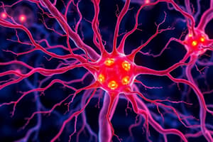
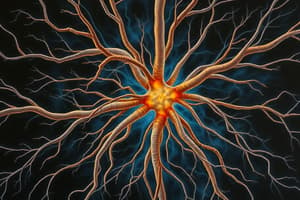
- The cell body (perikaryon or soma) contains the nucleus and surrounding cytoplasm.
- The cytoplasm of large neurons contains chromatophilic substance (Nissl substance), made of aggregated rough endoplasmic reticulum, free ribosomes, and polyribosomes.
- Cell bodies range in size from less than 10 µm to more than 100 µm in diameter
- The nucleus is typically spherical or ovoid and relatively euchromatic.
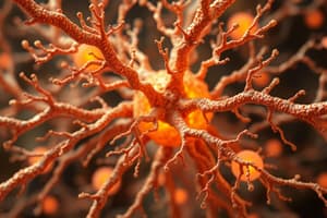
- Neurons have processes called dendrites and axons; dendrites extend out to receive input; axons conduct signals outwards.
- The axon hillock is a pale-staining region of the cell body where the axon originates.
Neuronal Structure
- Axon: Relatively long, cylindrical process arising from the axon hillock. Contains microtubules, neurofilaments, mitochondria, and smooth endoplasmic reticulum (sER)
- Dendrites: Highly branched, tree-like processes that receive synaptic input from other neurons. Contain microtubules, neurofilaments, mitochondria, and smooth endoplasmic reticulum (sER).
- Synaptic vesicles: Concentrated in axon terminals; contain neurotransmitters that are released into the synaptic cleft.
- Synaptic cleft: Space between two neurons across which neurotransmitters diffuse between presynaptic and postsynaptic neurons.
Neuroglia
- Neuroglia comprise over 90% of the cells in the nervous system.
- Neuroglia in the CNS include astrocytes, oligodendrocytes, microglia, and ependymal cells.
- Neuroglia in the PNS include ganglionic gliocytes and neurolemmocytes (Schwann cells)
Astrocytes
- Pale, ovoid nuclei
- Numerous processes containing glial fibrils
- White matter: Fibrous astrocytes (long, slender processes)
- Gray matter: Protoplasmic astrocytes (shorter, highly branched processes)
- Astrocyte processes form a glial limiting membrane
- Play vital roles in brain function.
Oligodendrocytes
- Small, spherical, densely stained nuclei
- Relatively few branches
- Form myelin sheaths around axons in white matter of the CNS
Microglia
- Small, elongated, chromophilic nuclei
- Act as macrophages, have phagocytic capabilities.
Ependymal Cells
- Line the central canal of the spinal cord and ventricles of the brain
- Cuboidal or columnar cells with cilia
- Involved in cerebrospinal fluid production
Ganglionic Gliocytes
- Encapsulate neuron cell bodies in the PNS.
- Form a tight capsule around sensory neuron cell bodies in sensory ganglia.
Neurolemmocytes
- Envelop and myelinate axons in the PNS
- Myelin sheath: Multiple layers of neurolemmocyte plasma membrane that surround and insulate axons.
- Nodes of Ranvier: Gaps in the myelin sheath.
- Saltatory conduction: Action potentials jump between nodes, resulting in faster transmission speed.
Nerves
- Bundles of axons and supporting cells (endoneurium, perineurium, and epineurium)
- Fascicles: Bundles of axons. Perineurium sheathes fascicles
- Epineurium: Sheaths the entire nerve.
- Endoneurium: surrounds individual axons
Ganglia
- Localized swellings in nerves containing neuron cell bodies.
- Sensory ganglia: Contain cell bodies of sensory neurons. Typically unipolar neurons except in sense organs.
- Autonomic ganglia: Contain multipolar neurons.
Efferent Neurons
- Convey signals from the CNS to muscles and glands (motor neurons)
- Somatic efferent neurons: Innervate skeletal muscle; each neuron with fibers is a motor unit.
- Visceral efferent neurons: innervate smooth and cardiac muscle and glands (autonomic nervous system)
Receptors
- Specialized cells or structures that detect stimuli.
- Nonencapsulated receptors: Free nerve endings, detect pain, temperature, touch, and pressure.
- Encapsulated receptors: Lamellar corpuscles (Pacini's corpuscles) are sensitive to transient pressures; they are widely distributed in the skin. Other examples include tactile corpuscles (Meissner's corpuscles), neurotendinous spindles, and muscle spindles (used for proprioception).
Meninges, Blood Vessels, and Cerebrospinal Fluid (CSF)
- The brain and spinal cord are covered by layers of connective tissue called meninges.
- Dura mater, arachnoid mater, and pia mater from superficial to deep.
- Arachnoid villi: Projections of arachnoid mater through which CSF is absorbed by blood vessels.
- Blood-brain barrier: A selective barrier between blood and brain tissue, composed of tight junctions between endothelial cells of brain capillaries. This barrier limits the passage of certain substances.
- CSF: Fluid produced by choroid plexuses of ventricles, circulating in the subarachnoid space, serving both cushioning and buffering functions.
Studying That Suits You
Use AI to generate personalized quizzes and flashcards to suit your learning preferences.