Podcast
Questions and Answers
What is the main function of the Nervous System?
What is the main function of the Nervous System?
The nervous system controls and coordinates all body functions.
What are the two primary subdivisions of the Nervous System?
What are the two primary subdivisions of the Nervous System?
The central nervous system (CNS) and the peripheral nervous system (PNS).
What is the function of the Sensory (afferent) division of the Nervous System?
What is the function of the Sensory (afferent) division of the Nervous System?
Carries information from Sensory receptors to the CNS.
What is the function of the Motor (efferent) division of the Nervous System?
What is the function of the Motor (efferent) division of the Nervous System?
What is the function of Astrocytes?
What is the function of Astrocytes?
What is the function of Microglia?
What is the function of Microglia?
What is the function of Ependymal cells?
What is the function of Ependymal cells?
What is the function of Oligodendrocytes?
What is the function of Oligodendrocytes?
What is the function of Schwann cells?
What is the function of Schwann cells?
Which of these is NOT a function of Satellite cells?
Which of these is NOT a function of Satellite cells?
Flashcards
Nervous System Functions
Nervous System Functions
The nervous system is responsible for collecting information from the environment, processing it, and sending out signals to initiate a response. This includes sensory input, integration, and motor output.
Central Nervous System (CNS)
Central Nervous System (CNS)
This is the control center of the nervous system and consists of the brain and spinal cord. It receives information from the peripheral system, processes it, and sends out commands.
Peripheral Nervous System (PNS)
Peripheral Nervous System (PNS)
This system contains all nerves that extend from the brain and spinal cord. It acts as the communication line between the CNS and the rest of the body.
Sensory (Afferent) Division
Sensory (Afferent) Division
Signup and view all the flashcards
Motor (Efferent) Division
Motor (Efferent) Division
Signup and view all the flashcards
Somatic Nervous System
Somatic Nervous System
Signup and view all the flashcards
Autonomic Nervous System
Autonomic Nervous System
Signup and view all the flashcards
Neuron
Neuron
Signup and view all the flashcards
Cell Body of a Neuron
Cell Body of a Neuron
Signup and view all the flashcards
Dendrites of a Neuron
Dendrites of a Neuron
Signup and view all the flashcards
Axon of a Neuron
Axon of a Neuron
Signup and view all the flashcards
Synapse
Synapse
Signup and view all the flashcards
Neurotransmitter
Neurotransmitter
Signup and view all the flashcards
Irritability
Irritability
Signup and view all the flashcards
Conductivity
Conductivity
Signup and view all the flashcards
Resting Neuron
Resting Neuron
Signup and view all the flashcards
Depolarization
Depolarization
Signup and view all the flashcards
Action Potential
Action Potential
Signup and view all the flashcards
Repolarization
Repolarization
Signup and view all the flashcards
Reflex
Reflex
Signup and view all the flashcards
Reflex Arc
Reflex Arc
Signup and view all the flashcards
Cerebrum
Cerebrum
Signup and view all the flashcards
Thalamus
Thalamus
Signup and view all the flashcards
Hypothalamus
Hypothalamus
Signup and view all the flashcards
Brain Stem
Brain Stem
Signup and view all the flashcards
Cerebellum
Cerebellum
Signup and view all the flashcards
Meninges
Meninges
Signup and view all the flashcards
Cerebrospinal Fluid (CSF)
Cerebrospinal Fluid (CSF)
Signup and view all the flashcards
Blood-Brain Barrier
Blood-Brain Barrier
Signup and view all the flashcards
Gray Matter in Spinal Cord
Gray Matter in Spinal Cord
Signup and view all the flashcards
White Matter in Spinal Cord
White Matter in Spinal Cord
Signup and view all the flashcards
Cauda Equina
Cauda Equina
Signup and view all the flashcards
Study Notes
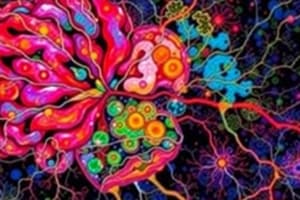
Nervous System
- Functions: Sensory input (gathering information), Integration (processing and interpreting sensory input), Motor output (activation of muscles or glands).
- Structural Classification: Central nervous system (CNS) - brain and spinal cord; Peripheral nervous system (PNS) - nerves extending from the brain and spinal cord.
- Support Cells (Neuroglia): Astrocytes (most abundant), Microglia (phagocytes), Ependymal cells (line cavities), Oligodendrocytes (wrap around nerve fibers in CNS), Satellite cells (protect neuron cell bodies in PNS), Schwann cells (form myelin sheath in PNS).
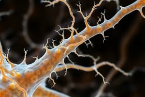
- Neurons: Specialized cells that transmit messages.
- Cell body: Nucleus and metabolic center.
- Processes: Dendrites (toward cell body), Axons (away from cell body).
Nervous Tissue: Support Cells
- General functions: Support, insulate, and protect neurons.
- Astrocytes: Abundant, star-shaped cells that brace neurons, form barrier between capillaries and neurons & control chemical environment.
- Microglia: Spiderlike phagocytes that dispose of debris.
- Ependymal cells: Line cavities of the brain and spinal cord; help circulate cerebrospinal fluid (CSF).
- Oligodendrocytes: Have processes that form myelin sheaths around CNS nerve fibers
- Satellite cells: Protect neuron cell bodies; surround neurons in PNS.
- Schwann cells: Form myelin sheath in peripheral nervous system.
Functional Classification of the Peripheral Nervous System
- Sensory (afferent) division: Carries information to the CNS.
- Motor (efferent) division: Carries impulses away from the CNS.
- Somatic nervous system (voluntary): Consciously controls skeletal muscles.
- Autonomic nervous system (involuntary): Automatically controls smooth and cardiac muscles and glands (sympathetic and parasympathetic subdivisions).
Nerve Impulses
- Resting neuron: Membrane is polarized (fewer positive ions inside the cell).
- Depolarization: Stimulus changes membrane permeability and sodium (Na+) ions rush inward, changing membrane potential.
- Action potential: If stimulus is strong enough, depolarization causes a complete reversal of membrane polarity.
- Propagation of action potential: The action potential travels along the entire axon.
- Repolarization: After the action potential, potassium (K+) ions rush out of the neuron, restoring the negative charge inside and positive charge outside.
- Transmission at synapses: Neurotransmitters are released to transmit the signal across the gap (synapse).
The Reflex Arc
- A direct route from a sensory neuron, to an interneuron, to an effector.
- A rapid, predictable, and involuntary response to a stimulus.
- Examples, patellar (knee-jerk) reflex & flexor (withdrawal) reflex.
Regions of the Brain, Cerebrum
- Cerebral hemispheres: Paired superior parts of the brain, include more than half of the brain mass; ridges (gyri) and grooves (sulci).
- Lobes of the cerebrum: Divided into Frontal, Parietal, Occipital, and Temporal lobes by fissures (deep grooves).
- Specialized areas in cerebrum: Primary somatic sensory area (receives impulses), Primary motor area (sends impulses to skeletal muscles), Broca's area (speech).
- Other areas: Gustatory areas (taste), Auditory areas (sound), Visual areas (vision), Olfactory areas (smell).
- Interpretation areas: Speech/language region, Language comprehension region, general interpretation area.
- Basal nuclei: Islands of gray matter buried deep within white matter.
Regions of the brain: Diencephalon
- Thalamus: Relay station for sensory impulses.
- Hypothalamus: Important autonomic nervous system center (regulates body-temp, water balance, metabolism, emotions; associated with pituitary gland); produces hormones.
- Epithalamus: Contains the pineal body (endocrine gland).
Regions of the Brain: Brain Stem
- Midbrain: Contains reflex centers for vision and hearing.
- Pons: Mostly composed of fiber tracts.
- Medulla oblongata: Lowest part of brain stem, includes important control centers (heart rate, blood pressure, breathing).
Protection of the Central Nervous System
- Scalp and skin
- Skull and vertebral column
- Meninges (dura mater, arachnoid mater, pia mater)
- Cerebrospinal fluid (CSF)
- Blood-brain barrier
Cerebrospinal Fluid (CSF)
- Similar to blood plasma
- Formed in choroid plexuses (capillaries in ventricles)
- Circulates in arachnoid space, ventricles and central canal.
Cerebrovascular Accident (CVA) or Stroke
- Results from ruptured blood vessel supplying a region of the brain.
- Loss of some functions may occur.
Traumatic Brain Injuries
- Concussion: Slight brain injury – no permanent brain damage.
- Contusion: Nervous tissue destruction occurs; does not regenerate.
Spinal Cord
- Extends from foramen magnum of the skull to first or second lumbar vertebra.
- Provides two-way conduction pathway from the brain to and from the brain.
- 31 pairs of spinal nerves arise from spinal cord.
- Cauda equina (collection of spinal nerves).
Spinal Cord Anatomy
- Internal gray matter: Primarily cell bodies.
- External white matter: Conduction tracts. (Dorsal, Lateral, Ventral columns).
Peripheral Nervous System (PNS)
- Nerves and ganglia (outside the central nervous system).
- Nerves: Bundles of neuron fibers (endoneurium, perineurium, epineurium).
- Classification of nerves: Mixed nerves (both sensory and motor fibres); sensory (afferent) nerves; motor (efferent) nerves.
- Cranial nerves: 12 pairs; primarily serve the head and neck.
- Spinal nerves: 31 pairs; named for the region from which they arise.
- Spinal nerve plexuses: Networks of nerves serving motor and sensory needs of the limbs (cervical, brachial, lumbar, sacral).
Autonomic Nervous System (ANS)
- Subdivision of PNS, involuntary control of visceral organs.
- Sympathetic division ("fight or flight")
- Parasympathetic division ("housekeeping")
- Both use two-neuron systems; preganglionic and postganglionic fibers.
Cranial Nerves
- 1. Olfactory: Smell.
- 2. Optic: Vision.
- 3. Oculomotor: Eye muscles.
- 4. Trochlear: Superior oblique muscle (eyeball).
- 5. Trigeminal: Sensory for face & chewing muscles.
- 6. Abducens: Lateral rectus muscle (eyeball).
- 7. Facial: Sensory for taste; motor for facial expressions.
- 8. Vestibulocochlear: Sensory for hearing and balance.
- 9. Glossopharyngeal: Sensory for taste; motor to the pharynx.
- 10. Vagus: Sensory and motor fibers for pharynx, larynx & viscera.
- 11. Accessory: Motor fibers to neck and upper back muscles.
- 12. Hypoglossal: Motor fibers to tongue muscles.
Studying That Suits You
Use AI to generate personalized quizzes and flashcards to suit your learning preferences.