Podcast
Questions and Answers
What is the primary role of the nervous system in an organism?
What is the primary role of the nervous system in an organism?
- Circulating blood throughout the body
- Producing hormones for growth
- Breaking down food for digestion
- Detecting changes in the environment and coordinating responses (correct)
Which of the following functions is NOT attributed to the nervous system?
Which of the following functions is NOT attributed to the nervous system?
- Muscle coordination
- Cognition
- Memory storage
- Blood sugar regulation directly (correct)
Why is the nervous system considered complex and adaptive?
Why is the nervous system considered complex and adaptive?
- It enables interaction with and adaptation to various environments. (correct)
- It solely processes sensory information.
- It functions independently of all other systems.
- It only controls basic reflexes.
What higher function is associated with the nervous system?
What higher function is associated with the nervous system?
Which statement about the nervous system's susceptibility to damage is correct?
Which statement about the nervous system's susceptibility to damage is correct?
What is a neurone commonly referred to as?
What is a neurone commonly referred to as?
Approximately how many neurones are there in the human body?
Approximately how many neurones are there in the human body?
What is one of the main functions of a neurone?
What is one of the main functions of a neurone?
How do neurones contribute to the body's response to stimuli?
How do neurones contribute to the body's response to stimuli?
Which of the following best describes the role of neurones in maintaining homeostasis?
Which of the following best describes the role of neurones in maintaining homeostasis?
What is the primary function of a neurone's cell membrane?
What is the primary function of a neurone's cell membrane?
How do neurones communicate with each other?
How do neurones communicate with each other?
Which statement best describes the function of the cell body (soma) of a neurone?
Which statement best describes the function of the cell body (soma) of a neurone?
What is the role of dendrites in a neurone?
What is the role of dendrites in a neurone?
What is the primary function of an axon in a neurone?
What is the primary function of an axon in a neurone?
What occurs at the nerve terminals (axon terminals) of a neurone?
What occurs at the nerve terminals (axon terminals) of a neurone?
Which statement about neurones is correct?
Which statement about neurones is correct?
What structure in a neurone is primarily responsible for receiving signals?
What structure in a neurone is primarily responsible for receiving signals?
What is the typical resting potential of a neurone?
What is the typical resting potential of a neurone?
What triggers the start of depolarization in a neurone?
What triggers the start of depolarization in a neurone?
What occurs during the repolarization phase of an action potential?
What occurs during the repolarization phase of an action potential?
What is the purpose of the sodium-potassium pump during the return to the resting potential?
What is the purpose of the sodium-potassium pump during the return to the resting potential?
What does the threshold value of around -55 mV represent in the context of action potentials?
What does the threshold value of around -55 mV represent in the context of action potentials?
What is the significance of the refractory period in an action potential?
What is the significance of the refractory period in an action potential?
How does the action potential propagate along an axon?
How does the action potential propagate along an axon?
What happens at the peak of an action potential?
What happens at the peak of an action potential?
What defines a synapse in the nervous system?
What defines a synapse in the nervous system?
What is the correct term for the gap between presynaptic and postsynaptic neurones?
What is the correct term for the gap between presynaptic and postsynaptic neurones?
How do neurotransmitters function in neuronal communication?
How do neurotransmitters function in neuronal communication?
What occurs when neurotransmitters bind to receptors on the postsynaptic membrane?
What occurs when neurotransmitters bind to receptors on the postsynaptic membrane?
What is the result of depolarization of the postsynaptic membrane?
What is the result of depolarization of the postsynaptic membrane?
What function do enzymes in the synaptic cleft serve?
What function do enzymes in the synaptic cleft serve?
What occurs during hyperpolarization of the postsynaptic membrane?
What occurs during hyperpolarization of the postsynaptic membrane?
What is the primary role of neurotransmitters in the synaptic cleft?
What is the primary role of neurotransmitters in the synaptic cleft?
What is the primary role of neurotransmitters in neuronal communication?
What is the primary role of neurotransmitters in neuronal communication?
What occurs when neurotransmitters bind to receptors on the postsynaptic membrane?
What occurs when neurotransmitters bind to receptors on the postsynaptic membrane?
What is the consequence of hyperpolarization in a postsynaptic neurone?
What is the consequence of hyperpolarization in a postsynaptic neurone?
What effect does the depolarization of the postsynaptic membrane have?
What effect does the depolarization of the postsynaptic membrane have?
What is the function of enzymes present in the synaptic cleft?
What is the function of enzymes present in the synaptic cleft?
Which statement accurately describes a synapse?
Which statement accurately describes a synapse?
What is the correct name for the gap between presynaptic and postsynaptic neurones?
What is the correct name for the gap between presynaptic and postsynaptic neurones?
What is a significant effect of neurotransmitter release into the synaptic cleft?
What is a significant effect of neurotransmitter release into the synaptic cleft?
What is specifically targeted by antibodies in Lambert-Eaton Myasthenic Syndrome?
What is specifically targeted by antibodies in Lambert-Eaton Myasthenic Syndrome?
What is the main consequence of impaired calcium influx in Lambert-Eaton Myasthenic Syndrome?
What is the main consequence of impaired calcium influx in Lambert-Eaton Myasthenic Syndrome?
Which group of muscles is primarily affected by Lambert-Eaton Myasthenic Syndrome?
Which group of muscles is primarily affected by Lambert-Eaton Myasthenic Syndrome?
In Myasthenia Gravis, antibodies primarily attack which component of the neuromuscular junction?
In Myasthenia Gravis, antibodies primarily attack which component of the neuromuscular junction?
What is the effect of the antibody-mediated attack on acetylcholine receptors in Myasthenia Gravis?
What is the effect of the antibody-mediated attack on acetylcholine receptors in Myasthenia Gravis?
Which symptom is commonly associated with Myasthenia Gravis?
Which symptom is commonly associated with Myasthenia Gravis?
Which type of muscles are predominantly affected in individuals with Myasthenia Gravis?
Which type of muscles are predominantly affected in individuals with Myasthenia Gravis?
What mechanism underlies the weakness experienced in Myasthenia Gravis?
What mechanism underlies the weakness experienced in Myasthenia Gravis?
What is the main function of afferent neurones?
What is the main function of afferent neurones?
What occurs when information from sensory neurones reaches a conscious level?
What occurs when information from sensory neurones reaches a conscious level?
Which type of neurone is responsible for transmitting impulses from the CNS to muscles or glands?
Which type of neurone is responsible for transmitting impulses from the CNS to muscles or glands?
When efferent neurones innervate skeletal muscles, they are classified as which type of neurone?
When efferent neurones innervate skeletal muscles, they are classified as which type of neurone?
What is the primary role of interneurones?
What is the primary role of interneurones?
Where are interneurones primarily located?
Where are interneurones primarily located?
What key function do interneurones serve in the nervous system?
What key function do interneurones serve in the nervous system?
Which of the following statements is true about the location of interneurones?
Which of the following statements is true about the location of interneurones?
What is the primary function of oligodendroglia in the central nervous system?
What is the primary function of oligodendroglia in the central nervous system?
Which neuroglial cell type is primarily responsible for providing structural support to neurones and regulating the extracellular environment?
Which neuroglial cell type is primarily responsible for providing structural support to neurones and regulating the extracellular environment?
Which of the following correctly describes the role of microglia in the central nervous system?
Which of the following correctly describes the role of microglia in the central nervous system?
What is one of the secondary functions of astrocytes in addition to forming the blood-brain barrier?
What is one of the secondary functions of astrocytes in addition to forming the blood-brain barrier?
What is the effect of the myelin sheath on action potential conduction in neurones?
What is the effect of the myelin sheath on action potential conduction in neurones?
Which germ layer is responsible for the formation of the neural tube?
Which germ layer is responsible for the formation of the neural tube?
What structure does the neural tube primarily develop into by the fifth week of gestation?
What structure does the neural tube primarily develop into by the fifth week of gestation?
Which three primary brain vesicles differentiate from the neural tube?
Which three primary brain vesicles differentiate from the neural tube?
By Week 7, the telencephalon develops into which of the following structures?
By Week 7, the telencephalon develops into which of the following structures?
In the diencephalon, which structure is key for relaying sensory and motor signals?
In the diencephalon, which structure is key for relaying sensory and motor signals?
What role is primarily associated with the metencephalon?
What role is primarily associated with the metencephalon?
What part of the brain is developed from the myelencephalon?
What part of the brain is developed from the myelencephalon?
Which function is primarily attributed to the cerebellum as it develops from the metencephalon?
Which function is primarily attributed to the cerebellum as it develops from the metencephalon?
Which structure is NOT a component of the brain stem?
Which structure is NOT a component of the brain stem?
What is the primary role of the brain stem?
What is the primary role of the brain stem?
How does the brain stem interact with the spinal cord?
How does the brain stem interact with the spinal cord?
What is the role of cerebrospinal fluid (CSF)?
What is the role of cerebrospinal fluid (CSF)?
Where is cerebrospinal fluid produced?
Where is cerebrospinal fluid produced?
Which ventricle is located within the diencephalon?
Which ventricle is located within the diencephalon?
What connects the lateral ventricles to the third ventricle?
What connects the lateral ventricles to the third ventricle?
What occurs to cerebrospinal fluid after it circulates through the ventricles?
What occurs to cerebrospinal fluid after it circulates through the ventricles?
What is one of the main protective functions of cerebrospinal fluid (CSF)?
What is one of the main protective functions of cerebrospinal fluid (CSF)?
Where is cerebrospinal fluid (CSF) produced?
Where is cerebrospinal fluid (CSF) produced?
Approximately how much cerebrospinal fluid (CSF) is present in the brain at any given time?
Approximately how much cerebrospinal fluid (CSF) is present in the brain at any given time?
How is cerebrospinal fluid (CSF) reabsorbed into the bloodstream?
How is cerebrospinal fluid (CSF) reabsorbed into the bloodstream?
What condition is characterized by an accumulation of CSF, leading to increased intracranial pressure?
What condition is characterized by an accumulation of CSF, leading to increased intracranial pressure?
What are some potential causes of hydrocephalus?
What are some potential causes of hydrocephalus?
What is one of the common treatments for hydrocephalus?
What is one of the common treatments for hydrocephalus?
What does an endoscopic third ventriculostomy (ETV) procedure involve?
What does an endoscopic third ventriculostomy (ETV) procedure involve?
What primarily composes grey matter in the nervous system?
What primarily composes grey matter in the nervous system?
Where is grey matter commonly found in the brain?
Where is grey matter commonly found in the brain?
What is one key function of grey matter in the brain?
What is one key function of grey matter in the brain?
What is the main component of white matter?
What is the main component of white matter?
What substance gives white matter its pale color?
What substance gives white matter its pale color?
What is the primary role of white matter in the brain?
What is the primary role of white matter in the brain?
Which of the following best describes the relationship between grey and white matter?
Which of the following best describes the relationship between grey and white matter?
What role does myelin play in white matter?
What role does myelin play in white matter?
What does the term 'nuclei' refer to in the context of the brain?
What does the term 'nuclei' refer to in the context of the brain?
Which statement about the functions of brain nuclei is true?
Which statement about the functions of brain nuclei is true?
Where are motor neurons that control related muscle groups typically organized in the brain?
Where are motor neurons that control related muscle groups typically organized in the brain?
Which of the following is true regarding the structure of brain nuclei?
Which of the following is true regarding the structure of brain nuclei?
What is an example of a function that a brain nucleus might serve?
What is an example of a function that a brain nucleus might serve?
What structures make up the central nervous system?
What structures make up the central nervous system?
What is the main function of the central nervous system?
What is the main function of the central nervous system?
Which role does the spinal cord play within the central nervous system?
Which role does the spinal cord play within the central nervous system?
What does the peripheral nervous system encompass?
What does the peripheral nervous system encompass?
What are cranial nerves responsible for?
What are cranial nerves responsible for?
Which spinal nerves are associated with the lower back and lower limbs?
Which spinal nerves are associated with the lower back and lower limbs?
What is the function of the sacral nerves in the peripheral nervous system?
What is the function of the sacral nerves in the peripheral nervous system?
Which type of spinal nerves innervate the neck and upper limbs?
Which type of spinal nerves innervate the neck and upper limbs?
What does the autonomic nervous system primarily regulate?
What does the autonomic nervous system primarily regulate?
Which division of the autonomic nervous system is known for its role in the 'rest and digest' state?
Which division of the autonomic nervous system is known for its role in the 'rest and digest' state?
Which system does the autonomic nervous system belong to?
Which system does the autonomic nervous system belong to?
What is the primary role of the sympathetic nervous system?
What is the primary role of the sympathetic nervous system?
How do the sympathetic and parasympathetic nervous systems work together?
How do the sympathetic and parasympathetic nervous systems work together?
Which of the following actions is enhanced by the parasympathetic nervous system?
Which of the following actions is enhanced by the parasympathetic nervous system?
Which of the following types of muscles does the autonomic nervous system innervate?
Which of the following types of muscles does the autonomic nervous system innervate?
What physiological response is typically associated with the sympathetic nervous system?
What physiological response is typically associated with the sympathetic nervous system?
What provides the outermost protection for the brain and spinal cord?
What provides the outermost protection for the brain and spinal cord?
Which meningeal layer adheres closely to the surface of the brain and spinal cord?
Which meningeal layer adheres closely to the surface of the brain and spinal cord?
What is the primary function of the cerebrospinal fluid (CSF) within the subarachnoid space?
What is the primary function of the cerebrospinal fluid (CSF) within the subarachnoid space?
Which layer of the meninges has a web-like structure?
Which layer of the meninges has a web-like structure?
What is the name of the space between the arachnoid mater and pia mater?
What is the name of the space between the arachnoid mater and pia mater?
Which arteries supply the anterior part of the brain?
Which arteries supply the anterior part of the brain?
The internal carotid arteries branch into which of the following cerebral arteries?
The internal carotid arteries branch into which of the following cerebral arteries?
From which artery do the vertebral arteries originate?
From which artery do the vertebral arteries originate?
What do the vertebral arteries form when they merge?
What do the vertebral arteries form when they merge?
Which part of the brain do the vertebral and basilar arteries primarily supply?
Which part of the brain do the vertebral and basilar arteries primarily supply?
What is the Circle of Willis?
What is the Circle of Willis?
What is the primary function of the Circle of Willis?
What is the primary function of the Circle of Willis?
Which arteries connect in the Circle of Willis to ensure blood supply to the brain?
Which arteries connect in the Circle of Willis to ensure blood supply to the brain?
What is unique about the veins of the brain compared to other veins in the body?
What is unique about the veins of the brain compared to other veins in the body?
What do deep cerebral veins primarily drain?
What do deep cerebral veins primarily drain?
Where are superficial veins located, and what is their function?
Where are superficial veins located, and what is their function?
What are dural venous sinuses?
What are dural venous sinuses?
Which of the following is a major dural venous sinus?
Which of the following is a major dural venous sinus?
Where does the blood collected in the dural venous sinuses eventually drain?
Where does the blood collected in the dural venous sinuses eventually drain?
What is the main function of the internal jugular vein?
What is the main function of the internal jugular vein?
Which structural feature distinguishes brain veins from other veins in the body?
Which structural feature distinguishes brain veins from other veins in the body?
Which part of the brain stem is primarily associated with visual and auditory processing?
Which part of the brain stem is primarily associated with visual and auditory processing?
What is the primary role of the pons within the brain stem?
What is the primary role of the pons within the brain stem?
Which structure in the brain stem is essential for regulating vital autonomic functions such as breathing?
Which structure in the brain stem is essential for regulating vital autonomic functions such as breathing?
What role does the thalamus serve in brain function?
What role does the thalamus serve in brain function?
What is the function of cranial nerves that originate from the brain stem?
What is the function of cranial nerves that originate from the brain stem?
What primary role does the brain stem serve in relation to the spinal cord and brain?
What primary role does the brain stem serve in relation to the spinal cord and brain?
Which part of the brain stem is primarily responsible for coordinating reflex actions such as swallowing?
Which part of the brain stem is primarily responsible for coordinating reflex actions such as swallowing?
Which structure is strategically located above the midbrain and is involved in transmitting signals to the cerebral cortex?
Which structure is strategically located above the midbrain and is involved in transmitting signals to the cerebral cortex?
What is a primary reason the medulla oblongata is considered vital for life?
What is a primary reason the medulla oblongata is considered vital for life?
Which function is NOT regulated by the medulla oblongata?
Which function is NOT regulated by the medulla oblongata?
How does the medulla oblongata assist in nervous system communication?
How does the medulla oblongata assist in nervous system communication?
Which section of the medulla is located closest to the spinal cord?
Which section of the medulla is located closest to the spinal cord?
Which cranial nerve is responsible for heart rate and digestive functions originating from the medulla?
Which cranial nerve is responsible for heart rate and digestive functions originating from the medulla?
What is the primary function of the hypoglossal nerve (CN XII)?
What is the primary function of the hypoglossal nerve (CN XII)?
Which part of the medulla is situated adjacent to the pons?
Which part of the medulla is situated adjacent to the pons?
The glossopharyngeal nerve (CN IX) is primarily involved in which functions?
The glossopharyngeal nerve (CN IX) is primarily involved in which functions?
Where is the pons located in the brain?
Where is the pons located in the brain?
What is the approximate length of the pons?
What is the approximate length of the pons?
What is the primary function of the pons?
What is the primary function of the pons?
Which cranial nerve originating from the pons is responsible for facial expressions?
Which cranial nerve originating from the pons is responsible for facial expressions?
What role does the pons play in respiration?
What role does the pons play in respiration?
Which cranial nerve from the pons is involved in hearing and balance?
Which cranial nerve from the pons is involved in hearing and balance?
The pons plays a role in which of the following functions?
The pons plays a role in which of the following functions?
What function does the abducens nerve (CN VI) serve, originating from the pons?
What function does the abducens nerve (CN VI) serve, originating from the pons?
Which function is NOT associated with the midbrain?
Which function is NOT associated with the midbrain?
What is the primary role of the trochlear nerve (CN IV)?
What is the primary role of the trochlear nerve (CN IV)?
What are the main cranial nerves that originate from the midbrain?
What are the main cranial nerves that originate from the midbrain?
Which structures in the midbrain are primarily responsible for visual reflexes?
Which structures in the midbrain are primarily responsible for visual reflexes?
What is NOT a function of the inferior colliculi?
What is NOT a function of the inferior colliculi?
Which function is primarily associated with the tegmentum of the midbrain?
Which function is primarily associated with the tegmentum of the midbrain?
What response is primarily coordinated by the superior colliculi?
What response is primarily coordinated by the superior colliculi?
What muscle is primarily controlled by the oculomotor nerve (CN III)?
What muscle is primarily controlled by the oculomotor nerve (CN III)?
What crucial roles does the reticular formation play in the body?
What crucial roles does the reticular formation play in the body?
Which statement best describes the effect of the reticular formation on the sleep-wake cycle?
Which statement best describes the effect of the reticular formation on the sleep-wake cycle?
What is NOT a function of the reticular formation?
What is NOT a function of the reticular formation?
In what way does the reticular formation contribute to survival?
In what way does the reticular formation contribute to survival?
What is the impact of dysfunction in the reticular formation?
What is the impact of dysfunction in the reticular formation?
Which statement accurately describes the location of the reticular formation?
Which statement accurately describes the location of the reticular formation?
What types of connections are characteristic of the reticular formation?
What types of connections are characteristic of the reticular formation?
What is a distinctive feature of the axons within the reticular formation?
What is a distinctive feature of the axons within the reticular formation?
Which essential function is primarily regulated by the reticular formation?
Which essential function is primarily regulated by the reticular formation?
How does the reticular formation impact the cardiovascular system?
How does the reticular formation impact the cardiovascular system?
What is the role of the reticular formation in the respiratory system?
What is the role of the reticular formation in the respiratory system?
Which statement about the connections made by the reticular formation is correct?
Which statement about the connections made by the reticular formation is correct?
In addition to consciousness, what other core function does the reticular formation participate in?
In addition to consciousness, what other core function does the reticular formation participate in?
What is a primary function of the thalamus?
What is a primary function of the thalamus?
Where is the thalamus situated within the brain?
Where is the thalamus situated within the brain?
Which statement accurately characterizes the size of the thalamus?
Which statement accurately characterizes the size of the thalamus?
Which part of the brain is the thalamus a significant component of?
Which part of the brain is the thalamus a significant component of?
What types of signals does the thalamus relay?
What types of signals does the thalamus relay?
How is the thalamus primarily supplied with blood?
How is the thalamus primarily supplied with blood?
What role does the thalamus play concerning consciousness?
What role does the thalamus play concerning consciousness?
Which of the following options describes a misconception about the thalamus?
Which of the following options describes a misconception about the thalamus?
Which of the following is a common cause of unilateral brain stem lesions?
Which of the following is a common cause of unilateral brain stem lesions?
What characterizes ipsilateral cranial nerve dysfunction?
What characterizes ipsilateral cranial nerve dysfunction?
What is a likely consequence of a unilateral brain stem lesion affecting motor pathways?
What is a likely consequence of a unilateral brain stem lesion affecting motor pathways?
What does the presence of an abnormal Babinski sign typically indicate?
What does the presence of an abnormal Babinski sign typically indicate?
What serious outcomes can result from bilateral brain stem lesions?
What serious outcomes can result from bilateral brain stem lesions?
Which vital centres are at risk during bilateral brain stem lesions?
Which vital centres are at risk during bilateral brain stem lesions?
What can hyperreflexia in a unilateral brain stem lesion indicate?
What can hyperreflexia in a unilateral brain stem lesion indicate?
What is the expected effect of unilateral brain stem lesions on muscle tone?
What is the expected effect of unilateral brain stem lesions on muscle tone?
Which structure serves as the primary hub for processing emotions such as fear and aggression in the limbic system?
Which structure serves as the primary hub for processing emotions such as fear and aggression in the limbic system?
What is the primary function of the hypothalamus within the limbic system?
What is the primary function of the hypothalamus within the limbic system?
Which statement best describes the role of the hippocampus in the limbic system?
Which statement best describes the role of the hippocampus in the limbic system?
Which of the following best characterizes the function of the basal ganglia in the limbic system?
Which of the following best characterizes the function of the basal ganglia in the limbic system?
Which structure connects to the endocrine system and influences hormone release within the limbic system?
Which structure connects to the endocrine system and influences hormone release within the limbic system?
What role does the amygdala play in emotional processing?
What role does the amygdala play in emotional processing?
In the context of memory, which statement accurately reflects the function of the hippocampus?
In the context of memory, which statement accurately reflects the function of the hippocampus?
Which structure in the limbic system is particularly associated with emotional learning?
Which structure in the limbic system is particularly associated with emotional learning?
What type of memory is mainly associated with the right amygdala?
What type of memory is mainly associated with the right amygdala?
How does the amygdala influence attentional processing?
How does the amygdala influence attentional processing?
Which statement accurately reflects the role of the amygdala in memory?
Which statement accurately reflects the role of the amygdala in memory?
In what way does the amygdala's function differ from that of the hippocampus in terms of memory?
In what way does the amygdala's function differ from that of the hippocampus in terms of memory?
Which best describes the overall contribution of the amygdala to emotional learning?
Which best describes the overall contribution of the amygdala to emotional learning?
What is the role of the amygdala in cognitive and emotional processes?
What is the role of the amygdala in cognitive and emotional processes?
How does the amygdala contribute to memory?
How does the amygdala contribute to memory?
Which emotions are the amygdala particularly associated with?
Which emotions are the amygdala particularly associated with?
What role does the amygdala play in social processing?
What role does the amygdala play in social processing?
What is the suggested function of the left amygdala?
What is the suggested function of the left amygdala?
What specific role is the right amygdala more strongly associated with?
What specific role is the right amygdala more strongly associated with?
How does the amygdala influence social behavior?
How does the amygdala influence social behavior?
Which type of memory is especially tied to the function of the amygdala?
Which type of memory is especially tied to the function of the amygdala?
What role does the hippocampus play in memory formation?
What role does the hippocampus play in memory formation?
Which aspect of memory does the hippocampus specifically facilitate?
Which aspect of memory does the hippocampus specifically facilitate?
What likely happens when the hippocampus is damaged?
What likely happens when the hippocampus is damaged?
How is the size of the hippocampus related to mental health conditions such as depression?
How is the size of the hippocampus related to mental health conditions such as depression?
Which neurological condition may benefit from oestrogen's effect on the hippocampus?
Which neurological condition may benefit from oestrogen's effect on the hippocampus?
What is the effect of oestrogen on the hippocampus?
What is the effect of oestrogen on the hippocampus?
What clinical significance is associated with the health of the hippocampus?
What clinical significance is associated with the health of the hippocampus?
What is the role of the hippocampus in navigation?
What is the role of the hippocampus in navigation?
How does oestrogen impact the hippocampus functionally?
How does oestrogen impact the hippocampus functionally?
In terms of navigation, what is a key function of the hippocampus?
In terms of navigation, what is a key function of the hippocampus?
What physiological aspect is primarily associated with the hippocampus?
What physiological aspect is primarily associated with the hippocampus?
What cognitive deficits may arise from hippocampal impairment?
What cognitive deficits may arise from hippocampal impairment?
Which cognitive function is NOT attributed to the hippocampus?
Which cognitive function is NOT attributed to the hippocampus?
Where are the basal ganglia located?
Where are the basal ganglia located?
Which of the following structures is NOT part of the basal ganglia?
Which of the following structures is NOT part of the basal ganglia?
What is the primary function of the basal ganglia?
What is the primary function of the basal ganglia?
Which structure in the midbrain, part of the basal ganglia, is significant for dopamine production?
Which structure in the midbrain, part of the basal ganglia, is significant for dopamine production?
What role does the basal ganglia play in behavior?
What role does the basal ganglia play in behavior?
Which component of the basal ganglia is located in the diencephalon?
Which component of the basal ganglia is located in the diencephalon?
What outcome do the basal ganglia enable when performing a task like reaching for an object?
What outcome do the basal ganglia enable when performing a task like reaching for an object?
Why is the substantia nigra important within the basal ganglia?
Why is the substantia nigra important within the basal ganglia?
What is the primary cause of motor difficulties in Parkinson’s disease?
What is the primary cause of motor difficulties in Parkinson’s disease?
Which symptom is least likely to occur in a patient with Parkinson’s disease?
Which symptom is least likely to occur in a patient with Parkinson’s disease?
In Huntington’s disease, which part of the brain is particularly affected?
In Huntington’s disease, which part of the brain is particularly affected?
What type of involuntary movements characterize Huntington's disease?
What type of involuntary movements characterize Huntington's disease?
How does Huntington's disease progress over time?
How does Huntington's disease progress over time?
What condition primarily results from a decline in dopamine levels due to basal ganglia damage?
What condition primarily results from a decline in dopamine levels due to basal ganglia damage?
What aspect of the basal ganglia is currently under exploration in relation to certain psychiatric conditions?
What aspect of the basal ganglia is currently under exploration in relation to certain psychiatric conditions?
Which of the following is NOT typically associated with Parkinson's disease?
Which of the following is NOT typically associated with Parkinson's disease?
What is the primary function of the hypothalamus?
What is the primary function of the hypothalamus?
Which of the following is NOT a function controlled by the hypothalamus?
Which of the following is NOT a function controlled by the hypothalamus?
What type of signals does the hypothalamus respond to in order to maintain homeostasis?
What type of signals does the hypothalamus respond to in order to maintain homeostasis?
What are the two types of inputs received by the hypothalamus?
What are the two types of inputs received by the hypothalamus?
Which of the following circulatory signals does the hypothalamus use to regulate homeostasis?
Which of the following circulatory signals does the hypothalamus use to regulate homeostasis?
How does the hypothalamus receive information about the body’s current metabolic state?
How does the hypothalamus receive information about the body’s current metabolic state?
What type of signal provides the hypothalamus with data about electrolyte balance?
What type of signal provides the hypothalamus with data about electrolyte balance?
Which aspect of the hypothalamus's function is essential for appetite control?
Which aspect of the hypothalamus's function is essential for appetite control?
What type of signal provides the hypothalamus with data about electrolyte balance?
What type of signal provides the hypothalamus with data about electrolyte balance?
Which statement best describes the hypothalamus's role in the body's sleep regulation?
Which statement best describes the hypothalamus's role in the body's sleep regulation?
Which of the following options does NOT contribute to the hypothalamus's role in homeostasis?
Which of the following options does NOT contribute to the hypothalamus's role in homeostasis?
Which physiological process is influenced by the chemical signals received by the hypothalamus?
Which physiological process is influenced by the chemical signals received by the hypothalamus?
What is the primary output of the hypothalamus related to sleep homeostasis?
What is the primary output of the hypothalamus related to sleep homeostasis?
What is the primary function of the hypothalamus?
What is the primary function of the hypothalamus?
Which function is controlled by the hypothalamus?
Which function is controlled by the hypothalamus?
What type of signals does the hypothalamus respond to in order to maintain homeostasis?
What type of signals does the hypothalamus respond to in order to maintain homeostasis?
What are the two types of input received by the hypothalamus?
What are the two types of input received by the hypothalamus?
Which circulatory signals does the hypothalamus use to regulate homeostasis?
Which circulatory signals does the hypothalamus use to regulate homeostasis?
How does the hypothalamus receive information about the current metabolic state of the body?
How does the hypothalamus receive information about the current metabolic state of the body?
Which of the following best describes the relationship between the hypothalamus and the autonomic nervous system?
Which of the following best describes the relationship between the hypothalamus and the autonomic nervous system?
What role do interoceptors play in the function of the hypothalamus?
What role do interoceptors play in the function of the hypothalamus?
What is the primary role of the pituitary gland in the body?
What is the primary role of the pituitary gland in the body?
How is the pituitary gland connected to the hypothalamus?
How is the pituitary gland connected to the hypothalamus?
Which of the following statements is true about the posterior pituitary?
Which of the following statements is true about the posterior pituitary?
What hormone released by the posterior pituitary helps regulate water balance in the body?
What hormone released by the posterior pituitary helps regulate water balance in the body?
Which hormone released by the anterior pituitary stimulates the adrenal glands to produce cortisol?
Which hormone released by the anterior pituitary stimulates the adrenal glands to produce cortisol?
What is the role of luteinising hormone (LH) in the body?
What is the role of luteinising hormone (LH) in the body?
Which hormone produced by the anterior pituitary promotes growth, cell repair, and metabolism?
Which hormone produced by the anterior pituitary promotes growth, cell repair, and metabolism?
What impact can a tumour in the hypothalamus or pituitary gland have on hormone production?
What impact can a tumour in the hypothalamus or pituitary gland have on hormone production?
What growth disorder can result from the overproduction of growth hormone?
What growth disorder can result from the overproduction of growth hormone?
What condition can develop from a tumour affecting the regulation of vasopressin?
What condition can develop from a tumour affecting the regulation of vasopressin?
How can a tumour in the hypothalamus or pituitary gland affect sexual development?
How can a tumour in the hypothalamus or pituitary gland affect sexual development?
Which disorder can result from the overproduction of adrenocorticotropic hormone (ACTH)?
Which disorder can result from the overproduction of adrenocorticotropic hormone (ACTH)?
What is a potential consequence of a pituitary adenoma pressing on the optic chiasma?
What is a potential consequence of a pituitary adenoma pressing on the optic chiasma?
Which eating disorders may be associated with hormonal disturbances due to hypothalamic or pituitary tumours?
Which eating disorders may be associated with hormonal disturbances due to hypothalamic or pituitary tumours?
What hormonal imbalance is often linked with hyperactivity of the pituitary gland?
What hormonal imbalance is often linked with hyperactivity of the pituitary gland?
Where is the cerebellum located in the brain?
Where is the cerebellum located in the brain?
What is the function of the inferior cerebellar peduncles?
What is the function of the inferior cerebellar peduncles?
Which cerebellar peduncles connect the cerebellum to the pons?
Which cerebellar peduncles connect the cerebellum to the pons?
What role does the cerebellum play in movement?
What role does the cerebellum play in movement?
Which of the following is a function of the cerebellum?
Which of the following is a function of the cerebellum?
What clinical condition may result from damage to the cerebellum?
What clinical condition may result from damage to the cerebellum?
How does the cerebellum influence posture?
How does the cerebellum influence posture?
What is a key component of the cerebellum's function in motor skills?
What is a key component of the cerebellum's function in motor skills?
What is a common consequence of a midline lesion in the cerebellum?
What is a common consequence of a midline lesion in the cerebellum?
What symptom is indicative of unilateral cerebellar hemispheric lesions?
What symptom is indicative of unilateral cerebellar hemispheric lesions?
Which condition is characterized by tremors occurring during voluntary movements?
Which condition is characterized by tremors occurring during voluntary movements?
What symptom is often seen with bilateral cerebellar dysfunction?
What symptom is often seen with bilateral cerebellar dysfunction?
Which of the following can result in cerebellar ataxia?
Which of the following can result in cerebellar ataxia?
What factors may lead to bilateral cerebellar dysfunction?
What factors may lead to bilateral cerebellar dysfunction?
What is nystagmus and how does it relate to cerebellar dysfunction?
What is nystagmus and how does it relate to cerebellar dysfunction?
What type of motor deficits are associated with lesions in the cerebellar hemispheres?
What type of motor deficits are associated with lesions in the cerebellar hemispheres?
What do the cerebral hemispheres primarily consist of?
What do the cerebral hemispheres primarily consist of?
Which structure is responsible for the higher cognitive processes of the brain?
Which structure is responsible for the higher cognitive processes of the brain?
What is the term for the grooves that separate the ridges in the cerebral cortex?
What is the term for the grooves that separate the ridges in the cerebral cortex?
How does the folding of the cerebral cortex impact cognitive function?
How does the folding of the cerebral cortex impact cognitive function?
What is the primary component found beneath the cerebral cortex?
What is the primary component found beneath the cerebral cortex?
Which statement accurately describes a function of white matter in the brain?
Which statement accurately describes a function of white matter in the brain?
Which of the following is NOT a function of the cerebral hemispheres?
Which of the following is NOT a function of the cerebral hemispheres?
What structural feature of the cerebral cortex contributes to its increased surface area?
What structural feature of the cerebral cortex contributes to its increased surface area?
How thick is the cerebral cortex?
How thick is the cerebral cortex?
What is a key characteristic of the cerebral cortex despite its thin structure?
What is a key characteristic of the cerebral cortex despite its thin structure?
What is one of the main functions of the cerebral cortex?
What is one of the main functions of the cerebral cortex?
Through which structure does sensory information primarily ascend to the cortex?
Through which structure does sensory information primarily ascend to the cortex?
What role does the cerebral cortex play in motor function?
What role does the cerebral cortex play in motor function?
Which function is associated with the cerebral cortex?
Which function is associated with the cerebral cortex?
What kind of perception is the cerebral cortex responsible for?
What kind of perception is the cerebral cortex responsible for?
Which statement best describes the structural organization of the cerebral cortex?
Which statement best describes the structural organization of the cerebral cortex?
Which lobe of the brain is involved in high-level decision making and problem-solving tasks?
Which lobe of the brain is involved in high-level decision making and problem-solving tasks?
What type of information is primarily processed by the sensory cortex?
What type of information is primarily processed by the sensory cortex?
Which lobe of the brain plays a crucial role in the processing of language and memory?
Which lobe of the brain plays a crucial role in the processing of language and memory?
What function is the occipital lobe primarily responsible for?
What function is the occipital lobe primarily responsible for?
Which structure controls involuntary functions such as breathing and heart rate?
Which structure controls involuntary functions such as breathing and heart rate?
What is the main function of the motor cortex?
What is the main function of the motor cortex?
Which lobe is most directly associated with the integration of sensory information?
Which lobe is most directly associated with the integration of sensory information?
Which option correctly identifies a primary function of the temporal lobe?
Which option correctly identifies a primary function of the temporal lobe?
What types of seizures can occur due to frontal lobe lesions?
What types of seizures can occur due to frontal lobe lesions?
What types of deficits can result from lesions in the frontal lobe?
What types of deficits can result from lesions in the frontal lobe?
What psychological changes may occur due to frontal lobe damage?
What psychological changes may occur due to frontal lobe damage?
What can a space-occupying focal lesion, such as a tumor, lead to?
What can a space-occupying focal lesion, such as a tumor, lead to?
What symptoms can raised intracranial pressure (ICP) cause?
What symptoms can raised intracranial pressure (ICP) cause?
Which brain function is particularly affected by lesions in the frontal lobe?
Which brain function is particularly affected by lesions in the frontal lobe?
What type of seizure is categorized by electrical activity that starts and remains in a limited brain region?
What type of seizure is categorized by electrical activity that starts and remains in a limited brain region?
Which of the following statements about intracranial pressure is true?
Which of the following statements about intracranial pressure is true?
Flashcards are hidden until you start studying
Study Notes
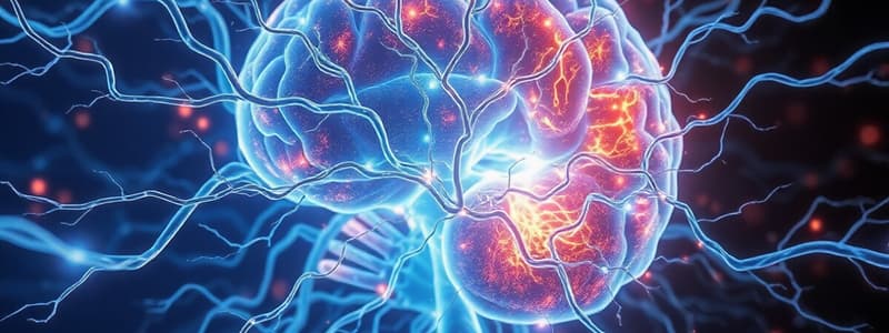
Nervous System Functions
- The nervous system is responsible for detecting environmental changes and coordinating responses within an organism.
- Cognition, muscle coordination, and memory storage are all functions attributed to the nervous system.
- The nervous system's ability to interact with and adapt to various environments is what makes it complex and adaptive.
- Learning and memory are higher functions associated with the nervous system.
- The nervous system can be impacted by diseases, trauma, or developmental issues.
- The nervous system contributes to maintaining homeostasis by coordinating muscle and gland responses to stimuli.
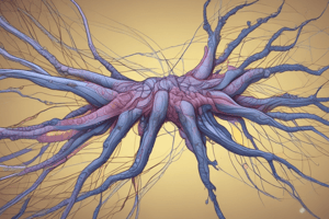
Neurones
- A neurone is commonly referred to as a nerve cell.
- The human body contains approximately 100 billion neurones.
- One of the main functions of a neurone is to receive and integrate information from sensory receptors or other neurones.
- Neurones contribute to the body's response to stimuli by transmitting signals to other neurones or effector organs.
- Neurones play a crucial role in maintaining homeostasis by transmitting signals that coordinate activities and responses in the body.
- Neurones process and integrate signals from sensory receptors or other neurones.
Neurone Function and Structure
- The cell membrane of a neurone is responsible for transmitting and maintaining electrical signals.
- Neurones communicate with each other through chemical synapses.
- The cell body (soma) of a neurone contains the nucleus and supports metabolic activities.
- Dendrites receive incoming signals from other neurones or sensory receptors.
- The primary role of an axon is to conduct electrical impulses away from the cell body.
- Nerve terminals (axon terminals) release neurotransmitters to communicate with adjacent cells.
- Neurones come in a variety of shapes and sizes but share core features.
Neuronal Resting Potential
- The typical resting potential of a neuron is -70 mV. This means the inside of the neuron is negatively charged compared to the outside.
Depolarization
- Sodium ions (Na⁺) rushing into the cell after a threshold is reached triggers depolarization. This influx of positive charge makes the inside of the neuron more positive.
- The threshold value is around -55 mV. Once this threshold is reached, an action potential is triggered.
Repolarization
- During repolarization, potassium channels open, allowing K⁺ to exit the cell. This outflow of positive charge makes the inside of the neuron more negative, bringing the membrane potential back towards its resting state.
Sodium-Potassium Pump
- The sodium-potassium pump actively transports sodium ions (Na⁺) out of the cell and potassium ions (K⁺) into the cell, restoring ion balance.
- This pump plays a crucial role in maintaining the resting potential and preparing the neuron for another action potential.
Refractory Period
- The refractory period follows an action potential, during which the neuron is less responsive to further stimuli.
- This period ensures that the action potential travels in one direction along the axon.
Action Potential Propagation
- An action potential propagates along an axon as a wave of depolarization and repolarization moving along the membrane.
- This wave of electrical activity is caused by the sequential opening and closing of sodium and potassium channels.
Peak of Action Potential
- The peak of an action potential occurs when the membrane potential reaches around +30 mV.
- At this point, the sodium channels close, and repolarization begins.
Synapses
- Synapses are the junctions between the axon terminal of one neuron and the dendrite or cell body of another neuron.
- The space between the presynaptic and postsynaptic neurons is called the synaptic cleft.
Neurotransmitters
- Neurotransmitters are chemical messengers released from vesicles in the presynaptic neuron.
- When neurotransmitters bind to receptors on the postsynaptic membrane, they can change the membrane potential of the postsynaptic cell.
Depolarization of the postsynaptic membrane
- Depolarization makes the membrane potential more positive and can initiate an action potential.
Enzymes in the synaptic cleft
- Enzymes in the synaptic cleft break down neurotransmitters to stop their action and clear the synapse.
Hyperpolarization of the postsynaptic membrane
- Hyperpolarization makes the membrane potential more negative, inhibiting the likelihood of an action potential.
Synapses
- Synapses are the junctions between the axon terminal of one neuron and the dendrite or cell body of another.
- The space between the presynaptic and postsynaptic neurons is called the synaptic cleft.
- Neurotransmitters are chemical messengers released from vesicles in the presynaptic neuron.
- Neurotransmitters can change the membrane potential of the postsynaptic cell when they bind to receptors on the postsynaptic membrane.
Action Potential
- Depolarization of the postsynaptic membrane makes the membrane potential more positive and can initiate an action potential.
- Hyperpolarization of the postsynaptic membrane makes the membrane more negative and inhibits the likelihood of an action potential.
Enzymes
- Enzymes in the synaptic cleft break down neurotransmitters to stop their action and clear the synapse.
Lambert-Eaton Myasthenic Syndrome (LEMS)
- Antibodies target voltage-gated calcium channels on the presynaptic nerve terminal
- Impaired calcium influx results in decreased release of acetylcholine (ACh)
- Primarily affects proximal muscles such as the shoulders and hips
Myasthenia Gravis (MG)
- Antibodies target acetylcholine receptors on the postsynaptic muscle membrane
- Antibody attack reduces the number of available receptors for acetylcholine
- Muscle weakness worsens with activity and improves with rest
- Most commonly affects muscles controlling eye movements, facial expressions, and swallowing
Sensory Neurones (Afferent Neurones)
- Transmit impulses from sensory receptors to the CNS
- Allow the brain to perceive sensations
Motor Neurones (Efferent Neurones)
- Transmit impulses from the CNS to muscles or glands
- Innervate skeletal muscles, also known as motor neurones
Interneurones
- Located entirely within the CNS
- Connect sensory and motor pathways within the CNS
- Process information and coordinate responses between sensory and motor neurones
Role of Neuroglia
- Neuroglia cells support and maintain the proper functioning of neurons.
Oligodendrocytes
- Oligodendrocytes are a type of neuroglia cell that forms the myelin sheath in the central nervous system (CNS).
- The myelin sheath increases the rate of action potential conduction along axons.
Astrocytes
- Astrocytes are another type of neuroglia cell that is involved in forming the blood-brain barrier.
- They also provide structural and metabolic support to neurons.
Microglia
- Microglia cells act as immune cells in the CNS, performing phagocytic roles.
- They maintain homeostasis and respond to tissue damage or pathogens.
Nervous System Development
- The ectoderm germ layer is responsible for the formation of the neural tube during early embryonic development, specifically in the third week.
- The neural tube develops into the central nervous system by the fifth week.
- The neural tube differentiates into three primary brain vesicles: prosencephalon, mesencephalon, and rhombencephalon.
- The prosencephalon develops into the telencephalon and diencephalon.
- The telencephalon forms the cerebral hemispheres by the seventh week.
- The diencephalon contains the thalamus which relays sensory and motor signals.
- The mesencephalon remains as the midbrain, responsible for visual and auditory processing.
- The rhombencephalon differentiates into the metencephalon and myelencephalon.
- The metencephalon develops into the pons and cerebellum.
- The pons aids in communication within the brain, while the cerebellum contributes to motor control and balance.
- The myelencephalon forms the medulla oblongata, which controls essential life functions like heart rate and breathing.
Brain Stem Structures
- The brain stem is comprised of the medulla oblongata, pons, and midbrain.
- The cerebellum is not part of the brain stem.
Brain Stem Function
- The brain stem's primary role is controlling automatic functions like heart rate and breathing.
Brain Stem and Spinal Cord
- The brain stem acts as a pathway for neural tracts, conveying signals between the brain and the spinal cord.
Cerebrospinal Fluid (CSF)
- CSF is produced by the choroid plexus in the ventricles.
- Its function is to cushion the brain, remove waste, and supply nutrients and hormones.
- CSF circulates through the ventricles and subarachnoid space and is ultimately reabsorbed into the bloodstream.
Ventricles
- The third ventricle is located in the diencephalon.
- The lateral ventricles are connected to the third ventricle by the interventricular foramen.
- The fourth ventricle is located between the brainstem and cerebellum.
Cerebrospinal Fluid (CSF) Protection
- CSF acts as a cushion and immune barrier for the brain, guarding against potential injuries and infections.
CSF Production
- CSF is produced by the choroid plexus, a network of capillaries located within the brain's ventricles.
CSF Volume
- The brain typically contains about 150 ml of CSF at any given time.
CSF Reabsorption
- CSF is reabsorbed back into the bloodstream through arachnoid villi, finger-like projections extending into the subarachnoid space.
Hydrocephalus
- Hydrocephalus is a condition where excess CSF accumulates, leading to increased intracranial pressure.
- This pressure can damage brain tissues and cause various neurological issues.
Causes of Hydrocephalus
- Hydrocephalus can arise from various factors, including:
- Congenital defects (present at birth)
- Tumors affecting CSF flow
- Infections
- Brain bleeding
Hydrocephalus Treatment
- Common treatments for hydrocephalus include:
- Placement of a shunt system to drain excess CSF
- Endoscopic third ventriculostomy (ETV) procedure, which creates an alternative pathway for CSF flow
Endoscopic Third Ventriculostomy (ETV)
- ETV aims to create a new drainage route for CSF by surgically connecting the third ventricle to the subarachnoid space.
Grey Matter
- Composed primarily of nerve cell bodies (soma) and dendrites.
- Found on the surface of the cerebral hemispheres, forming the cerebral cortex.
- Plays a crucial role in processing information, including sensory perception, memory, and decision-making.
White Matter
- Composed mainly of myelinated axons.
- Myelin sheath provides the white color.
- Serves as a communication network, connecting different grey matter areas and transmitting information between them.
Relationship Between Grey and White Matter
- Grey matter is responsible for processing information.
- White matter acts as the communication network between different grey matter areas.
Function of Myelin
- Insulates axons, which facilitates rapid transmission of nerve impulses.
Brain Nuclei
- Nuclei in the brain are clusters of nerve cell bodies (neurons) that share similar functions and connections.
- Function: They process and relay specific types of information, such as motor commands or sensory input from various parts of the body.
- Location: Motor neurons controlling related muscle groups are often organized within nuclei clusters.
- Importance: Nuclei play significant roles in the central nervous system (CNS) by processing and relaying information.
- Composition: They consist of nerve cell bodies that are functionally related, meaning they work together to achieve a specific task.
- Example: A nucleus might be responsible for relaying visual information from the eyes to the brain for interpretation.
Central Nervous System (CNS)
- Consists of the brain and spinal cord.
- Primarily responsible for processing and integrating sensory information, generating thoughts, and initiating motor responses.
- The spinal cord acts as a conduit for signals between the brain and the body, playing a role in reflex actions.
Peripheral Nervous System (PNS)
- Includes all nerves outside the CNS, connecting the brain and spinal cord to the body.
- Cranial nerves arise from the brain and transmit sensory and motor information for the head and neck.
- Spinal nerves are grouped based on their origin in the spinal cord and serve specific regions:
- Cervical nerves: Serve the neck and upper limbs.
- Thoracic nerves: Supply the chest and abdominal muscles.
- Lumbar nerves: Serve the lower back and parts of the lower limbs.
- Sacral nerves: Serve the pelvic region and lower limbs.
The Autonomic Nervous System (ANS)
- Regulates involuntary bodily functions such as heart rate, digestion, and breathing.
- Part of both the central nervous system (CNS) and the peripheral nervous system (PNS).
Sympathetic Nervous System
- Prepares the body for stressful or high-energy activities.
- Increases heart rate, dilates airways, and diverts blood flow to muscles.
- Often referred to as the "fight or flight" response.
Parasympathetic Nervous System
- Promotes energy storage, digestion, and relaxation.
- Slows heart rate, constricts airways, and promotes digestion.
- Complementary and often opposite effects to the sympathetic nervous system to maintain homeostasis (balance).
The Relationship Between the Sympathetic and Parasympathetic Systems
- Work together to maintain a balanced internal environment.
- The sympathetic system prepares the body for action, while the parasympathetic system promotes rest and recovery.
Innervation of Muscles and Glands
- The ANS innervates smooth muscle, cardiac muscle, and secretory glands, but not skeletal muscles.
Protection of the Brain and Spinal Cord
- The bones of the skull and vertebral column provide the outermost protection for the brain and spinal cord.
Meninges
- The meninges are a three-layered membrane that protects the brain and spinal cord.
- The three layers are:
- Dura mater: The tough, outermost layer of the meninges that attaches to the inner surface of the skull.
- Arachnoid mater: The middle layer with a web-like structure.
- Pia mater: The innermost layer that adheres closely to the surface of the brain and spinal cord.
Subarachnoid Space
- The space between the arachnoid mater and pia mater is called the subarachnoid space.
- The subarachnoid space contains cerebrospinal fluid (CSF).
Cerebrospinal Fluid (CSF)
- The primary function of CSF is to cushion the central nervous system (CNS) and provide a protective barrier.
Scalp
- The scalp is an additional protective structure that covers the skull.
Blood Supply to the Brain
- Internal carotid arteries are the primary arteries supplying the anterior part of the brain.
- The internal carotid arteries branch into the anterior cerebral artery and middle cerebral artery.
- The vertebral arteries originate from the subclavian arteries.
- The vertebral arteries merge to form the basilar artery.
- The vertebral and basilar arteries primarily supply the cerebellum and brainstem.
- The Circle of Willis is a circular network of arteries at the base of the brain.
- The Circle of Willis serves as a collateral blood flow pathway in case of an arterial blockage.
- The Circle of Willis is formed by the internal carotid and vertebral arteries.
Brain Veins
- Brain veins are unique because they lack valves, allowing blood to flow in either direction.
- This is unlike other veins in the body which have one-way valves to prevent backflow.
Deep Cerebral Veins
- Deep cerebral veins are responsible for draining blood from the forebrain and deeper structures of the brain.
Superficial Veins
- Superficial veins sit in the subarachnoid space and drain blood from the outer parts of the brain
Dural Veins
- Dural venous sinuses are channels located between the layers of the dura mater.
- These sinuses collect blood from the brain veins.
- The superior sagittal sinus is a primary example of a dural venous sinus.
Blood Drainage
- Blood collected in the dural venous sinuses drains into the internal jugular vein.
- The internal jugular vein then carries deoxygenated blood from the brain back to the heart.
Brain Stem Structure and Function
- The midbrain is a part of the brain stem involved in visual and auditory processing.
- The pons is also part of the brain stem and is responsible for coordinating facial movements and acting as a communication bridge between different brain areas.
- The medulla oblongata is another part of the brain stem and is responsible for regulating heartbeat and blood pressure, and manages reflexes like coughing and swallowing.
- The thalamus is located above the midbrain and acts as a relay station for sensory and motor signals to the cerebral cortex, contributing to consciousness.
- Cranial nerves originating from the brain stem control functions like eye movement, facial sensation, and swallowing.
- The brain stem serves as a pathway for sensory and motor tracts between the brain and spinal cord.
The Medulla Oblongata
- Vital Role: The medulla oblongata is crucial for life as it controls involuntary functions.
- Essential Functions: It regulates heart rate, breathing rhythm, and reflexes like coughing and sneezing.
- Non-Medulla Function: Complex problem-solving is not a function of the medulla oblongata.
- Neural Communication: The medulla acts as a relay center, transmitting information between the thalamus and spinal cord.
- Anatomical Location: The caudal medulla is closest to the spinal cord.
- Cranial Nerve Importance: The vagus nerve (CN X) originates from the medulla and governs heart rate and digestion.
- Hypoglossal Nerve Function: The hypoglossal nerve (CN XII) regulates tongue movements, vital for speech and swallowing.
- Medulla and Pons: The rostral medulla is adjacent to the pons.
- Glossopharyngeal Nerve Role: The glossopharyngeal nerve (CN IX) controls taste, swallowing, and salivation.
Location and Size
- The pons is situated between the medulla oblongata and the midbrain.
- The pons is approximately 2.5 cm in length.
Function
- The pons serves as a relay center, connecting the cerebellum and cerebrum.
- The pons plays a crucial role in regulating breathing patterns.
- The pons is involved in the sleep-wake cycle and REM sleep.
Cranial Nerves
- The facial nerve (CN VII) originates from the pons and controls facial expressions.
- The vestibulocochlear nerve (CN VIII) originates from the pons and is responsible for hearing and balance.
- The abducens nerve (CN VI) originates from the pons and controls lateral eye movement.
- The trigeminal nerve (CN V) originates from the pons and is responsible for facial sensation, chewing, and muscle control.
Midbrain Functions
- Primary function: Controlling eye movement and processing auditory and visual information
- Contains: Tectum (dorsal portion) and Tegmentum (ventral portion)
Tectum (Dorsal Portion)
- Superior colliculi: Responsible for coordinating head and eye movements in response to visual stimuli
- Inferior colliculi: Primary role in auditory processing and sound localization
Tegmentum (Ventral Portion)
- Associated with: Maintaining arousal and alertness
Cranial Nerves
- Originate from the midbrain:
- Oculomotor nerve (CN III): Controls most eye movements and pupil constriction
- Trochlear nerve (CN IV): Controls the superior oblique muscle, which rotates the eye downwards and outwards
Reticular Formation: Location and Connections
- The reticular formation extends throughout the brainstem, connecting the medulla to the midbrain.
- The reticular formation has both incoming (afferent) and outgoing (efferent) connections, reaching various parts of the central nervous system (CNS).
- Axons within the reticular formation are long, allowing for transmission of signals over significant distances.
Reticular Formation: Key Functions
- The reticular formation plays a vital role in regulating arousal and consciousness.
- It contributes to controlling heart rate and blood pressure through interactions with the autonomic nervous system.
- The reticular formation is involved in regulating the rate and depth of breathing, in coordination with the medulla oblongata.
- The reticular formation's influence on maintaining alertness and regulating the sleep-wake cycle makes it crucial for survival.
The Thalamus: A Key Relay Station in the Brain
- The thalamus processes and relays sensory and motor signals to the cerebral cortex, regulating consciousness, sleep, and alertness.
- It's situated between the cerebral hemispheres, resembling the size of a small hen's egg.
- The thalamus is the biggest component of the diencephalon, a part of the brain that also includes the hypothalamus.
- The thalamus receives and transmits both sensory and motor information, serving as a critical hub for communication within the brain.
- It's supplied with blood by branches of the posterior cerebral artery.
- Its central location within the brain allows it to easily relay information to and from different regions, crucial for its role in consciousness and alertness.
Unilateral Brainstem Lesions: Causes and Symptoms
-
Common Causes: Cerebrovascular accidents (CVAs), tumors, multiple sclerosis (MS)
-
Ipsilateral Cranial Nerve Dysfunction: Damage to cranial nerves on the same side as the lesion, leading to motor or sensory issues.
-
Contralateral Spastic Hemiparesis: A common outcome affecting the opposite side of the body, presenting as weakness and spasticity.
-
Abnormal Babinski Sign: Indicates upper motor neuron damage, characterized by an upward extension of the big toe when the sole of the foot is stroked.
Bilateral Brainstem Lesions: Severity and Impact
-
Severe Damage: Can lead to life-threatening outcomes such as coma or death due to damage to vital centers.
-
Vital Centers Affected: Centers that control respiration and circulation, essential for life.
-
Hyperreflexia: Exaggerated reflexes and increased spasticity, a potential outcome of unilateral brain stem lesion.
-
Contralateral Hemisensory Loss: Loss of sensation on the side opposite to the lesion.
The Limbic System Structures & Their Functions
- The amygdala plays a crucial role in processing emotions, particularly fear and aggression. This structure is directly involved in the fear response.
- The hippocampus is essential for forming and consolidating memories, including both short-term and long-term memories. It plays a vital role in memory formation and recall.
- The hypothalamus manages autonomic functions like hunger, thirst, body temperature, and the sleep-wake cycle. It connects to the endocrine system and influences hormone release.
- The basal ganglia is associated with motor control, coordinating movement and learning. It plays a crucial role in smooth and coordinated movements.
Amygdala: Role in Cognition and Emotion
- The amygdala plays a critical role in emotional and social behaviors, as well as in episodic-autobiographical memory (EAM), which involves recalling personal experiences tied to emotions.
- The amygdala is particularly associated with negative emotions like fear, anxiety, and aggression.
- The amygdala is involved in evaluating facial expressions and recognizing emotional cues in others, contributing to social processing.
- The left amygdala is suggested to be involved in both positive (happiness) and negative (fear, anxiety, sadness) emotions.
- The right amygdala is more strongly associated with negative emotions, particularly fear and sadness, and is involved in declarative memory, which is the conscious recall of facts and events.
- The amygdala plays a critical role in attentional processing, influencing how emotional stimuli are perceived and responded to.
The Hippocampus and Memory
- The hippocampus is a brain structure crucial for transferring short-term memories into long-term storage.
- This process is known as memory consolidation.
- The hippocampus is heavily involved in spatial memory, which is essential for navigation and understanding spatial relationships.
Hippocampus & Conditions
- Damage to the hippocampus often leads to the inability to form new memories and short-term memory loss.
- A shrunken hippocampus has been linked to cognitive and emotional regulation issues, including schizophrenia and severe depression.
Hormonal Influence on the Hippocampus
- Oestrogen has a positive impact on the hippocampus, potentially increasing neural connectivity.
Clinical Implications
- Health and function of the hippocampus are vital for cognitive health.
- Deterioration of the hippocampus is linked to many neurological and psychiatric conditions, including Alzheimer's.
- Oestrogen's positive effect on the hippocampus is seen as a potential treatment avenue for Alzheimer's disease.
- The hippocampus plays a key role in spatial memory, which is essential for navigation and understanding our surroundings.
The Basal Ganglia: An Overview
- The basal ganglia are a group of structures located deep within the cerebral hemispheres.
- These structures play a crucial role in facilitating movement, inhibiting unwanted movements, and regulating behavior.
Key Components of the Basal Ganglia
- The basal ganglia include the caudate nucleus, putamen, globus pallidus, subthalamic nucleus, and substantia nigra.
- The hippocampus, however, is not part of the basal ganglia.
The Substantia Nigra's Significance
- The substantia nigra, a structure in the midbrain, is part of the basal ganglia.
- It is particularly important for movement regulation as it is responsible for producing dopamine.
Functionality of the Basal Ganglia
- The basal ganglia enable coordinated movements by facilitating the desired action and inhibiting counter-movements.
- This is essential for tasks like smoothly reaching for an object.
- The basal ganglia contribute to habit formation and action selection, playing a significant role in behavior.
Location of the Subthalamic Nucleus
- The subthalamic nucleus, one of the components of the basal ganglia, is located in the diencephalon.
Parkinson's Disease
- Degeneration of dopaminergic neurons in the substantia nigra is the primary impairment
- Common symptom: Rigidity and bradykinesia (slow movement)
- Impaired ability to inhibit contradictory movements and initiate smooth movement
Huntington's Disease
- Caused by the unusual activity of the globus pallidus
- Characterized by jerky and writhing involuntary movements known as chorea
- Progressively worsens, affecting movement, cognition, and psychiatric health
Basal Ganglia and Other Conditions
- Reduced dopamine levels due to basal ganglia degeneration are associated with Parkinson's disease
- Scientists are investigating how disruptions or malfunctions in the basal ganglia's neural circuits may contribute to Tourette’s syndrome, schizophrenia, and OCD
Hypothalamus: Maintaining Homeostasis
- The hypothalamus is the brain region primarily responsible for maintaining homeostasis, the body's stable internal environment.
- It accomplishes this by regulating both autonomic and endocrine functions.
Functions of the Hypothalamus
- Regulation of Body Temperature: The hypothalamus monitors and adjusts body temperature through sweating, shivering, and other mechanisms.
- Thirst and Hunger Control: This region monitors blood nutrient levels and regulates hunger and thirst to ensure adequate nutrient intake.
- Sleep Regulation: The hypothalamus plays a critical role in regulating sleep-wake cycles, ensuring the body gets sufficient rest.
- Hormonal Control: The hypothalamus produces hormones that control the pituitary gland, which in turn regulates the endocrine system, impacting everything from growth to reproduction.
Hypothalamic Inputs
- Neural Inputs: Receives signals from various brain regions, providing information for decision-making.
- Circulatory Inputs: Monitored blood contains a wealth of information about the body's internal state:
- Blood Pressure: Hypothalamus responds to pressure fluctuations, regulating blood flow.
- Temperature: The hypothalamus uses this data to adjust body temperature.
- Nutrient Levels: Helps regulate hunger and thirst based on blood glucose and other nutrient levels.
- Hormonal Signals: Responds to various hormones in the blood, providing insight into the body's overall state.
Hypothalamus and Internal Signals
- The hypothalamus receives information about the body's internal state through various signals:
- Chemical Signals from Blood: Blood composition, including electrolyte balance, is essential for proper functioning.
- Hormonal Signals in the Blood: Provides the hypothalamus with insights into the body's current metabolic state.
Hypothalamus Function
- The hypothalamus is responsible for maintaining homeostasis - regulating autonomic and endocrine functions.
- It controls body temperature, thirst and hunger, sleep regulation.
- It receives information about the body's state through neural and circulatory inputs:
- Neural inputs from the brain provide information about sensory and emotional states.
- Circulatory inputs consist of blood pressure, temperature, nutrient levels, and hormones.
- Hormones in the blood provide the hypothalamus with information about the body's metabolic state, including electrolyte balance.
The Hypothalamus and Sleep
- The hypothalamus plays a crucial role in regulating sleep as part of its homeostatic functions.
The Pituitary Gland: Master Regulator
- The pituitary gland is often called the "master gland" due to its crucial role in regulating various hormonal functions across the body.
- It is located at the base of the brain, connected to the hypothalamus via a stalk-like structure.
- The pituitary gland is divided into two main lobes: the anterior pituitary (front) and the posterior pituitary (back).
Posterior Pituitary
- The posterior pituitary does not produce its own hormones but stores and releases hormones synthesized by the hypothalamus.
- Vasopressin (antidiuretic hormone), released from the posterior pituitary, plays a vital role in regulating water balance in the body.
Anterior Pituitary
- The anterior pituitary synthesizes and secretes a range of hormones that control crucial bodily functions.
- Adrenocorticotropic hormone (ACTH) stimulates the adrenal glands to produce cortisol, which is essential for stress response and energy regulation.
- Luteinizing hormone (LH) is responsible for stimulating ovulation in females and testosterone production in males.
- Growth hormone (GH) promotes growth, cell repair, and metabolism, playing a key role in development.
- Prolactin is responsible for stimulating milk production after childbirth in women.
- Thyroid-stimulating hormone (TSH) regulates the thyroid gland's release of thyroid hormones, essential for metabolism and growth.
Hypothalamus and Pituitary Tumours
- Tumours in the hypothalamus or pituitary gland can disrupt hormone production, leading to either overproduction or underproduction.
- Overproduction of Growth Hormone (GH): Results in gigantism.
- Overproduction of Adrenocorticotropic Hormone (ACTH): Leads to Cushing's disease.
- Underproduction of ACTH: Causes adrenal insufficiency.
- Tumours affecting Vasopressin Regulation: Can cause diabetes insipidus.
- Tumours impacting the Optic Chiasma: May cause bitemporal visual field loss (loss of peripheral vision in both eyes).
- Tumours affecting Sexual Development: Can lead to precocious puberty.
- Hypothalamic and Pituitary Tumours and Eating Disorders: May be associated with obesity and bulimia.
Cerebellum Location & Connections
- Located beneath the cerebral hemispheres and behind the brainstem.
- Connected to the brainstem via three pairs of cerebellar peduncles:
- Inferior Cerebellar Peduncles: Connect the cerebellum to the medulla.
- Middle Cerebellar Peduncles: Connect the cerebellum to the pons.
- Superior Cerebellar Peduncles: Connect the cerebellum to the midbrain.
Cerebellum Function
- Plays a crucial role in coordinating and fine-tuning movements, ensuring they are smooth and precise. It doesn't initiate movement, but it refines it, making it accurate and controlled.
- Maintains equilibrium (balance), contributing to our ability to stand, walk, and move without falling.
- Regulates muscle tone, ensuring muscles have the right level of stiffness for efficient movement.
- Adjusts and maintains posture during movement, keeping us upright and balanced.
Cerebellum Clinical Significance
- Damage to the cerebellum can lead to ataxia (loss of coordination), causing difficulty with movement, balance, and gait.
- Ataxia is characterized by jerky, uncoordinated movements, problems with balance, and unsteady gait.
Cerebellar Lesions and Motor Deficits
-
Midline lesions in the cerebellum often lead to loss of postural control and balance issues.
-
Unilateral cerebellar hemispheric lesions cause ipsilateral (same-side) motor deficits.
-
Intention tremors are tremors that occur during voluntary movements and are a common symptom of cerebellar dysfunction.
-
Bilateral cerebellar dysfunction can cause dysarthria, which is slurred speech.
-
Cerebellar ataxia is a condition that results from damage or degeneration of the cerebellum.
-
Bilateral cerebellar dysfunction can be caused by various factors such as alcoholic intoxication, hypothyroidism, inherited cerebellar degeneration, and multiple sclerosis (MS).
-
Nystagmus is a rapid, involuntary eye movement often associated with cerebellar disorders and MS.
-
An unsteady gait affecting walking stability is a typical result of incoordination in the leg due to a unilateral cerebellar lesion.
Cerebral Hemispheres
- The largest part of the forebrain
- Covered by the cerebral cortex, a superficial layer of grey matter
- Ridges and furrows on the cerebral cortex are known as gyri (ridges) and sulci (furrows)
- Folding pattern allows for a greater number of neurons, enhancing cognitive processing
- White matter, composed of axons, lies beneath the grey matter
- White matter connects different parts of the brain, facilitating communication
Cerebral Cortex
- Responsible for complex functions, including:
- Thought
- Memory
- Sensory perception
- Voluntary movement
- Divided into four lobes:
- Frontal
- Parietal
- Temporal
- Occipital
Cerebral Cortex Structure
- The cerebral cortex is a thin layer of brain tissue, only a few millimeters thick, yet it is densely packed with neurons and has multiple functional layers.
Cerebral Cortex Function
- The cerebral cortex is responsible for processing and interpreting sensory information.
- It plays a crucial role in motor function, generating commands that initiate voluntary movements and planning motor actions.
- It is involved in conscious perception and detailed interpretation of sensory stimuli.
- It processes both sensory and motor information.
Cerebral Cortex and Sensory Information
- Sensory information primarily ascends to the cortex through the thalamus.
Cerebral Cortex and Motor Function
- The cerebral cortex is the highest level of motor system representation.
- It initiates and plans motor actions.
- It is not directly involved in controlling reflexes.
Cerebral Lobes and Functions
- Frontal Lobe: Responsible for executive functions such as thinking, planning, organizing, and problem-solving. It also houses the motor cortex.
- Parietal Lobe: Primarily associated with sensation, perception, and integrating sensory information. It contains the sensory cortex.
- Occipital Lobe: Responsible for processing visual information.
- Temporal Lobe: Associated with memory, understanding, language, and auditory processing.
Motor and Sensory Cortexes
- Motor Cortex: Located in the frontal lobe, it processes voluntary movement commands.
- Sensory Cortex: Located in the parietal lobe, it receives and processes sensory information from the body.
Medulla Oblongata
- Medulla Oblongata: Located in the brainstem, it is vital for controlling essential functions like breathing, heart rate, and respiration.
Frontal Lobe Lesions
- Can lead to different types of seizures: Simple focal, complex partial, or generalized seizures
- May result in both motor and sensory deficits
- Can cause changes in behavior, decision-making, and emotional control
- Particularly affect executive functions, such as planning and decision-making
Space-Occupying Focal Lesions
- Can be caused by a tumor
- Lead to raised intracranial pressure (ICP)
- Prolonged raised ICP can have severe consequences
- Symptoms of raised ICP include headaches, nausea, and vomiting
Seizures
- Simple focal seizure: Electrical activity starts and remains in a limited brain region
Studying That Suits You
Use AI to generate personalized quizzes and flashcards to suit your learning preferences.