Podcast
Questions and Answers
Which of the following accurately describes the functional relationship between the central nervous system (CNS) and the peripheral nervous system (PNS)?
Which of the following accurately describes the functional relationship between the central nervous system (CNS) and the peripheral nervous system (PNS)?
- The CNS operates independently, while the PNS manages only reflex actions.
- The CNS and PNS work together, with the CNS processing information received from the PNS. (correct)
- The PNS is a self-regulating system that operates independently of the CNS.
- The CNS primarily handles sensory input, while the PNS is dedicated to motor output.
How do neurotransmitters facilitate communication between neurons?
How do neurotransmitters facilitate communication between neurons?
- By physically connecting adjacent neurons, creating a cytoplasmic bridge.
- By forming a continuous electrical pathway between neurons.
- By directly altering the genetic code of the postsynaptic neuron.
- By diffusing across the synapse and binding to receptors on the postsynaptic membrane. (correct)
How does the myelinated sheath contribute to the function of a neuron?
How does the myelinated sheath contribute to the function of a neuron?
- By increasing the speed of impulse conduction along the axon. (correct)
- By increasing the amplitude of the action potential.
- By preventing the neuron from forming connections with other neurons.
- By decreasing the energy required for neurotransmitter synthesis.
What role do glial cells play in the nervous system?
What role do glial cells play in the nervous system?
What is the primary function of the corpus callosum?
What is the primary function of the corpus callosum?
Which statement accurately describes the role of the hypothalamus in maintaining homeostasis?
Which statement accurately describes the role of the hypothalamus in maintaining homeostasis?
What is the blood-brain barrier's (BBB) primary function?
What is the blood-brain barrier's (BBB) primary function?
What is the role of the circle of Willis in cerebral circulation?
What is the role of the circle of Willis in cerebral circulation?
How do the ascending tracts in the spinal cord contribute to sensory perception?
How do the ascending tracts in the spinal cord contribute to sensory perception?
Which of the following accurately contrasts the sympathetic and parasympathetic nervous systems?
Which of the following accurately contrasts the sympathetic and parasympathetic nervous systems?
Flashcards
Central Nervous System (CNS)
Central Nervous System (CNS)
Includes the brain and spinal cord.
Functions of the Brain
Functions of the Brain
Controls motor, sensory, autonomic, cognitive, and behavioral activities. Monitors body processes and maintains homeostasis.
Function of Neurotransmitters
Function of Neurotransmitters
Communicate messages between neurons or from neurons to target cells (muscle or endocrine); manufactured and stored in synaptic vesicles; released into the synapse during electrical action potential.
Nerve Cell Bodies (Centers)
Nerve Cell Bodies (Centers)
Signup and view all the flashcards
Cerebral Cortex (Gray Matter)
Cerebral Cortex (Gray Matter)
Signup and view all the flashcards
Corpus Callosum
Corpus Callosum
Signup and view all the flashcards
Hypothalamus
Hypothalamus
Signup and view all the flashcards
Function of the Cerebellum
Function of the Cerebellum
Signup and view all the flashcards
Cerebrospinal Fluid (CSF)
Cerebrospinal Fluid (CSF)
Signup and view all the flashcards
Autonomic Nervous System (ANS)
Autonomic Nervous System (ANS)
Signup and view all the flashcards
Study Notes
- Anatomy and physiology of the nervous system
Major Parts of the Nervous System
- Central Nervous System (CNS): includes the brain and spinal cord.
- Peripheral Nervous System: comprises cranial nerves, spinal nerves, and the autonomic nervous system.
Functions of the Nervous System
- Controls motor, sensory, autonomic, cognitive, and behavioral activities.
- The brain contains over 100 billion cells linking motor and sensory pathways.
- Monitors body processes, responds to environments, and maintains homeostasis.
- Directs psychological, biological, and physical activity via chemical and electrical messages.
Cells of the Nervous System
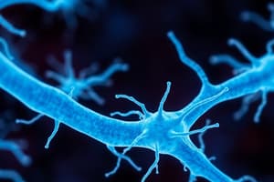
- Neurons are the basic functional unit of the brain.
- Neurons composed of dendrites, a cell body, and an axon.
- Dendrites receive electrochemical messages.
- Axons carry impulses away from the cell body.
- Some axons have a myelinated sheath for faster conduction.
- Nerve cell bodies: Clusters are called ganglia or nuclei; centers are clusters with similar functions (e.g., respiratory center).
- Glial cells: Support, protect, and nourish neurons; outnumber neurons by 50 times.
Neurotransmitters Function
- Communicate messages between neurons or from neurons to target cells (muscle or endocrine).
- Neurotransmitters are manufactured and stored in synaptic vesicles and released into the synapse during electrical action potential.
Neurotransmitters Mechanism
- Transported across the synapse, binding to receptors on the postsynaptic cell membrane.
- Neurotransmitters excitation or inhibition of target cell activity usually involves multiple neurotransmitters.
Neurotransmitters Regulation
- Enzymes destroy or reabsorb neurotransmitters after release for future use.
Neurologic Disorders
- Often linked to neurotransmitter imbalances (e.g., Parkinson's disease and dopamine deficiency, myasthenia gravis and acetylcholine impairment).
- Neurotransmitter receptor activity modulates all brain functions including memory and cognitive processes.
Major Neurotransmitters
- Acetylcholine source: Neurons in various brain areas; autonomic nervous system.
- Acetylcholine action: Usually excitatory; can be inhibitory (e.g., vagal nerve stimulation affects the heart).
- Serotonin source: Brain stem, hypothalamus, dorsal horn of the spinal cord.
- Serotonin action: Inhibitory; regulates mood and sleep and inhibits pain pathways.
- Dopamine source: Neurons in the substantia nigra and basal ganglia.
- Dopamine action: Usually inhibitory; influences behavior (attention, emotions) and fine motor control.
- Norepinephrine source: Brain stem, hypothalamus, postganglionic neurons of the sympathetic nervous system.
- Norepinephrine action: Usually excitatory; affects mood and overall activity.
- Gamma-aminobutyric acid (GABA) source: Nerve terminals in the spinal cord, cerebellum, basal ganglia, and some cortical areas.
- Gamma-aminobutyric acid (GABA) action: Inhibitory.
- Enkephalin and Endorphin source: Nerve terminals in the spine, brain stem, thalamus, hypothalamus, and pituitary gland.
- Enkephalin and Endorphin action: Excitatory; produces pleasurable sensations and inhibits pain transmission.
The Central Nervous System (CNS)
- The CNS components comprise the brain and spinal cord.
The Brain
- Weight: Accounts for approximately 2% of total body weight.
- Average young adult brain: ~1400 g; average older adult brain: ~1200 g.
Major areas of the Brain
- Cerebrum: Contains two hemispheres, thalamus, hypothalamus, and basal ganglia.
- Brain Stem: Includes the midbrain, pons, and medulla.
- Cerebellum: Located under the cerebrum and behind the brain stem.
Cerebrum
- Structure: Wrinkled surface due to convolutions called gyri, increasing surface area.
- Sulci or fissures separate each gyrus.
- The great longitudinal fissure divides the cerebrum into right and left hemispheres.
- Hemispheres joined by the corpus callosum.
- Layers: Cerebral cortex (gray matter): Outer layer (2 to 5 mm); contains billions of neuron cell bodies.
- Layers: White matter: Innermost layer; composed of myelinated nerve fibers and neuroglia cells, forming tracts connecting different brain areas.
- Frontal Lobe: Largest lobe, located at the front.
- Functions: concentration, abstract thought, memory, motor function, personality, judgment, and speech (Broca's area).
- Parietal Lobe: Located posterior to the frontal lobe.
- Primarily sensory; analyzes sensory information, body position awareness, and shape discrimination.
- Temporal Lobe: Located inferior to the frontal and parietal lobes.
- Contains auditory areas; involved in sound memory and understanding language/music.
- Occipital Lobe: Located posterior to the parietal lobe.
- Responsible for visual interpretation and memory.
The Corpus Callosum
- Thick collection of nerve fibers connecting the two brain hemispheres.
- Responsible for transmitting information (sensation, memory, learned discrimination) between hemispheres.
Cerebral Dominance
- Right-handed individuals and some left-handed individuals have left hemisphere dominance for:
- Verbal, linguistic, arithmetic, calculation, and analytic functions.
- Right hemisphere is responsible for geometric, spatial, visual, pattern, and musical functions.
Hypothalamus
- Located anterior and inferior to the thalamus, beneath and lateral to the third ventricle.
- Connected to the posterior pituitary gland via the infundibulum.
- Regulates pituitary hormone secretion influencing metabolism, reproduction, stress response, and urine production.
- Maintains fluid balance and temperature regulation.
- Houses centers for hunger, sleep-wake cycle, blood pressure, emotional responses, and controls the autonomic nervous system.
- Contains the optic chiasm and mammillary bodies (involved in olfactory reflexes and emotional responses).
Basal Ganglia
- Masses of nuclei deep in the cerebral hemispheres.
- Responsible for controlling fine motor movements, including those of the hands and lower extremities.
Brain Stem
- Components: Consists of the midbrain, pons, and medulla oblongata.
- Midbrain: Connects the pons and cerebellum with the cerebral hemispheres
- Contains sensory and motor pathways.
- Center for auditory and visual reflexes.
- Cranial nerves III and IV originate here.
- Pons: Located in front of the cerebellum, between the midbrain and medulla.
- Acts as a bridge between cerebellar halves and between the medulla and midbrain.
- Cranial nerves V through VIII originate here.
- Contains motor and sensory pathways and helps regulate respiration.
- Medulla Oblongata: Contains motor fibers from the brain to the spinal cord and sensory fibers from the spinal cord to the brain (most fibers decussate here).
- Cranial nerves IX through XII originate in the medulla.
- Houses reflex centers for respiration, blood pressure, heart rate, coughing, vomiting, swallowing, and sneezing.
- Reticular formation begins here, responsible for arousal and the sleep-wake cycle, connecting with higher brain structures.
Cerebellum
- Location: Positioned posterior to the midbrain and pons, and below the occipital lobe.
- Function:
- Integrates sensory information for smooth, coordinated movement.
- Controls fine movement, balance, and postural sense (proprioception).
Structures Protecting the Brain
- Skull: Rigid structure protecting the brain from injury.
- Major bones include frontal, temporal, parietal, occipital, and sphenoid bones.
- Bones join at suture lines.
- Indentations known as fossae: Anterior fossa contains the frontal lobe, Middle fossa contains the temporal lobe and Posterior fossa contains the cerebellum and brain stem.
- Meninges: Fibrous connective tissues covering the brain and spinal cord provide protection, support, and nourishment.
- Three layers:
- Dura Mater: Outermost layer; tough, thick, inelastic, fibrous, and gray.
- Major extensions: Falx cerebri folds between hemispheres.
- Tentorium folds between occipital lobe and cerebellum.
- Falx cerebelli is located between cerebellar hemispheres.
- Can be involved in brain herniation due to excess cranial pressure.
- Contains potential spaces: epidural space (between dura and skull) and subdural space (below dura) where blood or abscess can accumulate.
- Arachnoid: Middle membrane, delicate and spider-web-like.
- Contains cerebrospinal fluid (CSF) in the subarachnoid space.
- Arachnoid villi absorb CSF into the venous system and obstruction can lead to communicating hydrocephalus.
- Pia Mater: Innermost, thin, transparent layer that closely adheres to the brain and extends into its folds.
- Dura Mater: Outermost layer; tough, thick, inelastic, fibrous, and gray.
Cerebrospinal Fluid (CSF)
- Characteristics: Clear and colorless fluid produced in the choroid plexus of the ventricles.
- Circulates around the brain and spinal cord.
- Ventricles: Four ventricles: right and left lateral, third, and fourth ventricles.
- Lateral ventricles connect to the third ventricle via the interventricular foramen (foramen of Monro).
- Third and fourth ventricles connect via the aqueduct of Sylvius.
- Fourth ventricle drains CSF into the subarachnoid space, absorbed by arachnoid villi.
- Function:
- Important for immune and metabolic processes in the brain.
- Produced at a rate of about 500 mL/day; ventricles and subarachnoid space hold approximately 125 to 150 mL.
- Composition: Similar to extracellular fluids (e.g., blood plasma) but with different concentrations of constituents.
- Normal CSF contains minimal white blood cells and no red blood cells.
- Can be analyzed for color, specific gravity, protein count, cell count, glucose, electrolytes, and immunoglobulins.
- Sample obtained via lumbar puncture or intraventricular catheter.
Cerebral Circulation
- Blood supply: Brain receives about 15% of cardiac output (approximately 750 mL/min).
- Unique circulation characteristics: Arterial and venous vessels are not parallel.
- Collateral circulation through the circle of Willis allows blood flow redirection.
- Blood vessels have two layers, making them prone to rupture.
- Arterial Supply: Anterior brain supplied by common carotid artery, which bifurcates into internal carotid arteries.
- Internal carotid arteries branch into anterior and middle cerebral arteries, forming the circle of Willis.
- Posterior circulation supplied by vertebral arteries, which join to form the basilar artery, dividing into posterior cerebral arteries.
- Circle of Willis provides collateral circulation if vessels become occluded.
- Venous Drainage: Veins do not follow arterial pathways; they join larger veins and cross the subarachnoid space to empty into dural sinuses.
- Dural sinuses carry venous outflow to internal jugular veins, returning blood to the heart.
- Cerebral veins lack valves and rely on gravity and blood pressure for drainage.
Blood-Brain Barrier
- Function: Protects the CNS by restricting access to certain substances in blood plasma (e.g., dyes, medications).
- Formed by endothelial cells of brain capillaries with tight junctions, creating a barrier to macromolecules.
The Spinal Cord
- Structure: Continuous with the medulla; connects the brain to the periphery.
- Approximately 45 cm (18 inches) long and about the thickness of a finger.
- Extends from the foramen magnum at the base of the skull to the lower border of the first lumbar vertebra.
- Tapers to a fibrous band called the conus medullaris at the lower end.
- Below the conus, nerve roots extend, forming the cauda equina (resembling a horse's tail).
- Protection: Surrounded by meninges.
- Cross-Sectional Anatomy: Contains an H-shaped central core of gray matter (nerve cell bodies) surrounded by white matter (ascending and descending tracts).
- Lower portion of the H is broader, corresponding to the anterior horns.
Function of Horns
- Anterior Horns: Contain cells with fibers forming the anterior (motor) root and is essential for voluntary and reflex muscle activity.
- Posterior Horns: Contain thinner upper portion containing cells, with fibers entering through the posterior (sensory) root which acts as a relay station in the sensory/reflex pathway.
The Spinal Tracts Composition
- White matter consists of myelinated and unmyelinated nerve fibers.
- Fast-conducting myelinated fibers form bundles called tracts.
Ascending Tracts (6 Total)
- Fasciculus cuneatus and gracilis (posterior columns): Conduct sensations of deep touch, pressure, vibration, position, and passive motion.
- Fibers cross to the opposite side in the medulla before reaching the cerebral cortex.
- Anterior and posterior spinocerebellar tracts: Conduct sensory impulses from muscle spindles for coordinated muscle contraction.
- Ascend uncrossed and terminate in the cerebellum.
- Anterior and lateral spinothalamic tracts: Responsible for conduction of pain, temperature, proprioception, fine touch, and vibratory sense.
- Cross to the opposite side of the cord and then ascend to the brain, terminating in the thalamus.
Descending Tracts (8 Total)
- Anterior and lateral corticospinal tracts: Conduct motor impulses to anterior horn cells from the opposite side of the brain.
- Cross in the medulla and controls voluntary muscle activity.
- Vestibulospinal tracts (3 types): Descend uncrossed.
- Involved in autonomic functions (sweating, pupil dilation, circulation) and involuntary muscle control.
- Corticobulbar tract: Conducts impulses for voluntary head and facial muscle movement and crosses at the brain stem.
- Rubrospinal and reticulospinal tracts: Conduct impulses associated with involuntary muscle movement.
Vertebral Column
- Structure: Surrounds and protects the spinal cord and consists of 7 cervical, 12 thoracic, 5 lumbar vertebrae, sacrum (fused mass of 5 vertebrae) and coccyx.
- Function: Nerve roots exit through intervertebral foramina.
- Vertebrae are separated by discs, except for certain cervical, sacral, and coccygeal vertebrae.
The Peripheral Nervous System (PNS)
- Components: Includes cranial nerves, spinal nerves, and the autonomic nervous system.
Cranial Nerves
- Overview: Twelve pairs emerge from the lower surface of the brain and pass through openings in the skull.
- Classification: Sensory nerves (3): I (olfactory), II (optic), VIII (vestibulocochlear).
- Motor nerves (5): III (oculomotor), IV (trochlear), VI (abducens), XI (accessory), XII (hypoglossal).
- Mixed nerves (4): V (trigeminal), VII (facial), IX (glossopharyngeal), X (vagus).
- Function: Innervate the head, neck, and special sense structures.
Spinal Nerves
- Overview: Composed of 31 pairs: 8 cervical, 12 thoracic, 5 lumbar, 5 sacral and 1 coccygeal.
Autonomic Nervous System (ANS)
- Function: Regulates activities of internal organs (heart, lungs, blood vessels, digestive organs, glands).
- Responsible for maintaining and restoring internal homeostasis.
Divisions of the Autonomic Nervous System
- Sympathetic Nervous System: Produces predominantly excitatory responses (e.g., "fight-or-flight").
- Parasympathetic Nervous System: Controls primarily visceral functions.
- Regulation: Governed by centers in the spinal cord, brain stem, and hypothalamus.
- The hypothalamus is the major subcortical center for autonomic regulation, linking to the thalamus, cortex, olfactory apparatus, and pituitary gland.
- Controls visceral and somatic reactions related to emotional states (fear, anger, anxiety).
- Regulates metabolic processes, body temperature, arterial pressure, and gastrointestinal activities.
- Influences genital functions and sleep cycles.
- Co-innervation: Most tissues and organs are innervated by both sympathetic and parasympathetic systems.
- Example: Parasympathetic: Contraction of urinary bladder muscles and decreased heart rate.
- Sympathetic: Relaxation of urinary bladder and increased heart rate.
Sympathetic Division
- The sympathetic division is part of the autonomic nervous system.
- Primarily responsible for the body's fight-or-flight response.
- Activated under stress from physical or emotional causes.
- Effects include dilation of bronchioles for improved gas exchange, increased strength and speed of heart contractions, dilation of arteries to the heart and voluntary muscles for enhanced blood flow, constriction of peripheral blood vessels cooling the skin while redirecting blood to vital organs, dilation of pupils and release glucose from the liver for quick energy, slowed peristalsis (digestive movement), hair standing on end, and increased perspiration.
- Main sympathetic neurotransmitter: norepinephrine (noradrenaline).
- Sympathetic discharge also releases epinephrine (adrenaline), leading to the term "adrenergic" for this division.
Studying That Suits You
Use AI to generate personalized quizzes and flashcards to suit your learning preferences.