Podcast
Questions and Answers
Which muscle is responsible for dorsiflexion of the foot?
Which muscle is responsible for dorsiflexion of the foot?
- Gastrocnemius
- Fibularis longus
- Tibialis anterior (correct)
- Soleus
What is the primary function of the plantar group of muscles?
What is the primary function of the plantar group of muscles?
- Dorsiflexing the foot
- Plantarly flexing the foot (correct)
- Supporting the arches of the foot
- Everting the foot
Which component is part of the Central Nervous System (CNS)?
Which component is part of the Central Nervous System (CNS)?
- Spinal nerves
- Dorsal root ganglia
- Peripheral nerves
- Brain (correct)
What role do glial cells play in the nervous system?
What role do glial cells play in the nervous system?
Which of the following is an effect of the efferent neurons?
Which of the following is an effect of the efferent neurons?
Which muscle primarily assists in the eversion of the foot?
Which muscle primarily assists in the eversion of the foot?
What is a key function of sensory neurons?
What is a key function of sensory neurons?
Which of the following identifies the primary role of the integration function in the nervous system?
Which of the following identifies the primary role of the integration function in the nervous system?
What type of receptors adapt quickly to stimuli?
What type of receptors adapt quickly to stimuli?
Which of the following sensory receptors are specifically responsible for detecting pain?
Which of the following sensory receptors are specifically responsible for detecting pain?
What determines the intensity of a stimulus in the sensory system?
What determines the intensity of a stimulus in the sensory system?
Which type of receptors are located throughout the body and are sensitive to pressure and vibration?
Which type of receptors are located throughout the body and are sensitive to pressure and vibration?
Which type of receptor would respond to changes in solute concentrations within bodily fluids?
Which type of receptor would respond to changes in solute concentrations within bodily fluids?
What type of adaptation occurs when receptors become less sensitive over time to a constant stimulus?
What type of adaptation occurs when receptors become less sensitive over time to a constant stimulus?
Which category of receptors is primarily responsible for detecting light?
Which category of receptors is primarily responsible for detecting light?
Which type of receptor adapts slowly or not at all?
Which type of receptor adapts slowly or not at all?
What is the primary motor innervation provided by the Phrenic Nerve?
What is the primary motor innervation provided by the Phrenic Nerve?
Which of the following nerves provides sensory innervation to the posterior brachium?
Which of the following nerves provides sensory innervation to the posterior brachium?
What is true about the secondary neuron in sensory pathways?
What is true about the secondary neuron in sensory pathways?
Which nerve innervates the biceps femoris and is part of the sacral plexus?
Which nerve innervates the biceps femoris and is part of the sacral plexus?
Which of the following statements about reflexes is accurate?
Which of the following statements about reflexes is accurate?
Where does the decussation of the primary motor neuron occur?
Where does the decussation of the primary motor neuron occur?
Which nerve is responsible for the motor innervation of the anterior compartment of the thigh?
Which nerve is responsible for the motor innervation of the anterior compartment of the thigh?
The Ulnar Nerve primarily provides sensory innervation to which part of the hand?
The Ulnar Nerve primarily provides sensory innervation to which part of the hand?
What is the role of the lateral geniculate nucleus in visual processing?
What is the role of the lateral geniculate nucleus in visual processing?
Which structure is NOT part of the external ear?
Which structure is NOT part of the external ear?
Which nerve is NOT involved in transmitting taste information to the central nervous system?
Which nerve is NOT involved in transmitting taste information to the central nervous system?
What taste is activated by Na+ ions?
What taste is activated by Na+ ions?
Where does odorant molecule binding occur in the smell pathway?
Where does odorant molecule binding occur in the smell pathway?
Which of the following tastes is recognized by the least number of receptors?
Which of the following tastes is recognized by the least number of receptors?
What structure amplifies vibrations in the middle ear?
What structure amplifies vibrations in the middle ear?
Which area first processes taste information after the medulla oblongata?
Which area first processes taste information after the medulla oblongata?
Which type of hormone is lipid-soluble and can easily cross cell membranes?
Which type of hormone is lipid-soluble and can easily cross cell membranes?
How do peptide hormones differ from steroid hormones in terms of their storage and release?
How do peptide hormones differ from steroid hormones in terms of their storage and release?
Which of the following hormones is considered an amine hormone?
Which of the following hormones is considered an amine hormone?
Where are receptors for water-soluble hormones typically located?
Where are receptors for water-soluble hormones typically located?
Which statement accurately describes the general effects of the endocrine system compared to the nervous system?
Which statement accurately describes the general effects of the endocrine system compared to the nervous system?
Which type of hormone must bind to transport proteins to travel in the bloodstream?
Which type of hormone must bind to transport proteins to travel in the bloodstream?
What is a distinguishing characteristic of amine hormones compared to peptide hormones?
What is a distinguishing characteristic of amine hormones compared to peptide hormones?
Which of the following organs is NOT primarily involved in hormone production?
Which of the following organs is NOT primarily involved in hormone production?
Flashcards are hidden until you start studying
Study Notes
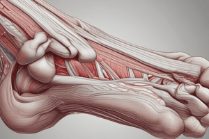
Muscles of the Feet
- Anterior compartment of the foot is responsible for dorsiflexion.
- Tibialis anterior, extensor hallucis longus, and extensor digitorum longus are the muscles in the compartment.
- Superior and inferior extensor retinaculum are responsible for anchoring tendons during contraction.
- Lateral compartment of the foot is responsible for eversion and plantar flexion.
- Fibularis longus and fibularis brevis are the two muscles in the compartment.
- Posterior compartment of the foot is responsible for plantar flexion.
- Gastrocnemius, soleus, plantaris, popliteus, flexor digitorum longus, flexor hallucis longus, and tibialis posterior are the muscles in the compartment.
- Superficial muscles of the posterior compartment insert onto the calcaneal tendon.
Muscles that Move the Toes
- The sole of the foot is supported by the plantar aponeurosis.
- Dorsal group: Extensor digitorum brevis.
- Plantar group: Flexor digitorum brevis, abductor hallucis, abductor digiti minimi brevis, flexor hallucis brevis.
Functions of the Nervous System
- Sensation: Receiving information from the environment through sensory receptors.
- Integration: Processing and interpreting sensory information, often combining it with higher cognitive functions.
- Response: Initiating and carrying out motor functions through effectors via both conscious and unconscious nervous pathways.
Central and Peripheral Nervous Systems
- The Central Nervous System (CNS) consists of the brain and spinal cord.
- The CNS is housed within the cranial cavity and vertebral cavity.
- The CNS is protected by bone.
- The Peripheral Nervous System (PNS) consists of nerves outside of the brain and spinal cord.
- The PNS is not protected by bone.
- Both the CNS and PNS are composed of nervous tissue.
- Neurons: Cells capable of communication.
- Glial cells: Cells that provide structure and support to neurons.
Functional Divisions of the Nervous System
- Sensory: Sends information towards the CNS.
- Integration: Occurs in the brain and spinal cord.
- Response: Communicates with effectors to initiate a response.
Major Spinal Nerves
- Cervical Plexus: C1-C5. Phrenic Nerve: C3-C5. Motor Innervation: Diaphragm. Cutaneous Innervation: Sensation to the central tendon of the diaphragm.
- Brachial Plexus: C5-T1. Radial Nerve: C5-T1. Motor Innervation: Triceps brachii, posterior compartment of the antebrachium. Cutaneous Innervation: Posterior brachium and dorsal aspect of the hand. Axillary Nerve: C5-C6. Motor Innervation: Teres minor and deltoids. Cutaneous Innervation: Lateral shoulder. Median Nerve: C5-T1. Motor Innervation: Anterior compartment of the antebrachium and thenar musculature. Cutaneous Innervation: Anterior and lateral aspect of the hand, dorsal and distal digits. Ulnar Nerve: C8-T1. Motor Innervation: Flexor carpi ulnaris, flexor digitorum profundus, and intrinsic muscles of the hand. Cutaneous Innervation: Medial aspect of the hand, digits 4 and 5.
- Lumbar Plexus: L1-L4. Femoral Nerve: L2-L4. Motor Innervation: Anterior compartment of the thigh. Cutaneous Innervation: Branches into the saphenous nerve. Saphenous Nerve: L3-L4. Motor Innervation: None. Cutaneous Innervation: Anterior surface of the lower leg.
- Sacral Plexus: L4-S4. Sciatic Nerve: L4-S3. Motor Innervation: Biceps femoris, semimembranosus, semitendinosus. Cutaneous Innervation: None. Tibial Nerve: L4-S3. Motor Innervation: Muscles of the posterior lower leg and intrinsic foot muscles. Cutaneous Innervation: Skin of the posterolateral side of the leg and the lateral aspect of the foot. Fibular Nerve: L4-S2. Motor Innervation: None. Cutaneous Innervation: Lateral aspect of the foot.
Sensory Pathways
- Sensory information is processed through a series of neurons.
- Sensory receptor/Primary neuron: The dendrites are in an organ and its axon transmits the action potential to the CNS.
- Secondary neuron: Synapses in the posterior horn and extends to the thalamus.
- Tertiary neuron: Extends from the thalamus to the primary somatosensory cortex in the parietal lobe.
- The thalamus processes and edits incoming sensory information.
Motor Pathways
- Motor responses are carried out by a series of neurons:
- Primary neuron: Located in the motor cortex of the parietal lobe, its axon extends through the brain, crossing over to the other side at the medulla oblongata, and synapses with the secondary neuron in the anterior horn of the spinal cord.
- Secondary neuron: Has its cell body in the anterior horn of the spinal cord and travels toward the muscle.
- Decussation (crossing over) leads to many brain injuries affecting the opposite side of the body.
Reflexes
- Reflexes are connections between sensory and motor neurons.
- Synapse between neurons occurs within the CNS.
- Reflexes do not include higher brain centers.
- Reflexes do not involve conscious or voluntary aspects of movement.
Categorizing Reflexes
- Spinal: Synapse within the spinal cord.
- To understand a stimulus, the brain needs three key pieces of information:
- Modality: Type of stimulus.
- Location: Where the stimulus is coming from.
- Intensity: How strong the stimulus is.
- Intensity of the stimulus is determined by the frequency of action potentials.
Characteristics of Sensory Receptors
- Sensory receptors transduce stimuli into an electrical charge interpreted by the brain.
- Adaptation: Allows receptors to become less sensitive to stimuli over time.
- Phasic receptors: Adapt quickly.
- Tonic receptors: Adapt slowly or not at all.
Adaptation
- Adaptation allows receptors to become less sensitive to stimuli over time.
- Action potential generation slows down.
- Phasic receptors adapt quickly.
- Tonic receptors do not adapt or so slowly.
Types of Receptors
- Types of stimuli:
- Chemoreceptors: Detect chemical stimuli.
- Osmoreceptors: Detect solute concentrations in bodily fluids.
- Thermoreceptors: Detect temperature.
- Mechanoreceptors: Detect physical stimuli like pressure and vibration.
- Baroreceptors: Detect pressure.
- Nociceptors: Detect pain.
- Photoreceptors: Detect photons of light. Only found in the eye.
- Source of stimuli:
- Exteroceptors: Stimuli from the external environment.
- Interoceptors: Stimuli from internal organs and tissues.
- Proprioceptors: Moving body parts.
- Distribution:
- General sense receptors: Found all over the body.
- Special sense receptors: Limited to the head. Used for vision, taste, hearing, olfaction, and balance.
General Senses
- General senses can be detected everywhere in/on the body.
- They include: pressure, vibration, light touch, tickle, itch, temperature, pain, and proprioception.
- General senses are found throughout the body in different forms, such as skin, muscles, tendons, joints, and organs.
- Unencapsulated receptors: Dendrites enmeshed by surrounding tissue.
- Encapsulated receptors: Dendrites wrapped to enable function.
Unencapsulated Receptors
- Free nerve endings: Detect pain and temperature.
- Merkel cells: Located in the skin, used for discriminatory touch.
- Hair follicle receptors: In the skin, detect movement of hair.
- Thermoreceptors: Detect temperature. Also respond to chemicals.
- Capsaicin: Activates thermoreceptors. Interpreted as an increase in temperature.
Encapsulated Receptors
- Lamellated corpuscles: In the dermis, detect deep pressure.
- Tactile corpuscles: In the dermis, detect light pressure.
- Bulbous corpuscles: Detect stretching of the skin.
Pathway of Visual Information
Lateral geniculate nucleus of the thalamus: Edits visual information.
- Information is then sent to the primary visual cortex in the occipital lobe.
- Some axons of the optic nerve cross to the opposite side of the brain at the optic chiasm.
- Right visual field: Processed by the left side of the occipital lobe.
- Left visual field: Processed by the right side of the occipital lobe.
Taste (Gustation)
- Taste is a chemical special sense.
- Recognized tastes include: sweet, salty, sour, bitter, umami, and fat.
- Papillae: On the tongue, contain taste buds.
- Taste buds: Contain taste receptors.
- Transduce: Chemicals in food into taste.
- Basal epithelial cells: Replace damaged taste receptors.
Detection of Tastes
- Saliva: Dissolves molecules so more chemicals reach taste receptor cells.
- Salty: Detected when Na+ ions activate taste cell receptors.
- Sour: Detected when H+ ions activate taste cell receptors.
- Sweet: Detected when glucose activates taste cell receptors. Other monosaccharides and artificial sweeteners can activate sweet receptors.
- Bitter: Detected by 23 different taste receptors. Varies with each individual.
- Umami: Detected when L-glutamate activates taste cell receptors. Monosodium glutamate (MSG) can also activate umami receptors.
Pathway of Taste Information
- Taste information travels to the CNS along cranial nerves: Glossopharyngeal, Facial, and Vagus nerves.
- Nerves converge at the medulla oblongata.
- Information is next sent to the thalamus.
- Thalamus: Sends information to the insula to be processed.
Smell (Olfaction)
- Smell is a chemical special sense.
- Odorant molecules: Bind to olfactory receptors in the olfactory epithelium.
- Olfactory neurons: Pass through the cribriform plate to form the olfactory bulb.
- Axons gather to form the olfactory tract: The olfactory tract projects to different regions of the brain:
- Primary olfactory cortex in the temporal lobe, limbic system, and thalamus.
External Ear
- Auricle, ear canal, and tympanic membrane: Make up the external ear.
- Auricle and auditory canal (external acoustic meatus): Funnel the sound waves toward the tympanic membrane.
- Tympanic membrane: Vibrates in response.
Middle Ear
- Middle ear: Contains the ossicles: Malleus, incus, and stapes.
- Ossicles: Attached to the tympanic membrane, amplify vibrations.
- Stapes: Rests against the oval window.
The Endocrine System
- Functions to regulate homeostasis by producing and releasing hormones.
- Endocrine glands are ductless. They release hormones directly into the bloodstream.
- Examples of endocrine glands include: the hypothalamus, thymus, heart, kidneys, stomach, small intestine, liver, adipose tissue, ovaries, and testes.
Target Cells
- Hormones travel through the bloodstream and can reach almost any cell in the body.
- Hormones only affect target cells, which are cells with a receptor for a particular hormone.
- Binding of a hormone to its receptor on a target cell initiates intracellular signaling.
Comparison of Nervous and Endocrine Systems
- Both nervous and endocrine systems allow control and communication of the body.
- They accomplish this in different ways:
- Neurotransmitters: Used by the nervous system.
- Hormones: Used by the endocrine system.
- The nervous system is generally faster to make a change.
- The endocrine system has more widespread effects.
- Endocrine system effects generally last longer.
Hormones
- Types of hormones:
- Steroid hormones: Cross cell membranes easily. Lipid-based hormones.
- Amine-based hormones: Modified amino acids. Water-soluble; cannot cross cell membranes.
- Peptide and Protein hormones: Made from chains of amino acids. Water-soluble; cannot cross cell membranes.
Steroid Hormones
- Produced from cholesterol molecules.
- Lipid-soluble hormones.
- Can pass through cell membranes.
- Require transport proteins to travel in the blood.
- Examples include testosterone and estrogens.
Amine Hormones
- Made from individual amino acids.
- Water-soluble hormones.
- Cannot freely pass through the cell membrane.
- Do not require transport proteins in the blood.
- Examples include melatonin, epinephrine, and norepinephrine.
Peptide and Protein Hormones
- Chains of amino acids.
- Water-soluble hormones.
- Cannot freely pass through the cell membrane.
- Do not require transport proteins in the blood.
- Examples include antidiuretic hormone and insulin.
Production of Hormones
- Steroid hormones: Made on demand by modifying cholesterol molecules. Cannot be stored. Not soluble in blood. Travel bound to transport proteins when in blood.
- Peptide hormones: Translated like other proteins. Modified and stored in vesicles until release. Soluble in blood. Travel in a "free" state.
Hormone Receptors
- Receptors can be intracellular or on the cell surface.
- Lipid-soluble hormone receptors: Usually intracellular (cytosol or nuclear). This is because lipid-soluble hormones can pass through cell membranes.
- Water-soluble hormone receptors: Usually on the surface of the cell.
Studying That Suits You
Use AI to generate personalized quizzes and flashcards to suit your learning preferences.