Podcast
Questions and Answers
What articulates with the anterior surface of the atlas?
What articulates with the anterior surface of the atlas?
- Occipital condyle
- Lateral mass of atlas
- Odontoid process (correct)
- Axis
What type of movement does the atlanto-axial joint allow?
What type of movement does the atlanto-axial joint allow?
- Rotational movement
- Up and down movement
- Forward and backward movement
- Side to side movement (correct)
What is the function of the transverse ligament?
What is the function of the transverse ligament?
- To strengthen the fibrous capsule
- To attach the basiocciput to the axis
- To attach the axis to the occipital bone
- To attach the atlas to the axis (correct)
What is the name of the ligament that extends from the tip of the odontoid process to the basilar part of the occipital bone?
What is the name of the ligament that extends from the tip of the odontoid process to the basilar part of the occipital bone?
What is the function of the membrana tectoria?
What is the function of the membrana tectoria?
What is the name of the ligament that connects the atlas with the axis and the occipital bone?
What is the name of the ligament that connects the atlas with the axis and the occipital bone?
What is the role of the fibrous capsule in the atlanto-axial joint?
What is the role of the fibrous capsule in the atlanto-axial joint?
What is the attachment site of the apical ligament of the dens?
What is the attachment site of the apical ligament of the dens?
What is the function of the alar ligaments?
What is the function of the alar ligaments?
What is the attachment site of the transverse ligament?
What is the attachment site of the transverse ligament?
Flashcards are hidden until you start studying
Study Notes
Muscles of Mastication
- Divided into two groups: principal muscles and accessory muscles
- Principal muscles:
- Origin: from skull
- Insertion: into ramus of mandible
- Nerve supply: mandibular nerve (from trigeminal nerve, 5th CN)
- Action: elevation and protraction of the mandible, except lateral pterygoid muscle which depresses the mandible
- Masseter:
- Origin: from inner surface and lower border of zygomatic arch
- Insertion: into outer surface of the ramus of mandible
- Nerve supply: branch from anterior division of mandibular nerve
- Action: elevation and protraction of the mandible
- Temporalis:
- Fan-shaped muscle
- Origin:
- Floor of the temporal fossa
- Temporal lines
- Insertion: into the coronoid process of the mandible
- Nerve supply: from the anterior division of the mandibular nerve
- Action: elevation and retraction of the mandible
- Lateral Pterygoid Muscle:
- Two heads (lower and upper)
- Origin:
- Lower head: from the lateral surface of the lateral pterygoid plate
- Upper head: from the greater wing of the sphenoid
- Insertion: into the pterygoid fovea
- Nerve supply: branch from the anterior division of the mandibular nerve
- Action:
- Depression of the mandible
- Protraction of the mandible
- Side-to-side movements of the mandible (chewing movement) with medial pterygoid muscle
- Medial Pterygoid Muscle:
- Two heads
- Origin:
- Superficial head: from the maxillary tuberosity
- Deep head: from the medial surface of the lateral pterygoid plate
- Insertion: into the inner surface of the ramus of mandible near its angle
- Nerve supply: branch from the main trunk of the mandibular nerve
- Action:
- Elevation and protraction of the mandible
- Side-to-side movement of the mandible with lateral pterygoid muscle
Tempromandibular Joint (TMJ)
- Type: synovial joint of condylar variety
- Articular surfaces:
- Mandibular fossa and articular tubercle of temporal bone (above)
- Head of mandible (below)
- Intraarticular disc (fibrocartilage disc inside the joint cavity)
- Capsule: attached to margins of articular surfaces and circumference of the intraarticular disc
- Ligaments:
- Lateral tempromandibular ligament (triangular in shape)
- Stylomandibular ligament (extends from the tip of the styloid process to the posterior border of ramus of mandible)
- Sphenomandibular ligament (extends from the spine of sphenoid to the lingula of mandible)
- Pterygomandibular ligament or raphe (extends from the pterygoid hamulus to the posterior end of mylohyoid line of mandible)
Movements of the Mandible
- Depression (Opening of Mouth): produced mainly by lateral pterygoid muscle, helped by gravity and assisted by digastric, geniohyoid, and mylohyoid muscles
- Elevation (Closing of Mouth): caused by medial pterygoid, masseter, and temporalis muscles
- Protraction: done by lateral and medial pterygoids and masseter
- Retraction: done by posterior fibers of temporalis
- Side-to-side (Chewing) Movements: performed by alternate contraction of medial and lateral pterygoids on each side
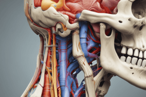
Atlanto-Occipital Joint
- Type: synovial joint of ellipsoid variety
- Articular surfaces:
- Occipital condyles (above)
- Superior articular facet of the atlas (below)
- Capsule: attached to the margins of the articular surfaces, lined by synovial membrane
- Ligaments:
- Anterior atlanto-occipital membrane (attached to the upper border of anterior arch of atlas and the anterior margin of foramen magnum)
- Posterior atlanto-occipital membrane (attached to the upper border of posterior arch of atlas and the posterior margin of foramen magnum)
- Movements: flexion and extension of the head (nodding), lateral flexion of the head
Atlanto-Axial Joints
- Three synovial joints:
- One median atlantoaxial joint
- Two lateral atlantoaxial joints
- Lateral Atlantoaxial Joints: plane synovial joints between inferior articular facet of atlas and superior articular facet of axis
- Median Atlantoaxial Joint:
- Type: synovial joint of pivot variety
- Articular surfaces:
- Odontoid process (dens) of axis
- Articular facet on the posterior surface of the anterior arch of atlas
- Ligaments:
- Fibrous capsule
- Transverse ligament
- Ligaments connecting the axis with the occipital bone
Studying That Suits You
Use AI to generate personalized quizzes and flashcards to suit your learning preferences.