Podcast
Questions and Answers
What is responsible for the voluntary action of skeletal muscles?
What is responsible for the voluntary action of skeletal muscles?
- Endocrine system
- Autonomic nervous system
- Somatic motor neurons (correct)
- Peripheral nervous system
Which of the following is NOT a function of the muscle system?
Which of the following is NOT a function of the muscle system?
- Communication (Verbal and Facial)
- Maintenance of posture
- Production of body heat (Thermogenesis)
- Digestion of food (correct)
Which property of muscle refers to its ability to return to its original shape after being stretched?
Which property of muscle refers to its ability to return to its original shape after being stretched?
- Excitability
- Contractility
- Extensibility
- Elasticity (correct)
Where is smooth muscle commonly found?
Where is smooth muscle commonly found?
What component of skeletal muscle is specifically responsible for generating pulling force?
What component of skeletal muscle is specifically responsible for generating pulling force?
What distinguishes smooth muscle from skeletal muscle in terms of control?
What distinguishes smooth muscle from skeletal muscle in terms of control?
How much of the body weight is made up of skeletal muscle?
How much of the body weight is made up of skeletal muscle?
What is a primary characteristic of single-unit smooth muscle?
What is a primary characteristic of single-unit smooth muscle?
In smooth muscle, which of the following substances directly causes contraction through passive diffusion?
In smooth muscle, which of the following substances directly causes contraction through passive diffusion?
Which layer of muscle fibers in hollow organs runs parallel to the organ's long axis?
Which layer of muscle fibers in hollow organs runs parallel to the organ's long axis?
What is the role of multi-unit smooth muscle in the body?
What is the role of multi-unit smooth muscle in the body?
Which of the following is not a function of smooth muscle?
Which of the following is not a function of smooth muscle?
What is the primary function of skeletal muscle?
What is the primary function of skeletal muscle?
Which structures are found within the myofibrils?
Which structures are found within the myofibrils?
What is the role of the M line in the sarcomere?
What is the role of the M line in the sarcomere?
What is the function of the neuromuscular junction?
What is the function of the neuromuscular junction?
Which component serves as a storage site for carbohydrates and amino acids in skeletal muscle?
Which component serves as a storage site for carbohydrates and amino acids in skeletal muscle?
Which part of the sarcomere contains only actin filaments?
Which part of the sarcomere contains only actin filaments?
What neurotransmitter is primarily involved at the neuromuscular junction?
What neurotransmitter is primarily involved at the neuromuscular junction?
Which protein complex is responsible for regulating the interaction of actin with myosin heads?
Which protein complex is responsible for regulating the interaction of actin with myosin heads?
What role do mitochondria play in skeletal muscle?
What role do mitochondria play in skeletal muscle?
What initiates the end plate potential in the muscle fiber?
What initiates the end plate potential in the muscle fiber?
What role does acetylcholinesterase play at the neuromuscular junction?
What role does acetylcholinesterase play at the neuromuscular junction?
Which structure is involved in the propagation of the action potential in skeletal muscle fibers?
Which structure is involved in the propagation of the action potential in skeletal muscle fibers?
What is the primary consequence of calcium ions being released from the sarcoplasmic reticulum?
What is the primary consequence of calcium ions being released from the sarcoplasmic reticulum?
What happens to the troponin-tropomyosin complex when calcium concentration increases in the muscle?
What happens to the troponin-tropomyosin complex when calcium concentration increases in the muscle?
What defines the threshold in relation to end plate potential?
What defines the threshold in relation to end plate potential?
What occurs when dihydropyridine receptors sense a voltage change?
What occurs when dihydropyridine receptors sense a voltage change?
During muscle relaxation, which factor contributes to a low concentration of calcium ions?
During muscle relaxation, which factor contributes to a low concentration of calcium ions?
What is the effect of sodium ion entry into the muscle cell?
What is the effect of sodium ion entry into the muscle cell?
What overall process does the release of calcium from the T tubule-sarcoplasmic reticulum system contribute to?
What overall process does the release of calcium from the T tubule-sarcoplasmic reticulum system contribute to?
What is the angle of the myosin head when it is at rest?
What is the angle of the myosin head when it is at rest?
What triggers the power stroke in muscle contraction?
What triggers the power stroke in muscle contraction?
How does temporal summation enhance muscle contraction?
How does temporal summation enhance muscle contraction?
Which of the following is a characteristic of smooth muscle cells?
Which of the following is a characteristic of smooth muscle cells?
What role do caveolae perform in smooth muscle cells?
What role do caveolae perform in smooth muscle cells?
What occurs to the myosin head after the power stroke?
What occurs to the myosin head after the power stroke?
At what stage does a single twitch produce low force?
At what stage does a single twitch produce low force?
Which of the following best describes the myofilament structure in smooth muscle?
Which of the following best describes the myofilament structure in smooth muscle?
What happens during the complete relaxation of a muscle fiber after a twitch?
What happens during the complete relaxation of a muscle fiber after a twitch?
What results from the absence of striations in smooth muscle cells?
What results from the absence of striations in smooth muscle cells?
Flashcards
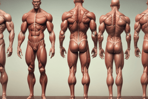
Skeletal Muscle
Skeletal Muscle
Muscle tissue attached to bones, responsible for movement, facial expressions, posture, and respiration. Controlled voluntarily by the somatic nervous system.
Smooth Muscle
Smooth Muscle
Muscle tissue found in the walls of organs and blood vessels; responsible for involuntary movements like digestion, blood flow regulation, and pupil dilation.
Muscle Excitability
Muscle Excitability
The ability of muscle tissue to respond to a stimulus.
Muscle Contractility
Muscle Contractility
Signup and view all the flashcards
Muscle Extensibility
Muscle Extensibility
Signup and view all the flashcards
Muscle Elasticity
Muscle Elasticity
Signup and view all the flashcards
Muscle Fiber
Muscle Fiber
Signup and view all the flashcards
Sarcoplasm Components
Sarcoplasm Components
Signup and view all the flashcards
Skeletal Muscle Function
Skeletal Muscle Function
Signup and view all the flashcards
Myofibril Protein: Actin
Myofibril Protein: Actin
Signup and view all the flashcards
Myofibril Protein: Tropomyosin-Troponin
Myofibril Protein: Tropomyosin-Troponin
Signup and view all the flashcards
Myofibril Protein: Myosin
Myofibril Protein: Myosin
Signup and view all the flashcards
Sarcomere Structure
Sarcomere Structure
Signup and view all the flashcards
Neuromuscular Junction Key Elements
Neuromuscular Junction Key Elements
Signup and view all the flashcards
Motor Unit Definition
Motor Unit Definition
Signup and view all the flashcards
Motor End Plate Function
Motor End Plate Function
Signup and view all the flashcards
Synaptic Gutter
Synaptic Gutter
Signup and view all the flashcards
Subneural Clefts
Subneural Clefts
Signup and view all the flashcards
Acetylcholine Receptors
Acetylcholine Receptors
Signup and view all the flashcards
Neuromuscular Junction
Neuromuscular Junction
Signup and view all the flashcards
End Plate Potential (EPP)
End Plate Potential (EPP)
Signup and view all the flashcards
T-tubules
T-tubules
Signup and view all the flashcards
Sarcoplasmic Reticulum
Sarcoplasmic Reticulum
Signup and view all the flashcards
Dihydropyridine Receptors
Dihydropyridine Receptors
Signup and view all the flashcards
Ryanodine Receptors
Ryanodine Receptors
Signup and view all the flashcards
Cross-bridge Cycle
Cross-bridge Cycle
Signup and view all the flashcards
Myosin head at rest
Myosin head at rest
Signup and view all the flashcards
Binding of Actin and Myosin
Binding of Actin and Myosin
Signup and view all the flashcards
What happens after Actin and Myosin bind?
What happens after Actin and Myosin bind?
Signup and view all the flashcards
Power Stroke
Power Stroke
Signup and view all the flashcards
Detachment of Myosin
Detachment of Myosin
Signup and view all the flashcards
What is a Twitch?
What is a Twitch?
Signup and view all the flashcards
Temporal Summation
Temporal Summation
Signup and view all the flashcards
Tetanus
Tetanus
Signup and view all the flashcards
Smooth Muscle Structure
Smooth Muscle Structure
Signup and view all the flashcards
What are Caveolae?
What are Caveolae?
Signup and view all the flashcards
Dense Bodies in Smooth Muscle
Dense Bodies in Smooth Muscle
Signup and view all the flashcards
Smooth Muscle Layers in Hollow Organs
Smooth Muscle Layers in Hollow Organs
Signup and view all the flashcards
Smooth Muscle Contraction Regulation
Smooth Muscle Contraction Regulation
Signup and view all the flashcards
Single-Unit Smooth Muscle vs. Multi-unit Smooth Muscle
Single-Unit Smooth Muscle vs. Multi-unit Smooth Muscle
Signup and view all the flashcards
Smooth Muscle Tone
Smooth Muscle Tone
Signup and view all the flashcards
Study Notes
Types of Muscle
- Skeletal muscle
- Attaches to bones
- Makes up 40% of body weight
- Responsible for movement, facial expressions, posture, respiration, and other body movements
- Voluntary movement; controlled by somatic motor neurons
- Smooth muscle
- Found in walls of hollow organs, blood vessels, eyes, glands, uterus, and skin
- Some functions include: moving urine, mixing food, dilating/constricting pupils, and regulating blood flow
- Involuntary movement; controlled by endocrine and autonomic nervous systems
- Cardiac muscle
- Found in the heart
- Involuntary movement
- Controlled by endocrine and autonomic nervous systems
Muscle Properties
- Excitability: Ability to respond to a stimulus
- Contractility: Ability to shorten and generate pulling force
- Extensibility: Ability to be stretched back to its original length
- Elasticity: Ability to recoil to its original resting length after being stretched
Muscle System Functions
- Body movement (locomotion)
- Maintenance of posture
- Respiration (diaphragm and intercostal contractions)
- Communication (verbal and facial expressions)
- Contraction of organs and blood vessels (e.g., peristalsis of intestines)
- Heartbeat
- Production of body heat (thermogenesis)
Skeletal Muscle Structure
- Muscle fibers: Basic cellular unit, the sarcomere
- Muscle tissue made of fascicles, bundles of muscle cells, which are themselves bundles of myofibrils
- Nuclei: Multiple, located at the periphery of the cell
- Sarcolemma: Tubular sheath that encases and defines each muscle fiber, containing locations for ion exchange
- T-tubules: Invaginations within the sarcolemma
- Sarcoplasm: Cytoplasm of a muscle fiber, containing sarcoplasmic reticulum, mitochondria, glycogen, and ions
- Myofibrils: Structural components within muscle fibers, containing actin and myosin filaments
Organization of Skeletal Muscle
- Skeletal muscle is composed of muscle fascicles
- Fascicles are comprised of individual muscle fibers
- Fibers are further subdivided into sarcolemma, t tubules, sarcoplasm , sarcoplasmic reticulum and myofibrils
- Myofibrils are composed of organized proteins, such as actin and myosin.
- These proteins organize into sarcomeres, the functional units of muscle contraction
Function of Skeletal Muscle
- Movement: Converts chemical energy to mechanical energy, generating force and power
- Body posture and position
- Storage of nutrients: Important for basal energy metabolism
- Maintenance of body temperature: Produced by muscular activity
Myofibrils
- Actin ("thin fibers"): Double-helical structure with polarized fibers; tropomyosin-troponin complex regulates interaction with myosin heads
- Myosin ("thick fibers"): Light meromyosin anchors myosin at the M line; heavy meromyosin S-1 portion (myosin head) binds actin, contains ATP sites, and initiates power stroke
- Supporting proteins: Titin, desmin, myomesin, C protein, nebulin, and plectin
Sarcomere
- Structural and functional unit of actin-myosin-linked muscle contraction
- Longitudinally arranged
- Include M line, Z discs, H band, A band, I band
- Z disc: Terminal boundary of the sarcomere
- I band: Contains only actin filaments
- A band: Contains the entirety of myosin and overlaps with actin
- H band: Contains only myosin filaments
- M line: Center of the sarcomere
Neuromuscular Junction
- Junction between motor neuron and skeletal muscle fiber
- Linked chemically
- Neurotransmitter at junction: Acetylcholine (ACh)
- Axon approaches muscle: Dividing into terminal branches, losing myelin sheath
- Axon terminal branch: Enlarged knob-like structure called the terminal button with vesicles of chemical transmitter
- Motor end-plate: Specialized area of muscle fiber membrane invaginated by motor nerve terminal
Motor end-plate
- The axon terminal contains vesicles that hold acetylcholine (ACh)
- Synaptic cleft: 20-30nm space separating the axon terminal and the muscle cell membrane
- Synaptic trough (synaptic gutter): Muscle membrane containing many folds called subneural clefts, where Ach receptors are located
Events at the Neuromuscular Junction
- Action potential to axon terminal
- Release and diffusion of acetylcholine
- Binding of acetylcholine to receptors
- Opening of gated Na+ channels
- Na+ influx, triggering end-plate potential (EPP)
- EPP initiates action potential in muscle fiber
- Acetylcholine breakdown by acetylcholinesterase
End-plate Potential
- Influx of Na+ inside muscle fiber, provoking a local potential at end-plate
- Initiator of the action potential
- Threshold potential
Action Potential
- Skeletal muscle fibers: No current flow deep within the fiber
- T tubule-sarcoplasmic reticulum system: Transmits action potentials
- Transmission of action potentials along transverse tubules
- Open in the exterior of the fiber
- Sarcoplasmic reticulum vesicular tubules with high concentration of Calcium
- T tubule action potentials release of calcium—causing contraction of the muscle
- Overall process called excitation-contraction coupling
Release of Ca2+ from the T-tubule sarcoplasmic reticulum system
- Action potential
- Dihydropyridine receptors (DHP receptors) sense voltage changes
- Pull the ryanodine receptor channels out in the sarcoplasmic reticular cisternae
- Calcium release causes contraction
- Calcium ions reuptake by calcium pumps (using ATP)
Cross-bridge Cycle
- Interaction between myosin heads and active sites on actin filaments
- Muscle relaxed: Low calcium, troponin-tropomyosin complex blocks actin sites
- Muscle contracted: Increased calcium, troponin-tropomyosin complex moves, exposing actin sites
- Myosin head attaches, moves, detaches, and reattaches
- Power stroke: Myosin head moves toward negative end of actin, pulling actin filaments
- Myosin detaches from actin: ATP binds to myosin
- Hydrolysis of ATP to ADP and Pi re-energizes the myosin head
Temporal Summation
- One action potential = one twitch (single muscle contraction)
- Amount of force produced very low
- Enhances contractile force: Repeated stimulation before muscle relaxes
- Partially fused twitches
- Tetanus
Pathophysiology of Neuromuscular Junction
- Disorders affecting neuromuscular junction, e.g.: Lambert-Eaton myasthenic syndrome (LEMS), Botulism, Myasthenia Gravis (MG)
Smooth Muscle
- Cells: Not striated, smaller than skeletal muscle cells, and usually cylindrical in shape; single nucleus; and lack striations or sarcomeres
- Caveolae: Indentations in the sarcolemma (may act like T tubules)
- Myofilaments: Thick myosin and thin filaments similar to actin, but lack troponin and tropomyosin
- Intermediate filaments: Cytoskeletal functions (non-contractile)
- Dense bodies: Membrane-associated structures instead of Z discs
- Group together into sheets in the walls of hollow organs
- Longitudinal layer
- Circular layer
Functions of Smooth Muscle
- Gastrointestinal tract: Propulsion of food,
- Cardiovascular: Regulation of blood flow,
- Renal: Regulation of urine flow,
- Genital: Muscle contractions during pregnancy, propulsion of sperm
- Respiratory: Regulation of bronchiole diameter
- Integument: Raises hairs with erector pili
- Sensory: Dilation and constriction of pupils, changing lens shape
Cell-to-cell Transmission and Electrical Activity in Smooth Muscle
- Single-unit: Often autorhythmic, and nerve innervations to cells are few. Coordinate contractions through electrical coupling via gap junctions
- Multi-unit: Each cell requires its own electrical impulse for activation (e.g., arrector pili muscles, large blood vessels, iris or eyes, airways)
Smooth Muscle Properties: Single Unit
- Gap junctions: Electrical coupling, potential change spreads to multiple cells
- Innervation: Primarily to few cells; coordinate contraction (e.g., GI tract)
- Slow, steady contractions, substance movement
- Tone: Basis level of contraction without stimulation
- Location: Walls of visceral organs (e.g., stomach, intestines, urinary bladder)
Smooth Muscle Properties: Multi-Unit
- No gap junctions
- More precise muscle control
- Produce asynchronous contractions
- Location: Large blood vessels, eyes, and respiratory airways
Direct and Receptor-Mediated Factors that Contract or Relax Smooth Muscle
- Direct Factors
- Increase intracellular calcium = contraction
- Passive diffusion of ligands to specific membrane receptors
- Receptor-Mediated Factors (chemical factors)
- Chemical constrictors (e.g., noradrenaline) - Relaxants (e.g., nitric oxide, CO2)
Contraction of Smooth Muscle
- Direct entry: passive calcium entry from leak channels, or voltage gated channels
- Second messenger system (IP3 induces release of calcium from sarcoplasmic reticulum)
Activation Cross-bridge Cycle in Smooth Muscle
- Lack troponin complex, regulated by myosin linked myosin
- Increased intracellular Ca2+ (depolarization activation, and/or Ca2+ release from SR)
- Calmodulin binds calcium
- Calcium-calmodulin complex activates myosin light-chain kinase (MLCK)
- MLCK phosphorylates light chains in myosin
- Increases myosin ATPase activity
- Myosin-P binds actin
Smooth Muscle Relaxation
- Reduces cytoplasmic calcium concentration (repolarization or calcium pump activity)
- Calcium unbinds from calmodulin
- Myosin light-chain phosphatase (MLCP) dephosphorylates myosin
- Dephosphorylated myosin: Low affinity for actin
- Cross-bridge cycle interruption = relaxation
Contractile Activity in Smooth Muscle
- Tonic contraction: Low, sustained tension. Ligan-gated calcium channels. Direct metabolic control.
- Phasic contraction: Momentary contraction with momentary activation of VG calcium channels. Similar to skeletal muscle twitch
Pathophysiology of Smooth Muscle
- Digestive system: Gastroparesis (loss of motility), Nerve dysfunction, collagen disease, muscular dystrophies
- Renal system: Chronic kidney disease (leads to end-stage renal disease), Ureters (nephrolithiasis), Bladde (neurogenic)
- Cardiovascular and respiratory systems: Atherosclerosis, pulmonary hypertension, asthma
Smooth, Skeletal, and Cardiac Muscle Comparison
- Properties like striations, level of control, neural input, hormonal control, calcium source, regulatory proteins, gap junctions, pacemaker activity, myosin ATPase, recruitment are compared
Studying That Suits You
Use AI to generate personalized quizzes and flashcards to suit your learning preferences.