Podcast
Questions and Answers
What is the primary function of the creatine phosphate (CP) cycle in muscle energy metabolism?
What is the primary function of the creatine phosphate (CP) cycle in muscle energy metabolism?
- Requires oxygen for ATP production
- Converts ADP back to ATP (correct)
- Utilizes fatty acids for energy
- Produces lactate
How long does the creatine phosphate cycle typically provide energy for muscle contractions?
How long does the creatine phosphate cycle typically provide energy for muscle contractions?
- Up to 30 seconds (correct)
- Unlimited duration
- Up to 1 minute
- Up to 5 seconds
Which of the following is the major end product of anaerobic glycolysis?
Which of the following is the major end product of anaerobic glycolysis?
- Lactate (correct)
- Carbon dioxide
- ATP
- Creatine
Which energy source is utilized first when muscle contraction begins?
Which energy source is utilized first when muscle contraction begins?
What is a defining characteristic of anaerobic glycolysis compared to the creatine phosphate cycle?
What is a defining characteristic of anaerobic glycolysis compared to the creatine phosphate cycle?
In which activity would the creatine phosphate cycle be the primary source of energy?
In which activity would the creatine phosphate cycle be the primary source of energy?
Which statement accurately describes the energy production rate of aerobic glycolysis compared to other systems?
Which statement accurately describes the energy production rate of aerobic glycolysis compared to other systems?
What is one of the limitations of energy generation through the creatine phosphate cycle?
What is one of the limitations of energy generation through the creatine phosphate cycle?
What role do T-tubules play in muscle contraction?
What role do T-tubules play in muscle contraction?
Which event occurs first during the process of muscle contraction?
Which event occurs first during the process of muscle contraction?
What initiates the release of calcium ions from the sarcoplasmic reticulum?
What initiates the release of calcium ions from the sarcoplasmic reticulum?
How does calcium regulate muscle contraction?
How does calcium regulate muscle contraction?
Which component is responsible for returning Ca2+ ions back into the sarcoplasmic reticulum after muscle contraction?
Which component is responsible for returning Ca2+ ions back into the sarcoplasmic reticulum after muscle contraction?
Which of the following describes the sliding filament model of muscle contraction?
Which of the following describes the sliding filament model of muscle contraction?
What happens to the myosin-binding sites on actin when muscle is in a relaxed state?
What happens to the myosin-binding sites on actin when muscle is in a relaxed state?
What triggers the process of muscle relaxation?
What triggers the process of muscle relaxation?
What effect does higher Ca2+ concentration have on caldesmon?
What effect does higher Ca2+ concentration have on caldesmon?
Which muscle type has no cross striations?
Which muscle type has no cross striations?
How do the calcium pumps of skeletal muscle compare to those of smooth muscle?
How do the calcium pumps of skeletal muscle compare to those of smooth muscle?
Which statement regarding hormone receptors is true?
Which statement regarding hormone receptors is true?
What type of regulatory protein is absent in smooth muscle?
What type of regulatory protein is absent in smooth muscle?
Which muscle type relies on extracellular fluid (ECF) calcium for contraction?
Which muscle type relies on extracellular fluid (ECF) calcium for contraction?
What is the role of phosphorylation by kinases in muscle contraction?
What is the role of phosphorylation by kinases in muscle contraction?
Which feature is specific to the arrangement of T tubules in cardiac muscle?
Which feature is specific to the arrangement of T tubules in cardiac muscle?
What role does the troponin-Ca++ complex play in muscle contraction?
What role does the troponin-Ca++ complex play in muscle contraction?
Which condition allows the myosin head to bind to the actin filament?
Which condition allows the myosin head to bind to the actin filament?
What happens to the muscle fiber as long as calcium ions are present in the sarcoplasm?
What happens to the muscle fiber as long as calcium ions are present in the sarcoplasm?
What is the function of Ca-ATPase in muscle relaxation?
What is the function of Ca-ATPase in muscle relaxation?
During muscle contraction, what occurs after the cross-bridge forms between actin and myosin heads?
During muscle contraction, what occurs after the cross-bridge forms between actin and myosin heads?
In the sliding filament theory, what is the primary cause of muscle contraction?
In the sliding filament theory, what is the primary cause of muscle contraction?
What must happen for relaxation of a muscle fiber to occur?
What must happen for relaxation of a muscle fiber to occur?
What role does titin play in muscle function?
What role does titin play in muscle function?
Which factor is essential for maintaining muscle contraction beyond the initial stimulus?
Which factor is essential for maintaining muscle contraction beyond the initial stimulus?
What is a consequence of dystrophin deficiency?
What is a consequence of dystrophin deficiency?
Which of the following is characteristic of smooth muscle cells?
Which of the following is characteristic of smooth muscle cells?
Which protein is responsible for linking actin filaments to the extracellular matrix?
Which protein is responsible for linking actin filaments to the extracellular matrix?
What primarily causes the thickened heart muscle in familial hypertrophic cardiomyopathy?
What primarily causes the thickened heart muscle in familial hypertrophic cardiomyopathy?
How does Ca++ regulate cardiac muscle contraction?
How does Ca++ regulate cardiac muscle contraction?
Which of the following is true regarding the sarcoplasmic reticulum in cardiac muscle cells?
Which of the following is true regarding the sarcoplasmic reticulum in cardiac muscle cells?
What initiates contraction in smooth muscle cells?
What initiates contraction in smooth muscle cells?
Flashcards are hidden until you start studying
Study Notes
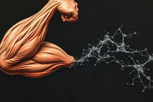
- Energy Sources for Muscle Contraction
- ATP, or adenosine triphosphate, is the primary source of energy for muscle contraction and serves as the cellular energy currency throughout the body. Its role in muscle contractions is essential, as it provides the necessary energy required for the various biochemical processes involved in muscle movement.
- Energy from ATP is released when the terminal phosphate group is cleaved through hydrolysis (ATP → ADP + Pi), resulting in the formation of adenosine diphosphate (ADP) and inorganic phosphate (Pi). This reaction is crucial because it allows myosin heads in muscle fibers to attach to actin filaments, facilitating muscle contraction.
- The creatine phosphate (CP) cycle is a rapid mechanism that provides immediate energy for short bursts of high-intensity activity, such as sprinting or weight lifting, lasting up to 30 seconds. This system is particularly important for activities that require quick, explosive movements.
- The reaction that occurs during the CP cycle is catalyzed by an enzyme known as creatine kinase (CK), which facilitates the transfer of a phosphate group from creatine phosphate to ADP, regenerating ATP quickly.
- Importantly, this process does not produce lactate or require oxygen, making it an anaerobic pathway that is effective in enhancing performance during brief, intense exercise.
- Anaerobic glycolysis serves as a source of energy for sustained, high-intensity activities lasting up to 60 seconds, such as repeated sprint efforts or high-rep weight training. During this metabolic pathway, glucose is broken down without the use of oxygen.
- The primary substrates for anaerobic glycolysis include muscle glycogen, which is stored within the muscle fibers, and blood glucose derived from the liver. This process results in the production of ATP in the absence of sufficient oxygen.
- As a byproduct of this energy production, lactate is generated, which can accumulate in the muscles and contribute to fatigue if not cleared efficiently.
- Aerobic glycolysis and oxidative phosphorylation are the primary sources of energy for sustained, low-intensity activities, such as distance running or cycling, like marathons. These metabolic pathways allow for prolonged physical endurance.
- Although highly efficient, these processes are much slower than anaerobic glycolysis because they require oxygen to produce ATP. Consequently, aerobic metabolism can produce significantly more ATP per glucose molecule than anaerobic glycolysis, supporting sustained exercise durations.
- Muscle Structure and Function
- Transverse tubules (T-tubules) are specialized structures that consist of invaginations of the sarcolemma, or muscle cell membrane. These tubules play a crucial role in conducting electrical impulses deep into the muscle fibers, ensuring a rapid and coordinated contraction.
- The sarcoplasmic reticulum (SR) is a unique organelle that stores and regulates calcium ion levels, which are vital for muscle contraction. When a muscle cell receives an electrical stimulus, the SR releases calcium ions into the cytosol, triggering the contraction process.
- Each T-tubule is flanked by two structures known as terminal cisternae, which are portions of the sarcoplasmic reticulum filled with high concentrations of calcium. This arrangement facilitates the effective release of calcium when the muscle is stimulated.
- Muscle contraction is initiated by a nerve impulse that depolarizes the sarcolemma. This electrical signal causes the opening of voltage-gated calcium channels in the SR, leading to an influx of calcium needed for muscle activation.
- Calcium release from the SR is the critical trigger for muscle contractions as it interacts with regulatory proteins, initiating the contractions and enabling the muscle fibers to shorten and generate force.
- Muscle relaxation occurs when calcium ions are actively pumped back into the SR by calcium pumps, allowing the muscle fibers to return to their resting state, ceasing contraction and recovering for the next stimulus.
- Sliding Filament Model
- In a relaxed muscle, the myosin binding sites on actin are blocked by a protein known as tropomyosin, which prevents cross-bridge formation between the actin and myosin filaments necessary for contraction.
- When calcium binds to troponin, it induces a conformational change that causes tropomyosin to move away from the myosin binding sites on actin. This movement is essential as it exposes the binding sites for myosin heads.
- This exposure allows the myosin heads to bind to actin, forming cross-bridges, which initiate the shortening of the muscle. These cross-bridges are fundamental in the contraction cycle, allowing the two filaments to slide past one another.
- During contraction, actin filaments are pulled by myosin heads towards the center of the sarcomere, the functional unit of muscle contraction, leading to visible muscle shortening and force generation within the muscle.
- The hydrolysis of ATP by the myosin heads is what powers the contraction. This ATP hydrolysis allows the myosin heads to return to an energized state, ready to bind to the next actin site and continue the contraction cycle.
- Muscle Proteins
- Titin is an essential structural protein that is responsible for the assembly and stabilization of the myosin myofilament within the sarcomere. It provides passive elasticity and contributes to the overall mechanical properties of skeletal muscle.
- Mutations in the titin gene, which plays a critical role in muscle elasticity and contraction, have been associated with familial hypertrophic cardiomyopathy, a condition characterized by abnormal thickening of the heart muscle.
- Calsequestrin is a calcium-binding protein located within the sarcoplasmic reticulum, which helps to store high concentrations of calcium ions. Its primary function is to facilitate the rapid release of calcium when the muscle is activated.
- Dystrophin is another vital muscle protein that links actin filaments to the extracellular matrix, providing structural stability to muscle fibers during contraction.
- Dystrophin deficiency is a genetic condition that causes Duchenne's muscular dystrophy, a severe form of muscle degeneration and weakness that primarily affects boys.
- Muscular dystrophy is a group of genetic disorders, with Duchenne's being one of the most prevalent, characterized by progressive muscle weakness and loss of muscle mass. It is an X-linked recessive disorder, resulting from mutations in the dystrophin gene, which can lead to a host of physical challenges for those affected.
- Cardiac Muscle
- Cardiac muscle, which makes up the heart, is unique in that it displays rhythmic contractions that allow for the continuous pumping of blood throughout the body. This rhythmicity is essential for maintaining circulatory function.
- The contraction in cardiac muscle cells is primarily controlled by extracellular calcium, which not only triggers muscle contraction but also regulates the strength and duration of the contraction, allowing the heart to respond to varying physiological demands.
- During rest periods, excess intracellular cytosolic calcium is removed and exported out of the cell by the calcium-sodium exchanger pump, helping to reset the muscle cell for the next contraction cycle.
- Cardiac muscle cells are characterized by a high number of mitochondria and myoglobin, providing them with the energy necessary for sustained contractions and ensuring efficiency in aerobic metabolism. This abundance of these components enables the heart to function effectively throughout a lifetime.
- Smooth Muscle
- Smooth muscle cells are distinct in that they are uninucleated and lack the organized myofibrils seen in skeletal and cardiac muscle. This structural organization also allows smooth muscle to function effectively in involuntary movements.
- In smooth muscle, troponin is absent, and the sarcoplasmic reticulum is poorly developed compared to other muscle types. Instead, smooth muscle contraction is primarily regulated by calcium levels, but through mechanisms different from striated muscle.
- Smooth muscle contraction is mediated by the phosphorylation of myosin light chains, which changes the myosin head conformation and allows for interaction with actin filaments.
- At elevated calcium concentrations, a calcium-calmodulin complex binds to caldesmon, a regulatory protein, causing it to dissociate from actin, allowing myosin to access and bind to actin.
- Caldesmon binds to actin in the absence of calcium, blocking the interaction sites for myosin. However, the binding of the calcium-calmodulin complex effectively allows myosin to associate with actin, ultimately leading to muscle contraction through this regulated mechanism.
- Muscle Types and Properties
- Skeletal muscle exhibits cross-striations due to the highly organized arrangement of sarcomeres, which are the repeating structural units responsible for contraction. This striation pattern is a hallmark of skeletal muscle and reflects its ability to contract forcefully and rapidly.
- Cardiac muscle also displays cross-striations and possesses a well-developed sarcoplasmic reticulum, enabling its unique rhythmic contractions. This specialized structure is essential for the heart's ability to contract efficiently and effectively without fatigue.
- Smooth muscle, in contrast, lacks visible striations and has a rudimentary sarcoplasmic reticulum. Since smooth muscle operates mainly through involuntary means, it is not reliant on nerve impulses for activation but can respond to various stimuli.
- Smooth muscle contraction can be triggered by multiple factors, including hormones, mechanical stretching, or other stimuli, allowing it to perform its functions in regulating blood flow, digestion, and other involuntary activities effectively.
Studying That Suits You
Use AI to generate personalized quizzes and flashcards to suit your learning preferences.