Podcast
Questions and Answers
The unstained object appears bright against the background when observed with a phase-contrast microscope.
The unstained object appears bright against the background when observed with a phase-contrast microscope.
False (B)
What does the condenser of a phase-contrast microscope produce?
What does the condenser of a phase-contrast microscope produce?
- A diffused light pattern
- A hollow cone of light (correct)
- A bright field of light
- A solid beam of light
The phase-contrast microscope converts slight differences in __________ and cell density into variations in light intensity.
The phase-contrast microscope converts slight differences in __________ and cell density into variations in light intensity.
refractive index
Name one application of the phase-contrast microscope.
Name one application of the phase-contrast microscope.
Match the following color filters with their respective light rays:
Match the following color filters with their respective light rays:
What type of microscope uses electron beams instead of visible light?
What type of microscope uses electron beams instead of visible light?
A Bright-field microscope is a type of electron microscope.
A Bright-field microscope is a type of electron microscope.
What is the measure of how greatly a substance slows the velocity of light called?
What is the measure of how greatly a substance slows the velocity of light called?
When light passes from air into glass, it is __________ and bent toward the normal.
When light passes from air into glass, it is __________ and bent toward the normal.
Match the following types of microscopes with their characteristics:
Match the following types of microscopes with their characteristics:
What is the primary use of a convex lens in microscopy?
What is the primary use of a convex lens in microscopy?
The distance from the center of a lens to the focal point is known as the refractive index.
The distance from the center of a lens to the focal point is known as the refractive index.
What happens to light as it exits glass and returns to air?
What happens to light as it exits glass and returns to air?
What is the primary purpose of using immersion oil in microscopy?
What is the primary purpose of using immersion oil in microscopy?
A bright-field microscope can distinguish between two dots that are 0.5 µm apart.
A bright-field microscope can distinguish between two dots that are 0.5 µm apart.
What is the maximum theoretical resolving power of an oil immersion objective with a numerical aperture of 1.25 using blue-green light?
What is the maximum theoretical resolving power of an oil immersion objective with a numerical aperture of 1.25 using blue-green light?
Match the following microscopy techniques with their descriptions:
Match the following microscopy techniques with their descriptions:
In dark-field microscopy, the field surrounding a specimen appears _____ while the object itself is brightly illuminated.
In dark-field microscopy, the field surrounding a specimen appears _____ while the object itself is brightly illuminated.
Which factor primarily affects the resolution of a microscope?
Which factor primarily affects the resolution of a microscope?
A shorter focal length lens results in lower magnification compared to a longer focal length lens.
A shorter focal length lens results in lower magnification compared to a longer focal length lens.
What is the Abbé equation used for in microscopy?
What is the Abbé equation used for in microscopy?
The resolution of a microscope increases as the distance (d) becomes __________.
The resolution of a microscope increases as the distance (d) becomes __________.
Match the following terms with their definitions:
Match the following terms with their definitions:
What happens to the resolution when light of longer wavelengths is used?
What happens to the resolution when light of longer wavelengths is used?
The maximum numerical aperture of a lens working in air can exceed 1.00.
The maximum numerical aperture of a lens working in air can exceed 1.00.
What is the refractive index of air?
What is the refractive index of air?
Which microscope uses differences in refractive indices and thickness to create images?
Which microscope uses differences in refractive indices and thickness to create images?
The Fluorescence Microscope produces images in full color against a light background.
The Fluorescence Microscope produces images in full color against a light background.
What is the primary advantage of using a DIC microscope compared to other types?
What is the primary advantage of using a DIC microscope compared to other types?
Fluorescein isothiocyanate emits a color of __________ when exposed to UV light.
Fluorescein isothiocyanate emits a color of __________ when exposed to UV light.
Match the following types of microscopy with their key features:
Match the following types of microscopy with their key features:
What substance allows Mycobacterium tuberculosis to emit a yellow glow under UV light?
What substance allows Mycobacterium tuberculosis to emit a yellow glow under UV light?
Electron microscopes can produce color images.
Electron microscopes can produce color images.
What is the purpose of electromagnetic lenses in electron microscopy?
What is the purpose of electromagnetic lenses in electron microscopy?
Flashcards are hidden until you start studying
Study Notes
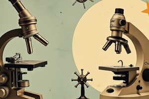
Microscope
- A crucial tool for identifying microorganisms.
- Essential for microbiological labs.
- Two main types: Light Microscope and Electron Microscope.
Light Microscope
- Utilizes glass lenses to focus light rays for magnified images.
- Four main types:
- Bright-field: Standard microscopy technique, image is bright against dark background.
- Dark-field: Illuminates specimens indirectly, creating bright objects against a black background.
- Phase-contrast: Enhances contrast between transparent structures by altering light phase.
- Fluorescence: Uses specific dyes that emit light under UV illumination, highlighting certain structures.
Electron Microscope
- Employs electron beams instead of light for enhanced resolution.
- Two main types:
- Scanning Electron Microscope (SEM): Provides detailed surface images of specimens.
- Transmission Electron Microscope (TEM): Shows internal structures of specimens.
Lenses and Light Bending
- Refraction: Light bending as it passes through different media.
- Refractive index: Measures the light velocity reduction in a medium.
- Lenses function like a collection of prisms, focusing light at a focal point.
- Focal length: Distance between the lens center and the focal point.
- Magnifying glass: Utilizes a convex lens to overcome the eye's limitation in focusing on close objects.
- Lens with a shorter focal length magnifies objects more than a lens with a longer focal length.
Bright-Field Microscope
- Resolution: The ability to distinguish between two closely spaced objects.
- Abbé equation:
- d = λ / (n sin θ)
drepresents the minimum resolvable distance between two points.λsymbolizes the wavelength of light.nsignifies the refractive index of the medium.θdenotes half the angle of the cone of light entering the objective.
- Smaller
dtranslates to better resolution. - Shortest wavelengths (around 450-500 nm) provide optimal resolution.
Numerical Aperture & Resolution
- Numerical Aperture (NA): Represents the light-gathering ability of an objective lens.
- Higher NA results in improved resolution.
- NA is influenced by the refractive index (n) of the medium surrounding the lens and the lens's design.
- Immersion oil with a refractive index similar to glass is used with high-power objectives to further improve light capture and resolution.
The Dark-Field Microscope
- Illuminates specimens indirectly using a hollow cone of light.
- Only rays that have been reflected or refracted by the specimen enter the objective, making the background appear dark and the specimen bright.
The Phase-Contrast Microscope
- Enhances contrast between transparent structures by altering light phase.
- A condenser with an annular stop creates a hollow cone of light.
- Direct and diffracted light rays interfere, enhancing image contrast.
- Makes unstained cells visible.
- Widely used for observing living cells, motility, and internal structures.
Differential Interference Contrast (DIC) Microscope
- Uses two beams of plane-polarized light to create a 3D-like image.
- Prism splits the light beam, creating color contrast for the specimen.
- High resolution microscopy for observing cell walls, endospores, and other structures.
The Fluorescence Microscope
- Utilizes fluorescent dyes that absorb UV light and emit visible light.
- Specimen appears bright against a dark background.
- Specific fluorescent dyes (fluorochromes) target distinct structures for visualization.
- Useful in ecological studies and for identifying specific microorganisms (e.g., Mycobacterium tuberculosis, Bacillus anthracis).
Electron Microscopy
- Uses a beam of electrons instead of light for superior resolution.
- Electrons have much shorter wavelengths than light, allowing for incredibly fine detail visualization.
- Images are always black and white.
- Electromagnetic lenses focus the electron beam on the specimen.
- Two main types:
- Scanning Electron Microscope (SEM): Images the surface of specimens with high resolution.
- Transmission Electron Microscope (TEM): Reveals internal structures of specimens.
Studying That Suits You
Use AI to generate personalized quizzes and flashcards to suit your learning preferences.