Podcast
Questions and Answers
What is the primary function of the magnetic lenses in a transmission electron microscope?
What is the primary function of the magnetic lenses in a transmission electron microscope?
- To convert electrons into visible light.
- To accelerate the electrons towards the specimen.
- To illuminate the specimen with electrons.
- To focus the electrons that have passed through the specimen. (correct)
In a transmission electron microscope, the final image is displayed by which method?
In a transmission electron microscope, the final image is displayed by which method?
- By projecting electrons directly onto a detector.
- By projecting electrons onto a fluorescent screen or photographic film. (correct)
- By using a series of mirrors to redirect the beam.
- By converting the electron beam into an audio signal.
What is the role of the specimen in a transmission electron microscope?
What is the role of the specimen in a transmission electron microscope?
- To magnify the electron beam before focusing.
- To absorb electrons, creating a shadow.
- To generate a beam of electrons.
- To allow electrons to pass through, scattering based on its structure. (correct)
What is the purpose of the electron beam initially in a transmission electron microscope?
What is the purpose of the electron beam initially in a transmission electron microscope?
What type of lens is used to focus electrons in a transmission electron microscope?
What type of lens is used to focus electrons in a transmission electron microscope?
What is the primary reason for using an electron microscope to view cellular components?
What is the primary reason for using an electron microscope to view cellular components?
Which of the following is LEAST likely to be visualized in detail using a standard light microscope?
Which of the following is LEAST likely to be visualized in detail using a standard light microscope?
If a scientist wished to study the structure of ribosomes within a cell, which type of microscopy would be MOST appropriate?
If a scientist wished to study the structure of ribosomes within a cell, which type of microscopy would be MOST appropriate?
Which structure requires the highest level of magnification to examine in detail?
Which structure requires the highest level of magnification to examine in detail?
Considering the typical sizes of biological entities, what relationship between cellular components and microscopy techniques can be inferred?
Considering the typical sizes of biological entities, what relationship between cellular components and microscopy techniques can be inferred?
What is the primary function of the ocular lens in a compound microscope?
What is the primary function of the ocular lens in a compound microscope?
Where is the ocular lens typically situated within the body tube of a compound microscope?
Where is the ocular lens typically situated within the body tube of a compound microscope?
Which component of a compound microscope is primarily responsible for making the image sharp and clear to the viewer?
Which component of a compound microscope is primarily responsible for making the image sharp and clear to the viewer?
What is the primary function of the objective lens in a microscope?
What is the primary function of the objective lens in a microscope?
In the context of image formation in a compound microscope, what is the direct role of the ocular?
In the context of image formation in a compound microscope, what is the direct role of the ocular?
Which material is most commonly used in the construction of objective lenses?
Which material is most commonly used in the construction of objective lenses?
If a compound microscope lacked an ocular lens, what would be the most immediate consequence?
If a compound microscope lacked an ocular lens, what would be the most immediate consequence?
What is the typical housing for the lens within an objective lens assembly?
What is the typical housing for the lens within an objective lens assembly?
What may be found inside a single objective lens cylinder?
What may be found inside a single objective lens cylinder?
In the context of microscopy, which of the following best describes the objective lens' function?
In the context of microscopy, which of the following best describes the objective lens' function?
What component directly holds the objective lenses in a standard microscope?
What component directly holds the objective lenses in a standard microscope?
What action is used to change the magnification of a microscope by switching lenses?
What action is used to change the magnification of a microscope by switching lenses?
In a standard microscope, what structural element is situated at the lower part of the microscope tube?
In a standard microscope, what structural element is situated at the lower part of the microscope tube?
What is the primary function of a revolving nosepiece in a microscope?
What is the primary function of a revolving nosepiece in a microscope?
Objective lenses are typically attached to which component of the microscope?
Objective lenses are typically attached to which component of the microscope?
Which objective lens is typically used for initial, low-magnification scanning of a specimen?
Which objective lens is typically used for initial, low-magnification scanning of a specimen?
A 50x objective lens would be classified under which category of magnification?
A 50x objective lens would be classified under which category of magnification?
When using the 100x objective lens, what substance should be used to enhance image clarity?
When using the 100x objective lens, what substance should be used to enhance image clarity?
If a scientist switches from a 10x lens to a 40x lens, how does this change the magnification?
If a scientist switches from a 10x lens to a 40x lens, how does this change the magnification?
A microscope has objective lenses with magnifications of 4x, 10x, 40x, and 100x. What is the primary function of the 10x objective lens?
A microscope has objective lenses with magnifications of 4x, 10x, 40x, and 100x. What is the primary function of the 10x objective lens?
Flashcards
Transmission Electron Microscope (TEM)
Transmission Electron Microscope (TEM)
A type of microscope that uses electrons to create an image of a sample.
Magnetic Lenses
Magnetic Lenses
In a TEM, these are used to focus the electron beam and create an image.
Electron Transmission
Electron Transmission
This process involves electrons passing through the sample, projecting an image onto a screen.
Fluorescent Screen
Fluorescent Screen
Signup and view all the flashcards
Photographic Film
Photographic Film
Signup and view all the flashcards
Electron Microscope
Electron Microscope
Signup and view all the flashcards
Organelle
Organelle
Signup and view all the flashcards
Viruses
Viruses
Signup and view all the flashcards
Molecule
Molecule
Signup and view all the flashcards
Sizes of Living Things
Sizes of Living Things
Signup and view all the flashcards
Ocular/Eyepiece
Ocular/Eyepiece
Signup and view all the flashcards
Body Tube
Body Tube
Signup and view all the flashcards
Objective Lenses
Objective Lenses
Signup and view all the flashcards
Revolving Nosepiece
Revolving Nosepiece
Signup and view all the flashcards
Stage
Stage
Signup and view all the flashcards
Lens Cylinder
Lens Cylinder
Signup and view all the flashcards
Glass
Glass
Signup and view all the flashcards
Collect Light
Collect Light
Signup and view all the flashcards
Sample
Sample
Signup and view all the flashcards
Selecting an Objective Lens
Selecting an Objective Lens
Signup and view all the flashcards
Microscope Tube
Microscope Tube
Signup and view all the flashcards
Objective Lens Attachment
Objective Lens Attachment
Signup and view all the flashcards
4x Objective Lens
4x Objective Lens
Signup and view all the flashcards
10x Objective Lens
10x Objective Lens
Signup and view all the flashcards
40x Objective Lens
40x Objective Lens
Signup and view all the flashcards
100x Oil Immersion Objective Lens
100x Oil Immersion Objective Lens
Signup and view all the flashcards
How to Calculate Magnification
How to Calculate Magnification
Signup and view all the flashcards
Study Notes
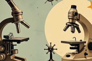
Microscope Structure and Types
- Microscopes are used to magnify small objects, enabling observation of detail invisible to the naked eye.
- Light microscopes use visible light and lenses to magnify images (2 types).
- Electron microscopes use beams of electrons and magnetic lenses to achieve higher magnification and resolution than light microscopes.
- The various types of microscopes include:
- Light microscopes (L/M)
- Electron microscopes (E/M)
- Transmission electron microscopes
- Scanning electron microscopes
- Fluorescence microscopes
- Super resolution microscopes
- X-ray microscopes
- Atomic Force microscopes
- Phase-Contrast Microscope
Types of Light Microscopes
- Simple Microscope: Uses a single lens for magnification.
- Compound Microscope: Uses multiple lenses (ocular and objective) for higher magnification.
Compound Light Microscope Components
- Ocular/Eyepiece: The lens you look through. Common magnifications: 5x, 10x, & 2x (magnification can vary). Some have a pointer.
- Objective Lenses: Lenses at the base of the microscope, mounted on a rotating nosepiece. Different objective lenses magnify at different levels, allowing for varying degrees of magnification. Common magnifications include 4x, 5x, 10x, 20x, 40x, 50x, 100x.
- Stage: A flat platform beneath the objectives; holds the specimen. Usually with a mechanical stage, allowing controlled movement of the specimen.
- Stage Clips: Hold the specimen slide in place on the stage.
- Diaphragm: Controls the amount of light passing through the specimen.
- Condenser: An optical device that focuses light onto the specimen.
- Illumination Source: A light source. Simple microscopes might use sunlight. Modern ones use an electric light bulb. A mirror is used with certain types.
- Coarse Adjustment Knob: Moves the stage up and down for initial focusing.
- Fine Adjustment Knob: Makes fine adjustments for precise focusing.
Additional Microscope-related Information
- Magnification: The amount the image is enlarged. Calculated by multiplying the magnification of the ocular lens by the magnification of the objective lens.
- Resolution: The ability of a microscope to distinguish two very close points.
- Metric System: A standardized system of units used in science, including measurements of length (nanometers, micrometers, millimeters, centimeters, meters).
Microscope Techniques
- Various chemical and staining techniques can be used in conjunction with microscopes to improve visualization of specific structures within a specimen.
Studying That Suits You
Use AI to generate personalized quizzes and flashcards to suit your learning preferences.