Podcast
Questions and Answers
Which cross-section of the medulla is associated with the sensory decussation?
Which cross-section of the medulla is associated with the sensory decussation?
- Caudal Aspect
- Pyramidal Decussation
- Pons-Medulla Junction
- Middle Portion (correct)
The hypoglossal nucleus is located in the caudal part of the medulla.
The hypoglossal nucleus is located in the caudal part of the medulla.
False (B)
What is the primary function of the medulla oblongata?
What is the primary function of the medulla oblongata?
Autonomic functions including breathing and heart rate
The ________ decussation is where corticospinal fibers cross.
The ________ decussation is where corticospinal fibers cross.
Match the following nuclei with their associated functions:
Match the following nuclei with their associated functions:
Which percentage of corticospinal fibers move laterally at the pyramidal decussation?
Which percentage of corticospinal fibers move laterally at the pyramidal decussation?
The dorsal nucleus of vagus is associated with cranial nerve XII.
The dorsal nucleus of vagus is associated with cranial nerve XII.
Name one key structure found in the middle portion of the medulla.
Name one key structure found in the middle portion of the medulla.
The medulla is located below the ________ and above the spinal cord.
The medulla is located below the ________ and above the spinal cord.
What is the function of the hypoglossal nucleus?
What is the function of the hypoglossal nucleus?
Which nucleus is involved in the autonomic functions and sensory processing?
Which nucleus is involved in the autonomic functions and sensory processing?
The dorsal spinocerebellar tract sends proprioceptive information from the upper body.
The dorsal spinocerebellar tract sends proprioceptive information from the upper body.
What is the primary function of the medial longitudinal fasciculus (MLF)?
What is the primary function of the medial longitudinal fasciculus (MLF)?
The __________ tract is responsible for transmitting pain and temperature sensations.
The __________ tract is responsible for transmitting pain and temperature sensations.
Match the following nuclei with their functions:
Match the following nuclei with their functions:
Which tract carries descending pathways that influence extensor muscles?
Which tract carries descending pathways that influence extensor muscles?
Fibers from the fasciculus cuneatus cross to the contralateral side after synapsing in the medulla.
Fibers from the fasciculus cuneatus cross to the contralateral side after synapsing in the medulla.
What percentage of corticospinal fibers form the lateral corticospinal tract after decussation?
What percentage of corticospinal fibers form the lateral corticospinal tract after decussation?
The __________ tract connects the inferior olivary nucleus to the spinal cord.
The __________ tract connects the inferior olivary nucleus to the spinal cord.
Which nuclei are part of the vestibular complex?
Which nuclei are part of the vestibular complex?
Which nucleus is primarily involved in motor functions of cranial nerve X?
Which nucleus is primarily involved in motor functions of cranial nerve X?
The ventral spinocerebellar tract is primarily engaged with providing proprioceptive information from the upper body.
The ventral spinocerebellar tract is primarily engaged with providing proprioceptive information from the upper body.
What is the main function of the medial lemniscus?
What is the main function of the medial lemniscus?
The __________ tract influences extensor muscles for posture and balance.
The __________ tract influences extensor muscles for posture and balance.
Match the following nuclei to their respective types of sensory information:
Match the following nuclei to their respective types of sensory information:
Which tract is involved in coordinating reflexive head and eye movements?
Which tract is involved in coordinating reflexive head and eye movements?
The arcuate nucleus contributes solely to sensory information processing.
The arcuate nucleus contributes solely to sensory information processing.
What is the significance of sensory decussation?
What is the significance of sensory decussation?
The __________ spinocerebellar tract carries proprioceptive information from the lower body.
The __________ spinocerebellar tract carries proprioceptive information from the lower body.
What is the function of the inferior olivary nucleus?
What is the function of the inferior olivary nucleus?
What is the primary role of the medulla oblongata?
What is the primary role of the medulla oblongata?
The dorsal nucleus of vagus is associated with cranial nerve XII.
The dorsal nucleus of vagus is associated with cranial nerve XII.
What structures are located in the middle portion of the medulla?
What structures are located in the middle portion of the medulla?
The hypoglossal nucleus is primarily responsible for movements of the ________.
The hypoglossal nucleus is primarily responsible for movements of the ________.
Match the following cross-section levels of the medulla with their characteristics:
Match the following cross-section levels of the medulla with their characteristics:
Which of the following best describes the function of the hypoglossal nucleus?
Which of the following best describes the function of the hypoglossal nucleus?
The pyramidal decussation is where 85% of the corticospinal fibers remain in the ventral column.
The pyramidal decussation is where 85% of the corticospinal fibers remain in the ventral column.
Name the primary function of the dorsal nucleus of vagus.
Name the primary function of the dorsal nucleus of vagus.
The __________ decussation occurs at the junction of the medulla and spinal cord.
The __________ decussation occurs at the junction of the medulla and spinal cord.
Which anatomical position is the medulla oblongata located relative to the pons?
Which anatomical position is the medulla oblongata located relative to the pons?
What is the primary function of the dorsal nucleus of vagus?
What is the primary function of the dorsal nucleus of vagus?
The pyramidal decussation is located at the rostral aspect of the medulla.
The pyramidal decussation is located at the rostral aspect of the medulla.
Which cranial nerve is associated with the hypoglossal nucleus?
Which cranial nerve is associated with the hypoglossal nucleus?
The ____ aspect of the medulla is where corticospinal fibers cross.
The ____ aspect of the medulla is where corticospinal fibers cross.
Match the following medullary nuclei with their associated functions:
Match the following medullary nuclei with their associated functions:
How many corticospinal fibers cross at the pyramidal decussation?
How many corticospinal fibers cross at the pyramidal decussation?
The sensory decussation is involved in motor control function.
The sensory decussation is involved in motor control function.
What significant structure is found in the middle portion of the medulla?
What significant structure is found in the middle portion of the medulla?
The medulla is located _____ the pons and above the spinal cord.
The medulla is located _____ the pons and above the spinal cord.
Which nuclei are located lateral to the hypoglossal nucleus?
Which nuclei are located lateral to the hypoglossal nucleus?
Which nucleus is involved in motor functions and innervates muscles in the pharynx and larynx?
Which nucleus is involved in motor functions and innervates muscles in the pharynx and larynx?
The medial lemniscus is responsible for transmitting auditory information.
The medial lemniscus is responsible for transmitting auditory information.
What is the primary role of the vestibular complex?
What is the primary role of the vestibular complex?
The ________ tract connects the spinal cord to the cerebellum and is involved in proprioception.
The ________ tract connects the spinal cord to the cerebellum and is involved in proprioception.
Match the following tracts with their primary functions:
Match the following tracts with their primary functions:
Where are the fasciculus gracilis and cuneatus located in the medulla?
Where are the fasciculus gracilis and cuneatus located in the medulla?
The inferior olivary nucleus has a circular appearance.
The inferior olivary nucleus has a circular appearance.
What percentage of corticospinal fibers form the ventral corticospinal tract after decussation?
What percentage of corticospinal fibers form the ventral corticospinal tract after decussation?
The ________ nucleus is associated with the regulation of salivation and related to the glossopharyngeal nerve.
The ________ nucleus is associated with the regulation of salivation and related to the glossopharyngeal nerve.
What is the function of the medial longitudinal fasciculus (MLF)?
What is the function of the medial longitudinal fasciculus (MLF)?
What percentage of corticospinal fibers remains in the ventral column during the pyramidal decussation?
What percentage of corticospinal fibers remains in the ventral column during the pyramidal decussation?
The dorsal nucleus of vagus is located medially to the hypoglossal nucleus.
The dorsal nucleus of vagus is located medially to the hypoglossal nucleus.
What is the main role of the medulla oblongata?
What is the main role of the medulla oblongata?
The ________ nucleus is responsible for tongue movements.
The ________ nucleus is responsible for tongue movements.
Which structure is involved in processing sensory information leading to the medial meniscus action?
Which structure is involved in processing sensory information leading to the medial meniscus action?
Match the following structures with their corresponding cranial nerves:
Match the following structures with their corresponding cranial nerves:
What is the inferior boundary of the medulla oblongata?
What is the inferior boundary of the medulla oblongata?
The sensory decussation is located at the most inferior aspect of the medulla.
The sensory decussation is located at the most inferior aspect of the medulla.
Which structure in the medulla is primarily involved in autonomic functions?
Which structure in the medulla is primarily involved in autonomic functions?
In the pyramidal decussation, ________% of corticospinal fibers move laterally.
In the pyramidal decussation, ________% of corticospinal fibers move laterally.
Which nucleus is primarily involved in auditory processing?
Which nucleus is primarily involved in auditory processing?
The medial lemniscus transmits visual information.
The medial lemniscus transmits visual information.
What structure facilitates communication between the medulla and cerebellum?
What structure facilitates communication between the medulla and cerebellum?
The tract that carries sensory information from the lower body is called the ________ gracilis.
The tract that carries sensory information from the lower body is called the ________ gracilis.
Which tract is involved in reflexive head and eye movement coordination?
Which tract is involved in reflexive head and eye movement coordination?
Match the following nuclei with their respective functions:
Match the following nuclei with their respective functions:
The descending sympathetic tract originates from the spinal cord.
The descending sympathetic tract originates from the spinal cord.
Which brain structure houses the reflexive pathways for motor functions in cranial nerve X?
Which brain structure houses the reflexive pathways for motor functions in cranial nerve X?
The ________ decussation refers to where sensory fibers cross to the opposite side of the body.
The ________ decussation refers to where sensory fibers cross to the opposite side of the body.
Which part of the medulla is associated with corticospinal fiber crossing?
Which part of the medulla is associated with corticospinal fiber crossing?
Which of the following nuclei is involved in tongue movements?
Which of the following nuclei is involved in tongue movements?
The medulla oblongata plays a role only in voluntary movement control.
The medulla oblongata plays a role only in voluntary movement control.
What is the primary function associated with the dorsal nucleus of vagus?
What is the primary function associated with the dorsal nucleus of vagus?
The ________ aspect of the medulla is where the pyramidal decussation occurs.
The ________ aspect of the medulla is where the pyramidal decussation occurs.
Match the following structures of the medulla with their associated characteristics:
Match the following structures of the medulla with their associated characteristics:
What percentage of corticospinal fibers move laterally at the pyramidal decussation?
What percentage of corticospinal fibers move laterally at the pyramidal decussation?
The sensory decussation is located in the most caudal part of the medulla.
The sensory decussation is located in the most caudal part of the medulla.
Name one key structure located in the middle portion of the medulla.
Name one key structure located in the middle portion of the medulla.
The ________ nucleus is associated with cranial nerve X.
The ________ nucleus is associated with cranial nerve X.
What is the role of the pyramidal decussation?
What is the role of the pyramidal decussation?
What is the primary role of the cerebellar connections?
What is the primary role of the cerebellar connections?
The medial lemniscus is responsible for transmitting pain and temperature sensations.
The medial lemniscus is responsible for transmitting pain and temperature sensations.
What connects the inferior olivary nucleus to the spinal cord?
What connects the inferior olivary nucleus to the spinal cord?
The __________ nucleus is involved in the regulation of salivation.
The __________ nucleus is involved in the regulation of salivation.
Match the following tracts with their primary functions:
Match the following tracts with their primary functions:
Which of the following nuclei is involved in processing sensory information from the face?
Which of the following nuclei is involved in processing sensory information from the face?
The arcuate nucleus has only contralateral fibers.
The arcuate nucleus has only contralateral fibers.
What percentage of corticospinal fibers descends as the ventral corticospinal tract after decussation?
What percentage of corticospinal fibers descends as the ventral corticospinal tract after decussation?
The __________ complex is critical for balance and spatial orientation.
The __________ complex is critical for balance and spatial orientation.
Which of the following statements is true about the pyramidal decussation?
Which of the following statements is true about the pyramidal decussation?
Flashcards are hidden until you start studying
Study Notes
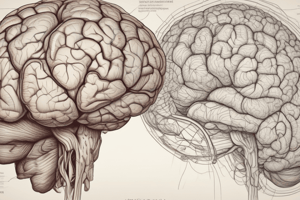
Medulla Oblongata Overview
- Median cross-sections are utilized to examine the medulla oblongata's internal structures, similar to the midbrain and pons.
- The medulla serves as a critical pathway connecting the brain to the spinal cord, located below the pons and above the spinal cord.
Cross-Section Levels
- Three primary cross-section levels of the medulla include:
- Junction at the pons-medulla: Most superior or rostral aspect.
- Middle portion: Known as the sensory decussation, where important sensory nuclei interact.
- Most inferior or caudal aspect: The pyramidal decussation, where corticospinal fibers cross.
Cross-Section Details
- Pons-Medulla Junction: The top cross-section showcases essential structures and is important for orientation.
- Sensory Decussation:
- Located centrally within the medulla.
- Contains the nucleus cuneatus and nucleus gracilis.
- Involved in processing sensory information leading to the medial meniscus action.
- Pyramidal Decussation:
- Located at the bottom portion.
- Facilitates the crossing of corticospinal fibers—85% move laterally and 15% remain in the ventral column.
Key Structures in Medulla
- Hypoglossal Nucleus:
- Paired structure located in the rostral portion of the medulla.
- Associated with cranial nerve XII (hypoglossal nerve), responsible for tongue movements.
- Dorsal Nucleus of Vagus:
- Positioned lateral to the hypoglossal nucleus.
- Pertains to cranial nerve X (vagus nerve), involved in autonomic functions.
Summary of Functions
- Medulla oblongata plays a vital role in autonomic functions, including breathing, heart rate, and reflexes.
- Consistency across cross-sections aids in the identification and understanding of structures within the medulla.### Neuroanatomy of the Medulla
- Hypoglossal nucleus is responsible for motor control of the tongue; located medially in the medulla.
- Dorsal nucleus of vagus plays a role in parasympathetic functions, linked to cranial nerve X.
- Nucleus of tractus solitarius is involved in autonomic functions and sensory processing; located laterally and anteriorly in respect to the hypoglossal and dorsal vagal nuclei.
Vestibular Nuclei
- Vestibular complex consists of medial and inferior vestibular nuclei; plays a critical role in balance and spatial orientation.
- Components of the vestibular complex include superior, lateral, medial, and inferior vestibular nuclei, with varying visibility in pons and medulla.
Cochlear Nuclei
- Dorsal cochlear nucleus and ventral cochlear nucleus are involved in auditory processing; dorsal lies more dorsally, while ventral is located ventrally.
Cerebellar Connections
- Inferior cerebellar peduncles serve as white matter tracts facilitating communication between the medulla and cerebellum.
Ponto-Bulbar Body
- Ponto-bulbar body connects structures within the medulla and pons, forming essential pathways for motor applications.
Ventral Spinocerebellar Tract
- Ventral spinocerebellar tract connects the spinal cord to the cerebellum, involved in proprioception and coordination.
Inferior Olivary Nuclei
- Inferior olivary nuclear complex has a U-shaped appearance and is involved in motor learning and coordination.
Arcuate Nucleus
- Arcuate nucleus has contralateral and ipsilateral fibers contributing to coordination between body movements and connections to the ponto-bulbar body.
- Anterior external arcuate fibers allow communication between the arcuate nucleus and the ponto-bulbar body, contributing to motor function.
Stria Medullaris
- Stria medullaris forms part of the posterior wall of the fourth ventricle and is crucial for integrating sensory information.
Corticospinal Tracts
- Corticospinal tracts are descending motor pathways originating from the cortex, responsible for voluntary motor control.
Medial and Spinal Lemniscus
- Medial lemniscus carries proprioceptive and touch information from the body, whereas spinal lemniscus is involved in transmitting pain and temperature sensations.
Trigeminal Nerves and Nuclei
- The spinal nucleus and tract of the trigeminal nerve (cranial nerve V) process sensory information from the face and has three components across the brainstem.
Descending Motor Tracts
- Tectospinal tract coordinates reflexive head and eye movements in response to visual and auditory stimuli.
- Rubrospinal tract transmits descending motor signals; critical for flexor movements and utilizes the red nucleus in the midbrain.
Medial Longitudinal Fasciculus (MLF)
- MLF connects cranial nerve nuclei for eye movement coordination, playing a vital role in conjugate eye movements.
Nucleus Ambiguous
- Nucleus ambiguous is involved in the motor functions of cranial nerve X and contributes to the innervation of muscles in the pharynx and larynx.
Inferior Salivatory Nucleus
- This nucleus is related to the glossopharyngeal nerve and involved in the regulation of salivation.
Descending Sympathetic Tract
- The descending sympathetic tract originates from the hypothalamus and participates in autonomic functions.
Sensory Decussation
- The sensory decussation marks the point where sensory fibers cross to the opposite side, crucial for sensory perception.
Fasciculus Gracilis and Cuneatus
- Fasciculus gracilis carries lower extremity sensory information, while fasciculus cuneatus carries upper extremity sensory information to the respective nuclei.
Dorsal and Ventral Spinocerebellar Tracts
- Dorsal spinocerebellar tract sends proprioceptive information from lower body, while ventral spinocerebellar tract is more involved with coordination from the spinal cord.
Olivospinal Tract
- The olivospinal tract connects the inferior olivary nucleus to the spinal cord, playing a role in motor processing.
Vestibulospinal Tract
- Vestibulospinal tract carries descending pathways that control posture and balance by influencing extensor muscles.### Medulla Anatomy and Functions
- The nucleus of the tractus solitarius plays a significant role in processing sensory information.
- Sensory decussation is a key concept where sensory pathways cross to the opposite side of the body.
- Proprioceptive sensory information from legs and arms follows distinct pathways into the spinal cord:
- Information from the legs arrives medially (fasciculus gracilis), while from the arms it arrives laterally (fasciculus cuneatus).
- Both fasciculi terminate in the medulla:
- The nucleus gracilis for leg sensations (medially).
- The nucleus cuneatus for arm sensations (laterally).
- After synapsing, fibers from both nuclei cross to the contralateral side, integrating into the medial lemniscus.
Pyramidal Decussation
- Located at the most caudal portion of the medulla, where corticospinal fibers cross to descend into the spinal cord.
- Approximately 85% of corticospinal fibers form the lateral corticospinal tract after decussation.
- Remaining 15% descend as the ventral corticospinal tract, crossing at the spinal cord level they innervate.
External Anatomy of the Medulla
- The anterior median fissure, anterior lateral sulcus, and posterior lateral sulcus are key landmarks.
- Junctions between the pons and medulla contain several cranial nerves:
- Cranial Nerve VI: Abducens nerve.
- Cranial Nerve VII: Facial nerve.
- Cranial Nerve VIII: Vestibulocochlear nerve (comprising cochlear and vestibular branches).
- Cranial Nerve IX: Glossopharyngeal nerve (located at the posterior lateral sulcus).
- Cranial Nerve X: Vagus nerve (more caudally located).
- Cranial part of Cranial Nerve XI: Accessory nerve, which has both cranial and spinal components.
Key Pathways
- Dorsal and ventral spinocerebellar tracts support sensory integration.
- Descending motor pathways include:
- Vestibulospinal tract responsible for extensor muscle control.
- Medial longitudinal fasciculus and rubrospinal tracts, crucial for coordinated movement.
Summary of Cranial Nerves at Ponto-Medullary Junction
- Functionally paired cranial nerves emerge at specified sulci:
- Anterior median fissure: Significant for motor pathways.
- Anterior lateral and posterior lateral sulci: Important for sensory and cranial nerve emergence.
Medulla Oblongata Overview
- Comprises internal structures examined through median cross-sections, linking brain and spinal cord.
- Positioned inferior to the pons and superior to the spinal cord, playing a vital role in communication.
Cross-Section Levels
- Key levels include the junction at the pons-medulla, sensory decussation, and pyramidal decussation.
- Pons-medulla junction is the most superior section, while pyramidal decussation marks the lowest point where corticospinal fibers cross.
Cross-Section Details
- Pons-Medulla Junction showcases essential nuclei; important for anatomical orientation.
- Sensory Decussation features nucleus cuneatus and nucleus gracilis for sensory processing, leading to medial meniscus communication.
- Pyramidal Decussation enables 85% of corticospinal fibers to cross laterally, with 15% remaining in the ventral column.
Key Structures in Medulla
- Hypoglossal Nucleus: Associated with cranial nerve XII, controls tongue movement; located in rostral medulla.
- Dorsal Nucleus of Vagus: Related to cranial nerve X, governs autonomic functions; positioned lateral to the hypoglossal nucleus.
Summary of Functions
- Regulates autonomic functions like breathing, heart rate, and reflexes.
- Cross-sectional consistency aids in identifying medullary structures.
Neuroanatomy of the Medulla
- Hypoglossal nucleus contributes to tongue motor control; dorsal nucleus of vagus manages parasympathetic functions.
- Nucleus of tractus solitarius processes sensory and autonomic functions; located laterally and anteriorly.
Vestibular Nuclei
- Comprises medial and inferior vestibular nuclei; essential for balance and spatial awareness.
- Additional vestibular nuclei (superior, lateral, medial, inferior) support various balance functions.
Cochlear Nuclei
- Dorsal and ventral cochlear nuclei facilitate auditory processing, varying in dorsal and ventral positioning.
Cerebellar Connections
- Inferior cerebellar peduncles connect the medulla to the cerebellum for motor coordination.
Ponto-Bulbar Body
- Serves as a linkage between medulla and pons, forming pathways critical for motor function.
Ventral Spinocerebellar Tract
- Transmits proprioceptive signals from the spinal cord to the cerebellum, aiding in coordination.
Inferior Olivary Nuclei
- U-shaped complex involved in motor learning and coordination.
Arcuate Nucleus
- Has contralateral and ipsilateral fibers for movement coordination; presents connections to the ponto-bulbar body.
Stria Medullaris
- Part of the fourth ventricle's posterior wall; crucial for sensory information integration.
Corticospinal Tracts
- Descending motor pathways originating from the cerebral cortex; essential for voluntary movements.
Medial and Spinal Lemniscus
- Medial lemniscus relays proprioceptive and touch info; spinal lemniscus transmits pain and temperature sensations.
Trigeminal Nerves and Nuclei
- Processes facial sensory information via the spinal nucleus and tract of cranial nerve V, across three brainstem regions.
Descending Motor Tracts
- Tectospinal tract coordinates reflexive head/eye movements; rubrospinal tract facilitates flexor signals through the red nucleus.
Medial Longitudinal Fasciculus (MLF)
- Connects cranial nerve nuclei for coordinated eye movements, vital for conjugate eye motion.
Nucleus Ambiguous
- Involved in motor functions of cranial nerve X, innervating pharynx and larynx muscles.
Inferior Salivatory Nucleus
- Associated with the glossopharyngeal nerve; plays a role in salivation regulation.
Descending Sympathetic Tract
- Originates from the hypothalamus; influences autonomic functions throughout the body.
Sensory Decussation
- Point where sensory fibers cross to the opposite side, essential for sensory perception integration.
Fasciculus Gracilis and Cuneatus
- Fasciculus gracilis transmits lower body sensory info; cuneatus conveys upper body sensations, both terminating in relevant medullary nuclei.
Dorsal and Ventral Spinocerebellar Tracts
- Dorsal tract relays lower body proprioception; ventral tract is more involved in spinal cord coordination.
Olivospinal Tract
- Connects the inferior olivary nucleus with the spinal cord; contributes to motor processing.
Vestibulospinal Tract
- Controls posture and balance via descending influence on extensor muscles.
Medulla Anatomy and Functions
- Nucleus of tractus solitarius is key for sensory information processing.
- Sensory pathways cross at decussation, with specific paths for proprioceptive signals from both legs and arms.
Pyramidal Decussation
- Site for corticospinal fiber crossing; approximately 85% descend as lateral corticospinal tracts, with 15% forming ventral corticospinal tracts.
External Anatomy of the Medulla
- Anterior median fissure and sulci serve as important landmarks; cranial nerves emerge at specific points, with functionally paired nerves at various sulci.
Key Pathways
- Dorsal and ventral spinocerebellar tracts facilitate sensory integration; descending motor pathways are essential for coordinated movements.
Studying That Suits You
Use AI to generate personalized quizzes and flashcards to suit your learning preferences.