Podcast
Questions and Answers
What is a primary application of scanning electron micrographs (SEMs) in medicine?
What is a primary application of scanning electron micrographs (SEMs) in medicine?
- To develop new materials for industrial use
- To analyze the chemical composition of tissues
- To diagnose diseases based on tissue analysis (correct)
- To study the microstructure of living organisms
What is the purpose of using maggots in wound treatment?
What is the purpose of using maggots in wound treatment?
- To introduce anti-bacterial chemicals into the wound
- To stimulate the immune response of the patient
- To feed on dead tissue and leave healthy tissue untouched (correct)
- To promote the growth of healthy tissue
What is the effect of LGO-treatment on E.coli cells, as observed under a transmission electron microscope (TEM)?
What is the effect of LGO-treatment on E.coli cells, as observed under a transmission electron microscope (TEM)?
- The cytoplasm becomes more regularly distributed
- The cell wall appears disrupted, with variable periplasmic spaces (correct)
- The plasma lemma moves away from the cell wall
- The cell wall becomes thicker and more regular
What is the function of the anti-bacterial chemicals in the saliva of maggots?
What is the function of the anti-bacterial chemicals in the saliva of maggots?
What is a common application of scanning electron micrographs (SEMs) outside of medicine?
What is a common application of scanning electron micrographs (SEMs) outside of medicine?
What is the primary function of the positively biased grid or detector in Scanning Electron Microscopy (SEM)?
What is the primary function of the positively biased grid or detector in Scanning Electron Microscopy (SEM)?
What is the typical range of magnifications required for most applications in Scanning Electron Microscopy (SEM)?
What is the typical range of magnifications required for most applications in Scanning Electron Microscopy (SEM)?
What is the acceleration voltage of up-to-date routine Transmission Electron Microscope (TEM) instruments?
What is the acceleration voltage of up-to-date routine Transmission Electron Microscope (TEM) instruments?
What is the role of the condenser lens system in a conventional Transmission Electron Microscope (TEM)?
What is the role of the condenser lens system in a conventional Transmission Electron Microscope (TEM)?
What is the analogy of a Transmission Electron Microscope (TEM) according to Philips?
What is the analogy of a Transmission Electron Microscope (TEM) according to Philips?
Flashcards are hidden until you start studying
Study Notes
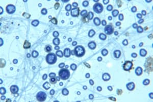
Microstructure
- Maggots, the larvae of a bluebottle fly, are used medicinally to clean wounds by feeding on dead tissue and leaving healthy tissue untouched.
- The maggots' saliva contains anti-bacterial chemicals that maintain sterility in the wound area.
- Maggots are used to treat ulcers and deep wounds, particularly diabetic ulcers on the feet, away from organs or body cavities.
SEM and TEM Applications
- SEM and TEM are used in:
- Medicine and veterinary medicine for diagnosis and medical research, including developing and testing new drugs.
- Study of materials in other sciences such as archeology, metallurgy, botany, and zoology.
- Development and testing of new materials for industrial use.
Scanning Electron Microscopy (SEM)
- SEM is used to inspect topographies of specimens at very high magnifications, up to 300,000 X.
- Most applications require magnifications of less than 3,000 X.
- SEM uses a focused beam of electrons to dislodge electrons from the specimen, which are then collected and translated into a signal.
- The electron beam is swept across the area being inspected, producing many signals that are amplified, analyzed, and translated into images of the topography.
Transmission Electron Microscopy (TEM)
- TEM is analogous to a slide projector.
- In a conventional TEM, a thin specimen is irradiated with an electron beam of uniform current density.
- Electrons are emitted from the electron gun and illuminate the specimen through a condenser lens system.
- The electron intensity distribution behind the specimen is magnified with a lens system and viewed on a fluorescent screen.
- The image can be recorded by direct exposure of a photographic emulsion or digitally by a CCD camera.
- The acceleration voltage of modern TEM instruments is up to 200 kV.
Studying That Suits You
Use AI to generate personalized quizzes and flashcards to suit your learning preferences.