Podcast
Questions and Answers
Which artery is the largest branch of the femoral artery?
Which artery is the largest branch of the femoral artery?
- Obturator artery
- Profunda femoris (correct)
- Lateral femoral circumflex artery
- Medial femoral circumflex artery
Where does the profunda femoris artery pass in relation to the femur?
Where does the profunda femoris artery pass in relation to the femur?
- Superiorly above the femoral ligament
- Posteriorly, lying behind the femoral artery and vein (correct)
- Anteriorly along the shaft of the femur
- Laterally alongside the femoral vein
What does the obturator artery primarily accompany through the obturator canal?
What does the obturator artery primarily accompany through the obturator canal?
- Lateral femoral circumflex artery
- Obturator nerve (correct)
- Profunda femoris artery
- Femoral artery
Into which vein does the profunda femoris vein drain?
Into which vein does the profunda femoris vein drain?
Which branches does the obturator artery divide into?
Which branches does the obturator artery divide into?
What is the primary nerve responsible for supplying the adductor muscles of the thigh?
What is the primary nerve responsible for supplying the adductor muscles of the thigh?
Which structures pass through the adductor canal?
Which structures pass through the adductor canal?
The anterior division of the obturator nerve supplies which of the following muscles?
The anterior division of the obturator nerve supplies which of the following muscles?
What is the correct outcome of a nerve lesion affecting the obturator nerve?
What is the correct outcome of a nerve lesion affecting the obturator nerve?
Which nerve has a minor role in supplying the adductor muscles aside from the obturator nerve?
Which nerve has a minor role in supplying the adductor muscles aside from the obturator nerve?
Which structure primarily innervates the Adductor longus muscle?
Which structure primarily innervates the Adductor longus muscle?
What is the main action of the Gracilis muscle?
What is the main action of the Gracilis muscle?
Which artery is primarily responsible for the blood supply to the medial thigh compartment?
Which artery is primarily responsible for the blood supply to the medial thigh compartment?
Where does the Pectineus muscle insert?
Where does the Pectineus muscle insert?
Which of the following muscles originates from the body and inferior ramus of the pubis?
Which of the following muscles originates from the body and inferior ramus of the pubis?
The primary action performed by the Adductor magnus is:
The primary action performed by the Adductor magnus is:
What common feature do all the muscles of the medial thigh compartment share?
What common feature do all the muscles of the medial thigh compartment share?
Which nerve is primarily responsible for innervating the medial thigh region?
Which nerve is primarily responsible for innervating the medial thigh region?
Flashcards
What is the Adductor Canal?
What is the Adductor Canal?
A channel in the thigh containing the femoral artery, vein, saphenous nerve, lymph vessels, and nerves to the vastus medialis.
What is the Adductor Hiatus?
What is the Adductor Hiatus?
The adductor canal opens into the adductor hiatus, a space between the adductor magnus and the femur.
What is the Obturator Nerve?
What is the Obturator Nerve?
The main nerve supplying most of the adductor muscles, originating from the lumbar plexus (L2-L4).
What are the Anterior and Posterior Obturator Branches?
What are the Anterior and Posterior Obturator Branches?
Signup and view all the flashcards
What is the Medial Femoral Cutaneous Nerve?
What is the Medial Femoral Cutaneous Nerve?
Signup and view all the flashcards
Medial thigh muscles
Medial thigh muscles
Signup and view all the flashcards
Adduction
Adduction
Signup and view all the flashcards
Obturator Nerve
Obturator Nerve
Signup and view all the flashcards
Profunda Femoris Artery
Profunda Femoris Artery
Signup and view all the flashcards
Gracilis muscle
Gracilis muscle
Signup and view all the flashcards
Pectineus muscle
Pectineus muscle
Signup and view all the flashcards
Adductor longus muscle
Adductor longus muscle
Signup and view all the flashcards
Adductor magnus muscle
Adductor magnus muscle
Signup and view all the flashcards
Obturator Artery
Obturator Artery
Signup and view all the flashcards
Profunda Femoris Vein & Obturator Vein
Profunda Femoris Vein & Obturator Vein
Signup and view all the flashcards
Branches of the Profunda Femoris Artery
Branches of the Profunda Femoris Artery
Signup and view all the flashcards
What is the main artery supplying blood to the medial compartment of the thigh?
What is the main artery supplying blood to the medial compartment of the thigh?
Signup and view all the flashcards
Study Notes
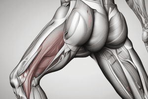
Medial Thigh Anatomy
- The medial thigh comprises muscles, nerves, and vascular structures.
- The muscles of the medial thigh include Gracilis, Pectineus, Adductor longus, Adductor brevis, and Adductor magnus.
- Obturator externus is also discussed in the context of the surrounding gluteal region.
Muscle Details
- Gracilis: Originates from the pubic body, inserts on the medial tibia, and functions in hip adduction, knee flexion, and medial rotation. Innervated by the Obturator nerve (L2-L3).
- Pectineus: Originates from the pectineal surface of the pubis, inserts between the lesser trochanter and the linea aspera. It adducts the thigh at the hip. Innervated by the femoral nerve (L2-L3) and possibly the obturator nerve.
- Adductor longus: Originates from the pubic crest, inserts on the linea aspera (mid-third). Functions in hip adduction. Innervated by the obturator nerve (L2-L4).
- Adductor brevis: Originates from the body and inferior ramus of pubis, inserts on the linea aspera. It adducts the thigh. Innervated by the obturator nerve (L2-L4).
- Adductor magnus: Originates from the ischiopubic ramus and ischial tuberosity. It inserts on the adductor portion, posterior part of the femur from gluteal tuberosity to the medial supra-condylar ridge, and the adductor tubercle. It functions in hip adduction and thigh extension. Innervated by the obturator (L3-L4) and sciatic nerve (L4-L5).
Adductor Canal and Hiatus
- The adductor canal is a passageway for femoral artery, vein, saphenous nerve, and lymph vessels.
- It begins at the inferior apex of the femoral triangle and ends at the adductor hiatus.
- The canal's borders are the sartorius, adductor longus and magnus, and vastus medialis and intermedius.
- Importantly, the great saphenous vein is NOT in the canal.
Nerves
- The major nerve to the medial thigh muscles is the obturator nerve (L2-L4).
- This nerve has both anterior and posterior divisions which supply the muscles.
- Additional nerves include branches from the femoral nerve (medial femoral cutaneous) and possibly a bit of the ilioinguinal nerve providing sensory innervation for the skin.
- Nerve lesions can lead to pain, paraesthesia, loss of sensation, and loss of hip adduction.
Vascular Structures
- Arterial Supply: Profunda femoris and medial/lateral circumflex, and four perforating arteries contribute.
- Profunda femoris artery: Major branch arising on the lateral thigh, 3-5 cm below the inguinal ligament. It curves over adductor brevis and behind the femoral artery and vein to reach the medial thigh. Perforating branches pass between the femur and the tendon of adductor magnus.
- Obturator artery: Arises from the internal iliac artery, travels through the obturator canal, dividing into medial and lateral branches for muscles and hip joint supply.
- Venous Drainage: The obturator vein (ObV) drains into the internal iliac vein. The profunda femoris vein (PFV) drains into the femoral vein.
Summary of Study Points
- Students should be able to identify and describe the actions of the medial thigh muscles.
- Students should know which muscles and nerves are supplied by which nerves.
- The vascular structures important in the medial thigh should be recognised and understood.
- Note the clinical correlations with nerve lesion.
Further Study
- Review of vascular structures and nerves of the medial thigh.
- Preparation for next week's lecture on the gluteal region.
Studying That Suits You
Use AI to generate personalized quizzes and flashcards to suit your learning preferences.