Podcast
Questions and Answers
What is the primary function of the lower limb?
What is the primary function of the lower limb?
- Facilitates digestion
- Regulates body temperature
- Supports body weight (correct)
- Enhances vision
Which of the following bones is not part of the lower limb?
Which of the following bones is not part of the lower limb?
- Humerus (correct)
- Femur
- Tibia
- Fibula
At what age does the complete fusion of the hip bone occur?
At what age does the complete fusion of the hip bone occur?
- 20-25 years (correct)
- 25-30 years
- 15-17 years
- 18-20 years
What is the role of the acetabulum in the hip joint?
What is the role of the acetabulum in the hip joint?
Which lower limb region is primarily involved in weight-bearing and balance?
Which lower limb region is primarily involved in weight-bearing and balance?
What are the hip bones collectively known as?
What are the hip bones collectively known as?
Which part of the pelvis articulates with the sacrum?
Which part of the pelvis articulates with the sacrum?
The obturator foramen is closed by which structure?
The obturator foramen is closed by which structure?
What is a characteristic of the femur's shaft?
What is a characteristic of the femur's shaft?
What is the function of the deep fascia of the thigh?
What is the function of the deep fascia of the thigh?
Which of the following illustrates a typical location for femoral fractures?
Which of the following illustrates a typical location for femoral fractures?
What does the saphenous opening allow to pass through?
What does the saphenous opening allow to pass through?
What shapes the structure of the thigh's fascial compartments?
What shapes the structure of the thigh's fascial compartments?
Which of the following bones contributes to the proximal end of the femur?
Which of the following bones contributes to the proximal end of the femur?
What is a significant outcome of calcium loss affecting the femur's shaft?
What is a significant outcome of calcium loss affecting the femur's shaft?
What do the lateral and medial intermuscular septa primarily separate?
What do the lateral and medial intermuscular septa primarily separate?
What is the primary action of the quadriceps femoris muscle group?
What is the primary action of the quadriceps femoris muscle group?
Which nerve innervates the muscles of the anterior compartment of the thigh?
Which nerve innervates the muscles of the anterior compartment of the thigh?
Which of the following muscles allows for a 'cross-leg' position?
Which of the following muscles allows for a 'cross-leg' position?
What is the origin of the pectineus muscle?
What is the origin of the pectineus muscle?
Which compartment's muscles are primarily responsible for extending the hip and flexing the knee?
Which compartment's muscles are primarily responsible for extending the hip and flexing the knee?
What is the action of the iliopsoas muscle group?
What is the action of the iliopsoas muscle group?
How many heads does the quadriceps femoris muscle have?
How many heads does the quadriceps femoris muscle have?
What is a sesamoid bone in the context of the quadriceps femoris?
What is a sesamoid bone in the context of the quadriceps femoris?
What is the primary action of the iliopsoas muscle?
What is the primary action of the iliopsoas muscle?
Which structure forms the roof of the femoral triangle?
Which structure forms the roof of the femoral triangle?
What is located in the medial compartment of the femoral triangle?
What is located in the medial compartment of the femoral triangle?
From which spinal nerves does the femoral nerve originate?
From which spinal nerves does the femoral nerve originate?
Which structure is NOT a content of the femoral sheath?
Which structure is NOT a content of the femoral sheath?
What is the clinical significance of the femoral canal?
What is the clinical significance of the femoral canal?
Which muscle forms the lateral boundary of the femoral triangle?
Which muscle forms the lateral boundary of the femoral triangle?
What contributes to the formation of the femoral sheath?
What contributes to the formation of the femoral sheath?
What is the diameter of the adductor canal in the middle thigh?
What is the diameter of the adductor canal in the middle thigh?
Which nerve continues anterior to the medial malleolus at the ankle joint?
Which nerve continues anterior to the medial malleolus at the ankle joint?
What is the chief artery supplying the thigh?
What is the chief artery supplying the thigh?
Which of the following structures is NOT included in the boundaries of the adductor canal?
Which of the following structures is NOT included in the boundaries of the adductor canal?
Which vein does the femoral vein receive tributaries from?
Which vein does the femoral vein receive tributaries from?
Which nerve is responsible for innervating the vastus medialis?
Which nerve is responsible for innervating the vastus medialis?
At which anatomical landmark does the femoral artery begin?
At which anatomical landmark does the femoral artery begin?
Which artery is directly responsible for supplying the head and neck of the femur?
Which artery is directly responsible for supplying the head and neck of the femur?
Flashcards are hidden until you start studying
Study Notes
Lower Limb Function
- The lower limb is essential for weight support, locomotion, and balance.
- It is divided into six regions: gluteal, femoral, knee, calf, leg, ankle, and foot.
Regions
- The lower limb regions include the gluteal region, thigh, knee, calf, leg, ankle, and foot.
- The sole is the bottom surface of the foot.
Bones
- The lower limb bones include the pelvic girdle, femur, tibia/fibula, tarsals, and phalanges.
Pelvic Girdle
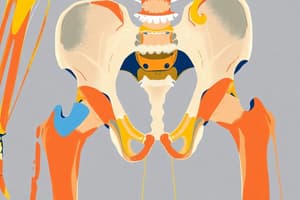
- The pelvic girdle is composed of the sacrum and both hip bones.
- Each hip bone consists of the ilium, ischium, and pubis.
Hip Bone Development
- The three bones of the hip bone are joined by hyaline cartilage at birth.
- The Y-shaped triradiate cartilage forms at puberty.
- Fusion of these bones begins at 15-17 years of age and is complete at 20-25 years.
Ilium
- The ilium is the largest part of the hip bone and provides attachment for numerous muscles.
- The outer aspect of the ilium has gluteal lines which separate the three gluteal muscles.
- The inner aspect of the ilium has an auricular surface that articulates with the sacrum.
Ischium and Pubis
- The ischium is the posteroinferior part of the hip bone.
- The pubis is the anteromedial part of the hip bone.
- The ischium and pubis contribute to the formation of the obturator foramen which is closed by the obturator membrane except for the obturator canal.
- The obturator nerve and vessels pass through the obturator canal.
Acetabulum
- The acetabulum is a cup-shaped cavity on the lateral hip bone.
- It articulates with the head of the femur to form the hip joint.
- The acetabulum has an incomplete inferior margin called the acetabular notch.
- The acetabular fossa is covered by articular cartilage.
Femur
- The femur is the longest and heaviest bone in the body, accounting for about ¼ of a person's height.
- It has a shaft, a proximal end, and a distal end.
- The proximal end of the femur includes the head, neck, and two trochanters.
Femur: Shaft
- The shaft of the femur provides attachment for many muscles.
- It is affected by calcium loss, as seen in rickets.
Femur: Correlates (Clinical Relevance)
- Hip bone fractures (pelvic fractures) can occur.
- Coxa vara and coxa valga are conditions characterized by changes in the angle of inclination of the femoral neck.
- Femoral fractures commonly occur at the neck, which is the narrowest and weakest point.
Fascial Compartments of the Thigh
- The superficial fascia lies deep to the skin and contains fat, superficial vessels, cutaneous nerves, and superficial lymphatics and nodes.
- The deep fascia of the thigh, also known as fascia lata, surrounds all thigh muscles and acts as an elastic stocking, separating groups of muscles.
Thigh Muscle Compartments
- Fascia lata forms three septa: lateral intermuscular septum, medial intermuscular septum, and posterior intermuscular septum, which divide the thigh muscles into three compartments:
- Anterior compartment
- Medial compartment
- Posterior compartment
- These septa attach to the linea aspera of the femur.
Anterior Compartment (Thigh)
- Muscles in the anterior compartment are innervated by the femoral nerve and responsible for extending the knee and flexing the hip.
Medial Compartment (Thigh)
- Muscles in the medial compartment are innervated by the obturator and femoral nerves and adduct the thigh.
Posterior Compartment (Thigh)
- Muscles in the posterior compartment are innervated by the sciatic nerve and extend the hip and flex the knee.
Anterior Thigh Muscles
- The anterior thigh muscles include the sartorius, quadriceps femoris, psoas major, iliacus, and pectineus.
- All of these muscles are innervated by the femoral nerve, flex the hip, and extend the knee.
Pectineus Muscle
- Origin: Superior ramus of pubis
- Insertion: Pectineal line of femur
- Action: Adducts and flexes the thigh, medially rotates the thigh
- Nerve: Femoral nerve +/- obturator nerve
Sartorius Muscle (Tailor's Muscle)
- Origin: Anterior superior iliac spine
- Insertion: Medial surface of the tibia
- Action: Abducts the thigh at the hip joint, laterally rotates the thigh, flexes the hip and knee joints, enabling the "cross-leg" position.
- Innervation: Femoral nerve
- Note: The sartorius muscle is the longest muscle in the body.
Quadriceps Femoris Muscle
- The quadriceps femoris has four heads:
- Rectus femoris: Originates from the ASIS and ilium.
- Vastus lateralis: Originates from the greater trochanter and lateral lip of the linea aspera of the femur.
- Vastus medialis: Originates from the intertrochanteric line and medial lip of the linea aspera of the femur.
- Vastus intermedius: Originates from the anterolateral shaft of the femur.
- All four heads of the quadriceps femoris form the quadriceps tendon, which inserts into the patella and from there into the tubercle of the tibia.
- The patella is a sesamoid bone in the tendon of the quadriceps femoris.
- Action: Extends the leg at the knee joint, flexes the thigh, and steadies the hip joint.
Iliopsoas Muscle
- Psoas minor:
- Origin: Sides of T12-L5 vertebrae and intervertebral discs
- Insertion: Pectineal line and iliopectineal eminence
- Psoas major:
- Origin: Sides and intervertebral discs of T12-L5, transverse processes of all lumbar vertebrae
- Insertion: Lesser trochanter of femur
- Iliacus:
- Origin: Iliac crest and fossa, ala of sacrum
- Insertion: Lesser trochanter of femur
- Action: Flexes, abducts, and laterally rotates the thigh at the hip joint, flexes the leg at the knee joint.
Femoral Triangle
- The femoral triangle is a fascial space located in the superoanterior 1/3 of the thigh.
- Boundaries:
- Medially: Lateral border of the adductor longus muscle
- Laterally: Medial border of the sartorius muscle
- Superiorly: Inguinal ligament
- Floor:
- Laterally: Iliopsoas
- Medially: Pectineus
- Roof:
- Fascia lata
- Cribriform fascia
- Subcutaneous tissue
- Skin
- The saphenous opening is located in the upper part of the femoral triangle.
Femoral Triangle Contents
- The femoral triangle contains the following structures, from lateral to medial:
- Femoral nerve and its branches
- Femoral sheath, which encloses:
- Femoral artery and branches
- Femoral vein and tributaries
- Deep inguinal lymph nodes and vessels
Femoral Sheath
- The femoral sheath is formed by the fusion of the transversalis fascia and psoas fascia below the inguinal ligament.
- It encloses the femoral artery, femoral vein, and femoral canal.
- The femoral nerve lies outside the sheath on its lateral aspect.
Femoral Canal
- The femoral canal occupies the medial compartment of the femoral triangle.
- It opens into the abdominal cavity superiorly at the femoral ring.
- Contains deep inguinal lymph nodes (gland of Cloquet).
- It is a site for a hernia of a gut loop, resulting in a femoral hernia.
Nerves of the Anterior Compartment
- Femoral Nerve:
- Formed from the lumbar plexus (L2, L3, and L4).
- Branches:
- Muscular branches: Innervate the anterior thigh muscles.
- Anterior femoral cutaneous branches: Supply the skin of the anterior thigh.
- Lateral femoral cutaneous branches: Supply the skin of the lateral thigh.
- Saphenous nerve branch:
- Note: Muscular branches of the femoral nerve innervate the anterior thigh muscles.
- Note: The lateral femoral cutaneous nerve courses through the thigh and supplies the skin of the lateral thigh.
Saphenous Nerve
- Course: Travels with the great saphenous vein.
- Termination: Continues anterior to the medial malleolus at the ankle joint, which it innervates.
Femoral Artery
- Origin: A continuation of the external iliac artery.
- Beginning: At the mid-inguinal ligament.
- Branches:
- Profunda femoris: The chief thigh artery; gives 3-4 perforating arteries.
- Circumflex femoral arteries: Supply the head and neck of the femur, especially the medial circumflex artery.
Femoral Vein
- Origin: Continuation of the popliteal vein.
- Tributaries: Receives tributaries that follow the arteries.
- Receives: The great saphenous vein.
- Termination: Becomes the external iliac vein at the inguinal ligament.
Adductor Canal (Hunter's Canal or Subsartorial Canal)
- Location: A 15 cm passageway in the middle of the thigh.
- Course: Extends from the apex of the femoral triangle to the adductor hiatus in the tendon of adductor magnus.
- Contents:
- Femoral artery
- Femoral vein
- Saphenous nerve
- Nerve to vastus medialis
- Boundaries:
- Anteriorly and laterally: Vastus medialis
- Posteriorly: Adductor longus and magnus
- Medially: Sartorius
Studying That Suits You
Use AI to generate personalized quizzes and flashcards to suit your learning preferences.


