Podcast
Questions and Answers
What percentage of blood flow to the liver is supplied by the hepatic artery?
What percentage of blood flow to the liver is supplied by the hepatic artery?
- 20-30% (correct)
- 40-50%
- 10-15%
- 60-70%
Which of the following accurately describes the ultrasound appearance of the liver?
Which of the following accurately describes the ultrasound appearance of the liver?
- Smooth, homogeneous echotexture (correct)
- Isoechoic to the pancreas
- Hypoechoic to surrounding tissues
- Heterogeneous echotexture
What is the primary function of albumin produced by the liver?
What is the primary function of albumin produced by the liver?
- Transporting oxygen in the blood
- Converting glycogen to glucose
- Regulating osmotic pressure (correct)
- Breaking down dietary fibers
What is converted to nontoxic urea in the detoxification process?
What is converted to nontoxic urea in the detoxification process?
In the storage function of the liver, which of the following nutrients is NOT stored?
In the storage function of the liver, which of the following nutrients is NOT stored?
What is the diameter of the main portal vein considered to be if it is normal?
What is the diameter of the main portal vein considered to be if it is normal?
Which description correctly characterizes the right portal vein?
Which description correctly characterizes the right portal vein?
How do the walls of portal veins compare to the walls of hepatic veins?
How do the walls of portal veins compare to the walls of hepatic veins?
What direction does blood flow in the portal veins?
What direction does blood flow in the portal veins?
Which statement about the bifurcation of the main portal vein is accurate?
Which statement about the bifurcation of the main portal vein is accurate?
What is the average weight of the liver in males?
What is the average weight of the liver in males?
Which lobe of the liver is the smallest?
Which lobe of the liver is the smallest?
What separates the right lobe from the left lobe on the diaphragmatic surface?
What separates the right lobe from the left lobe on the diaphragmatic surface?
Which structure provides approximately 70-80% of the blood supply to the liver?
Which structure provides approximately 70-80% of the blood supply to the liver?
What is the primary function of the hepatic veins?
What is the primary function of the hepatic veins?
Which lobe is located anterior to the pancreas?
Which lobe is located anterior to the pancreas?
What is the role of the ligamentum venosum in relation to the liver?
What is the role of the ligamentum venosum in relation to the liver?
What anatomical feature divides the liver into right and left portions based on venous drainage?
What anatomical feature divides the liver into right and left portions based on venous drainage?
What is considered a normal diameter range for hepatic veins?
What is considered a normal diameter range for hepatic veins?
What anatomical structure separates the quadrate lobe from the ligamentum teres?
What anatomical structure separates the quadrate lobe from the ligamentum teres?
Flashcards are hidden until you start studying
Study Notes
Liver Anatomy
- Located intraperitoneally
- Male liver weighs approximately 1600 grams, female liver weighs approximately 1200 grams
- Dimensions
- Transverse: 20-22.5 cm
- Anterior to Posterior: 10-12.5 cm
- Length (Right Lobe): 15-17 cm
- Covered by Glisson’s Capsule - a broelastic connective tissue that contains blood vessels, lymphatics, and nerves
- Covered by peritoneum (except for the falciform ligament, porta hepatis, IVC, GB fossa, and "bare area")
Lobes
- Right Lobe: Largest lobe, separated from the left lobe by the main lobar fissure, anterior/posterior division by the right hepatic vein
- Left Lobe: Located anterior to the pancreas, medial/lateral division by the left portal vein
- Caudate Lobe: Smallest lobe, located posterior to the left lobe, separated from the left lobe by the ligamentum venosum
- Quadrate Lobe: Lies on the medial aspect of the left lobe, anterior to the porta hepatis, between the GB fossa and ligamentum Teres
Lobar Division
- Traditional Lobar Anatomy (Divides liver into four lobes: right, left, caudate, and quadrate based on external landmarks)
- Functional Lobar Anatomy (Divides liver into three lobes: right, left, and caudate based on vascular supply and drainage)
- Couinaud's Anatomy (Divides the liver into eight sections for hepatic lesion localization, each section has its own portal vein, hepatic artery, and bile duct)
Ligaments and Fissures
- Main Lobar Fissure: Divides the liver into right and left lobes, joins the GB fossa and Right Portal Vein
- Falciform Ligament: Separates the right and left lobes on the diaphragmatic surface, gives rise to the ligamentum teres
- Ligamentum Teres (Round Ligament): Continuation of the falciform ligament, remnant of the umbilical vein
- Ligamentum Venosum: Separates the left lobe from the caudate lobe, results from the ductous venosus after birth
Normal Variations
- Reidel’s Lobe: A “tongue-like” projection extending inferiorly past the lower pole of the right kidney, more common in females
- Situs Inversus: Organ positioning is reversed
- Pseudo fissures: Indentations of the diaphragm
- Papillary Process: An inferior extension of the caudate lobe
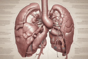
Vascular Supply
- Hepatic Veins: Carry blood from the liver to the inferior vena cava (IVC), increase in diameter closer to the IVC, intersegmental and interlobar, have a normal diameter of 4-7 mm, hepatofugal flow
- Portal Veins: Carry blood from the intestines to the liver, account for 70-80% of the liver’s blood supply, main portal vein originates just to the right of midline at the junction of the splenic vein and superior mesenteric vein
- Hepatic Artery: Supplies blood from the aorta to the liver, accounts for 20-30% of the liver’s blood supply, branches off of the celiac axis
Hepatic Veins (Cont’d)
- Three Major Hepatic Veins:
- Right Hepatic Vein: Drains the right lobe, divides the right lobe of the liver into anterior and posterior portions
- Middle Hepatic Vein: Drains the caudate lobe, divides the liver into right and left portions
- Left Hepatic Vein: Drains the left lobe, divides the left lobe of the liver into medial and lateral segments
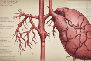
Portal Veins (Cont’d)
- Main portal vein: Bifurcates into the right and left portal veins at the porta hepatis, has a diameter of less than 13 mm
- Right Portal Vein: Larger than the left portal vein, runs centrally and horizontally in the right lobe, branches divide the right lobe into anterior and posterior segments
- Left Portal Vein: Smaller than the right portal vein, divides the left lobe into medial and lateral aspects
Distinguishing characteristics of hepatic and portal veins
- Portal Veins: Blood flow into the liver (hepatopetal), more echogenic walls
- Hepatic Veins: Blood flow out/away from the liver (hepatofugal), less echogenic than portal vein walls
Hepatic Artery (Cont’d)
- Low resistance arterial waveform, supplies 20-30% of the blood to the liver
- Bifurcates into the gastroduodenal artery, supraduodenal artery, right gastric artery, and hepatic artery proper
Ultrasound Appearance of the Liver
- Smooth, homogeneous echotexture
- Slightly hyperechoic or isoechoic to the kidney
- Isoechoic to the spleen
Liver Physiology
- Metabolism:
- Carbohydrates: Converts dietary sugar into glucose, stores excess sugar as glycogen, converts stored glycogen into glucose when dietary sugar is deficient
- Fats: Converts dietary fat into lipoproteins, which are stored or used by other organs
- Proteins: Produces albumin (regulates osmotic pressure), produces blood coagulation proteins (fibrinogen and prothrombin)
Digestion
- Nutrients from food absorbed by the small intestine enter the portal venous system
- Bile manufactured in the liver emulsifies fat in the intestines and removes waste products excreted by the liver
Detoxification
- Converts ammonium to nontoxic urea and excreted by the kidneys
- Removes bilirubin (a by-product of red blood cell breakdown) from the bloodstream, stores it in hepatocytes, converts it to bile, and releases it through the bile ducts
- Breaks down hormones, medications, and foreign chemicals
Storage
- Glycogen
- Amino acids
- Fats
- Vitamins A, D, and B complex
- Iron and copper
- Blood reservoir
Pertinent Lab Values
- AST and ALT: Elevated in cases of severe hepatocellular disease
Studying That Suits You
Use AI to generate personalized quizzes and flashcards to suit your learning preferences.