Podcast
Questions and Answers
What is the magnification of the ocular lenses?
What is the magnification of the ocular lenses?
- 10X (correct)
- 15X
- 20X
- 5X
What connects to the base and supports the microscope head?
What connects to the base and supports the microscope head?
Arm
What device controls the intensity of light coming from the bulb?
What device controls the intensity of light coming from the bulb?
Light Intensity Knob
What knob is used for precise focusing of the object?
What knob is used for precise focusing of the object?
What is used for rapid focusing on the specimen?
What is used for rapid focusing on the specimen?
What holds the slide to be observed?
What holds the slide to be observed?
What provides a steady light source for the microscope?
What provides a steady light source for the microscope?
Which components direct light from the lamp through the slide specimen?
Which components direct light from the lamp through the slide specimen?
What is adjusted to control the amount of light striking the specimen?
What is adjusted to control the amount of light striking the specimen?
What are the types of light microscopy?
What are the types of light microscopy?
What is the relationship between resolution and limit of resolution?
What is the relationship between resolution and limit of resolution?
What is the total magnification of the oil immersion lens?
What is the total magnification of the oil immersion lens?
What does the lens paper clean?
What does the lens paper clean?
What begins image formation in microscopy?
What begins image formation in microscopy?
What are parfocal lenses known for?
What are parfocal lenses known for?
Flashcards
Ocular Lens Magnification
Ocular Lens Magnification
The ocular lens magnifies the image by 10X.
Arm Function
Arm Function
The arm supports the microscope head and serves as a carrying handle.
Coarse Focus Adjustment
Coarse Focus Adjustment
Rapid focusing for low power lenses.
Fine Focus Adjustment
Fine Focus Adjustment
Signup and view all the flashcards
Mechanical Stage
Mechanical Stage
Signup and view all the flashcards
Microscope Resolution
Microscope Resolution
Signup and view all the flashcards
Limit of Resolution
Limit of Resolution
Signup and view all the flashcards
Numerical Aperture
Numerical Aperture
Signup and view all the flashcards
Total Magnification
Total Magnification
Signup and view all the flashcards
Scanning Objective Lens
Scanning Objective Lens
Signup and view all the flashcards
Low Power Objective Lens
Low Power Objective Lens
Signup and view all the flashcards
High Power Objective Lens
High Power Objective Lens
Signup and view all the flashcards
Oil Immersion Objective
Oil Immersion Objective
Signup and view all the flashcards
Depth of Field
Depth of Field
Signup and view all the flashcards
Working Distance
Working Distance
Signup and view all the flashcards
Study Notes
Light Microscope Components and Their Functions
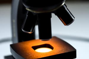
- Ocular Lenses: Magnification of 10X; equipped with a pointer for identifying specimen features.
- Arm: Supports the microscope head; connects to the base and serves as a carrying handle.
- Light Intensity Knob: Controls light intensity and power to the bulb; serves as an on/off switch.
- Fine Focus Adjustment Knob: Allows for precise focusing, particularly with high magnification lenses; aids in assessing specimen depth.
- Coarse Focus Adjustment Knob: Provides rapid focusing for scanning and low power lenses; used only to get the specimen roughly in focus.
- Mechanical Stage Adjustment Knobs: Two knobs that enable slide motion; one side-to-side, the other forward and backward, allowing for precise positioning of the slide.
- Base: Provides stability and support for the entire microscope.
- Lamp: Supplies steady illumination for observing specimens.
- Condenser Lens: Directs light from the lamp through the specimen, enhancing clarity.
- Iris Diaphragm: Adjusts light entering the specimen for better visibility; located on the condenser.
- Stage: Platform that holds slides; has an aperture for light to illuminate specimens.
- Stage Clips: Securely fasten slides onto the stage.
- Objective Lenses: Produce real images from specimens; available at magnifications of 4X, 10X, 40X, and 100X.
- Revolving Nosepiece: Rotatable turret holding objective lenses; facilitates easy switching between lenses.
Microscopy Concepts
- Resolution: Defines image clarity; improves with better optics.
- Limit of Resolution: Minimum distance at which two points are distinguishable.
- Numerical Aperture: Quantifies a lens's ability to gather light from the specimen for image formation.
- Bright-field Microscopy: Displays a shadowy specimen against a bright background; often requires stains for contrast.
- Dark-field Microscopy: Utilizes reflected light; results in a brightly lit specimen against a dark background.
- Phase Contrast Microscopy: Enhances contrast based on refractive index differences; produces shades of brightness.
- Fluorescence Microscopy: Employs fluorescent dyes; highlights structures under UV lighting.
- Parfocal Lenses: Maintain specimen focus when switching objectives.
Practical Information
- Depth of Field: Refers to the thickness of specimen in focus; decreases with increased magnification.
- Working Distance: Distance from the lens to the specimen; diminishes as magnification rises.
- Total Magnification: Calculated by multiplying ocular magnification (10X) with the objective magnification.
- Types of Objective Lenses:
- Scanning Lens: 4X magnification, total 40X
- Low Power Lens: 10X magnification, total 100X
- High Power Lens: 40X magnification, total 400X
- Oil Immersion Lens: 100X magnification, total 1000X
- Light Microscopy with Stains: Crucial for identifying microbes in clinical samples.
- Immersion Oil Benefits: Reduces light loss, increases numerical aperture, improves resolution.
- Lens Paper Use: Essential for cleaning lenses smoothly to avoid damage.
Relationships
- Resolution vs. Limit of Resolution: As the limit of resolution decreases, resolution improves.
- Image Formation Process: Initiates with light from an internal or external source.
Studying That Suits You
Use AI to generate personalized quizzes and flashcards to suit your learning preferences.
Description
Explore the essential components of a light microscope through these detailed flashcards. Each card defines key parts such as ocular lenses and the arm, explaining their functions in the magnification process. Perfect for students looking to enhance their understanding of microscopy.


