Podcast
Questions and Answers
The internal elastic lamina is composed of ______, with holes allowing better diffusion of substances from blood deeper into the wall.
The internal elastic lamina is composed of ______, with holes allowing better diffusion of substances from blood deeper into the wall.
elastin
In arteries, the media may have a thin external ______, separating it from the outermost tunic.
In arteries, the media may have a thin external ______, separating it from the outermost tunic.
elastic lamina
Interposed among the muscle fibers are variable amounts of elastic fibers and ______, all of which are produced by the smooth muscle cells.
Interposed among the muscle fibers are variable amounts of elastic fibers and ______, all of which are produced by the smooth muscle cells.
elastic lamellae
The tunica media consists chiefly of concentric layers of helically arranged smooth muscle cells and variable amounts of elastic fibers and ______.
The tunica media consists chiefly of concentric layers of helically arranged smooth muscle cells and variable amounts of elastic fibers and ______.
The adventitia consists principally of type I collagen and ______.
The adventitia consists principally of type I collagen and ______.
The media may have a thin ______, separating it from the outermost tunic.
The media may have a thin ______, separating it from the outermost tunic.
The media may contain up to 40 layers of large smooth muscle cells interspersed with a variable number of ______ (depending on the size of the vessel).
The media may contain up to 40 layers of large smooth muscle cells interspersed with a variable number of ______ (depending on the size of the vessel).
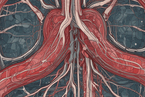
An external elastic lamina, the last component of the media, is present only in the larger muscular arteries. The adventitia consists of connective tissue. Lymphatic capillaries, vasa vasorum, and nerves are also found in the adventitia, and these structures may penetrate to the outer part of the media. Smooth muscle cell nuclei Media Endothelium Endothelium Smooth muscle cell nuclei Intima Internal ______ Internal elastic lamina Externa E A A Fuscin (Gallego's method) A A A - Muscular Arteries; E – Elastic artery. A V L L V A - Artery, V – Vein, L – Adipocyte, C – Capilary. C Arterioles Muscular arteries branch repeatedly into smaller and smaller arteries, until reaching a size with three or four medial layers of smooth muscle.
An external elastic lamina, the last component of the media, is present only in the larger muscular arteries. The adventitia consists of connective tissue. Lymphatic capillaries, vasa vasorum, and nerves are also found in the adventitia, and these structures may penetrate to the outer part of the media. Smooth muscle cell nuclei Media Endothelium Endothelium Smooth muscle cell nuclei Intima Internal ______ Internal elastic lamina Externa E A A Fuscin (Gallego's method) A A A - Muscular Arteries; E – Elastic artery. A V L L V A - Artery, V – Vein, L – Adipocyte, C – Capilary. C Arterioles Muscular arteries branch repeatedly into smaller and smaller arteries, until reaching a size with three or four medial layers of smooth muscle.
The smallest arteries branch as arterioles (0.1 mm), which have only one or two smooth muscle layers; these indicate the beginning of an organ’s microvasculature where exchanges between blood and tissue fluid occur. The subendothelial layer is very thin, ______ are absent, and the media consists of the circularly arranged smooth muscle cells. In both small arteries and arterioles, the adventitia is very thin and inconspicuous. Arterioles almost always branch to form anastomosing networks or beds of capillaries that surround the parenchymal cells of the organ.
The smallest arteries branch as arterioles (0.1 mm), which have only one or two smooth muscle layers; these indicate the beginning of an organ’s microvasculature where exchanges between blood and tissue fluid occur. The subendothelial layer is very thin, ______ are absent, and the media consists of the circularly arranged smooth muscle cells. In both small arteries and arterioles, the adventitia is very thin and inconspicuous. Arterioles almost always branch to form anastomosing networks or beds of capillaries that surround the parenchymal cells of the organ.
The internal elastic lamina is similar to the elastic laminae of the next layer, making it difficult to identify because of its _____.
The internal elastic lamina is similar to the elastic laminae of the next layer, making it difficult to identify because of its _____.
Elastic arteries, such as the aorta and the pulmonary artery, are also known as conducting arteries as their primary function is to carry blood to ____ arteries.
Elastic arteries, such as the aorta and the pulmonary artery, are also known as conducting arteries as their primary function is to carry blood to ____ arteries.
In elastic arteries, the thick media consists of elastic lamellae alternating with layers of smooth ____ fibers.
In elastic arteries, the thick media consists of elastic lamellae alternating with layers of smooth ____ fibers.
The adult aorta typically has around 50 elastic ____ in its structure.
The adult aorta typically has around 50 elastic ____ in its structure.
Carotid sinuses, which act as important baroreceptors, are slight dilations located where the internal carotid arteries branch from the common carotid arteries, and they monitor ____ blood pressure.
Carotid sinuses, which act as important baroreceptors, are slight dilations located where the internal carotid arteries branch from the common carotid arteries, and they monitor ____ blood pressure.
In the carotid sinuses, the media is thinner to allow greater distension when blood pressure rises, while the adventitia contains many sensory nerve endings from cranial nerve IX, the ____ nerve.
In the carotid sinuses, the media is thinner to allow greater distension when blood pressure rises, while the adventitia contains many sensory nerve endings from cranial nerve IX, the ____ nerve.
Flashcards are hidden until you start studying