Podcast
Questions and Answers
Which structure runs along the medial side of the knee?
Which structure runs along the medial side of the knee?
- Iliotibial band
- Biceps femoris tendon
- Fibular collateral ligament (LCL)
- Medial collateral ligament (MCL) (correct)
The vastus intermedius is superficial to the rectus femoris.
The vastus intermedius is superficial to the rectus femoris.
False (B)
Name the muscle that attaches posteriorly from the outside of the pubis and runs diagonally down the back side of the hamstring.
Name the muscle that attaches posteriorly from the outside of the pubis and runs diagonally down the back side of the hamstring.
Biceps femoris long head
In knee range of motion testing, the stabilizing surface should be against the ________ to prevent hip movement.
In knee range of motion testing, the stabilizing surface should be against the ________ to prevent hip movement.
Match each muscle to its primary action at the knee:
Match each muscle to its primary action at the knee:
During knee flexion range of motion testing, what is the average end range?
During knee flexion range of motion testing, what is the average end range?
During manual muscle testing, a grade of '3' indicates that the patient can move through full ROM against gravity with no added resistance.
During manual muscle testing, a grade of '3' indicates that the patient can move through full ROM against gravity with no added resistance.
What anatomical landmark is used as the fulcrum when measuring knee flexion and extension?
What anatomical landmark is used as the fulcrum when measuring knee flexion and extension?
The common hand placement used during manual muscle testing to resist knee extension involves placing the hand on the ________ end of the thigh.
The common hand placement used during manual muscle testing to resist knee extension involves placing the hand on the ________ end of the thigh.
Which of the following special tests helps determine collateral ligament injuries?
Which of the following special tests helps determine collateral ligament injuries?
Flashcards
Medial Collateral Ligament (MCL)
Medial Collateral Ligament (MCL)
Medial side of the knee, prevents valgus stress. Connects the medial side of the femur to the medial side of the tibia.
Lateral Collateral Ligament (LCL)
Lateral Collateral Ligament (LCL)
Lateral side of the knee, resists varus stress. Connects the lateral side of the femur to the fibular head.
Iliotibial (IT) Band
Iliotibial (IT) Band
A thick band of fibrous tissue that runs along the outside of the thigh, from the hip to the knee. It provides lateral knee stabilization.
Biceps femoris
Biceps femoris
Signup and view all the flashcards
Semitendinosus
Semitendinosus
Signup and view all the flashcards
Semimembranosus
Semimembranosus
Signup and view all the flashcards
Rectus femoris
Rectus femoris
Signup and view all the flashcards
Vastus lateralis
Vastus lateralis
Signup and view all the flashcards
Vastus medialis
Vastus medialis
Signup and view all the flashcards
Vastus intermedius
Vastus intermedius
Signup and view all the flashcards
Study Notes
- Notes on a knee joint evaluation
Knee Joint Evaluation: History
- Determine the duration of the injury or symptoms.
- Identify the mechanism of injury.
- Characterize the type of pain.
- Identify activities that increase or relieve pain.
- Inquire about any clicking, catching, snapping, popping, or grinding sensations.
Knee Joint Evaluation: Observation
- Scars should be noted.
- Assessment of the knee joint's alignment is important, including quadriceps angle.
- Genu varum (bowlegged), genu valgum (knock-kneed), and genu recurvatum (hyperextended knee) should also be noted.
Knee Joint Evaluation: Palpation of Anterior Structures
- Palpate the quadriceps and patellar tendons.
- Note the relationship of the patella to these structures.
Joint Line Structures
- The medial side possesses more surface area than the lateral.
- The posterior side of the patella has less surface area on the lateral side compared to the medial side.
- Ligaments, tendons, and other relevant structures should be palpated.
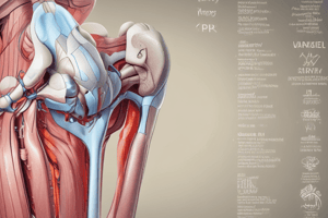
Medial Structures
- Medial Collateral Ligament (MCL) is located on the medial side of the knee.
Lateral Structures
- Lateral Collateral Ligament (LCL) is located on the lateral side of the knee.
- The Iliotibial Band runs from the ilium to the lateral side of the knee.
- Biceps femoris long head attaches posteriorly from the outside of the pubis, running diagonally down the back of the hamstring before connecting to the lateral side of the knee.
Posterior Structures
- Structures sit on top of the semimembranosus.
- The semitendinosus connects from the posterior side of the midpoint.
- The semimembranosus runs down the posterior part of the hamstring towards the medial side of the thigh, connecting to the medial side of the knee.
- Posterior to the quadriceps, the biceps femoris connects to the lateral side of the knee structure.
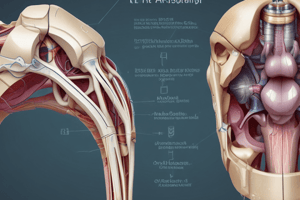
Quadriceps
- The quadriceps is composed of
- Vastus Lateralis: Lateral side of the quad
- Vastus Medialis: Medial side of the quad
- Vastus Intermedius: Sits deep to rectus femoris
- From deepest to superficial, the order of the quadriceps muscles is vastus intermedius, vastus medialis, rectus femoris, and vastus lateralis.
Manual Muscle Testing (MMT)
- Semitendinosus/Semitendinosus: Flexion and medial rotation of the knee is tested with the patient prone. Flex the knee between 50° and 70°, medially rotating the thigh and leg. Stabilize the thigh and resist at the proximal ankle.
- Biceps Femoris: Flexion and lateral rotation of the knee is tested in a prone position. Flex the knee between 50° and 70°, laterally rotating the thigh and leg. Stabilize the thigh and resist at the proximal ankle.
- Quadriceps Femoris: Extension of the knee is tested with the patient sitting with knees over the side of the table. Apply resistance above the ankle in the direction of flexion.
MMT Grading Scale
- 5/5: Full ROM against gravity with full manual resistance (Normal).
- 4/5: Full ROM against gravity with moderate resistance (Good).
- 3/5: Full ROM against gravity, but unable to go through full ROM when resistance is applied (Fair).
- 2/5: Full ROM with gravity eliminated (Poor).
- 1/5: Evidence of muscle contraction but no joint movement (Trace).
- 0/5: No evidence of muscle contraction (Zero).
Knee Flexion Measurement
- Start Position: Prone with hip in neutral, foot off the table, bolster under the thigh.
- Stabilization: Stabilize the femur to prevent hip movement.
- Fulcrum: Lateral epicondyle of the femur.
- Stationary Arm: Parallel to the lateral midline of the femur, towards the greater trochanter.
- Moveable Arm: Parallel to the lateral midline of the fibula, towards the lateral malleolus.
- End position: Maximum knee flexion.
- Range: 120-145°.
Knee Extension Measurement
- Start Position: Prone with bolster under the thigh.
- Stabilization: Stabilize the femur to prevent hip movement.
- Fulcrum: Lateral epicondyle of the femur.
- Stationary Arm: Parallel to the lateral midline of the femur, towards the greater trochanter.
- Moveable Arm: Parallel to the lateral midline of the fibula, towards the lateral malleolus.
- End position: Maximum knee extension.
- Range: 0-15°.
Studying That Suits You
Use AI to generate personalized quizzes and flashcards to suit your learning preferences.