Podcast
Questions and Answers
In the knee joint, what is the combined number of osseous structures and distinct joints?
In the knee joint, what is the combined number of osseous structures and distinct joints?
- 4
- 8
- 6 (correct)
- 10
Which of the following structures is NOT part of the four osseous structures of the knee joint?
Which of the following structures is NOT part of the four osseous structures of the knee joint?
- Proximal tibia
- Distal femur
- Patella
- Calcaneus (correct)
How do geometry and shape of the two condyles of the distal femur impact knee joint function?
How do geometry and shape of the two condyles of the distal femur impact knee joint function?
- They facilitate obligatory axial rotation. (correct)
- They minimize articular cartilage contact.
- They ensure equal force distribution.
- They prevent axial rotation.
What is the primary function of the intercondylar notch in the distal femur?
What is the primary function of the intercondylar notch in the distal femur?
How does the dimension of the medial femoral condyle compare to the lateral femoral condyle, and what is a characteristic of its shape?
How does the dimension of the medial femoral condyle compare to the lateral femoral condyle, and what is a characteristic of its shape?
What structural feature makes the tibia suited to act as a pivot point during axial rotation?
What structural feature makes the tibia suited to act as a pivot point during axial rotation?
Which statement best describes the superior view profile and central part of the lateral tibial condyle?
Which statement best describes the superior view profile and central part of the lateral tibial condyle?
In what way does the shape of the medial tibial condyle contribute to knee stability?
In what way does the shape of the medial tibial condyle contribute to knee stability?
What is the main functional role of the menisci within the knee joint?
What is the main functional role of the menisci within the knee joint?
What action illustrates the function of menisci of creating a congruous joint surface?
What action illustrates the function of menisci of creating a congruous joint surface?
Which of the following statements accurately describes the shape of the medial meniscus?
Which of the following statements accurately describes the shape of the medial meniscus?
What primary movement does the anterior cruciate ligament (ACL) prevent in the knee joint?
What primary movement does the anterior cruciate ligament (ACL) prevent in the knee joint?
The anterior cruciate ligament (ACL) attaches to which specific part of the lateral femoral condyle?
The anterior cruciate ligament (ACL) attaches to which specific part of the lateral femoral condyle?
How does the tension within the fiber bundles of the ACL change throughout the flexion range?
How does the tension within the fiber bundles of the ACL change throughout the flexion range?
How does the attachment location of the posterior cruciate ligament (PCL) differ from that of the ACL on the tibial plateau?
How does the attachment location of the posterior cruciate ligament (PCL) differ from that of the ACL on the tibial plateau?
What is the specific term for the PCL bundle that is taut during knee extension?
What is the specific term for the PCL bundle that is taut during knee extension?
Besides the ACL, what other structure provides primary resistance to anterior translation in the knee?
Besides the ACL, what other structure provides primary resistance to anterior translation in the knee?
What is the secondary role for support against anterior translation?
What is the secondary role for support against anterior translation?
The superficial medial ligament of the tibial collateral ligament is also referred to as which of the following?
The superficial medial ligament of the tibial collateral ligament is also referred to as which of the following?
The deep medial ligament of the tibial collateral ligament can be alternatively described as which of the following?
The deep medial ligament of the tibial collateral ligament can be alternatively described as which of the following?
What is the function of the posterior oblique ligament (POL) in relation to the medial collateral ligament (MCL)?
What is the function of the posterior oblique ligament (POL) in relation to the medial collateral ligament (MCL)?
When the knee flexes, how do the anterior and posterior bundles within the superficial ligament of the MCL respond?
When the knee flexes, how do the anterior and posterior bundles within the superficial ligament of the MCL respond?
What specific type of stress is the superficial ligament of the medial collateral ligament (MCL) particularly influential in resisting?
What specific type of stress is the superficial ligament of the medial collateral ligament (MCL) particularly influential in resisting?
What part of the fibular collateral ligament contributes to its function?
What part of the fibular collateral ligament contributes to its function?
What specific type of loading does the lateral collateral ligament resist?
What specific type of loading does the lateral collateral ligament resist?
What type of movement does the lateral collateral ligament resist?
What type of movement does the lateral collateral ligament resist?
Which statement best describes the role of the joint capsule in the knee?
Which statement best describes the role of the joint capsule in the knee?
In the context of knee joint biomechanics, what action is described as translation along the z-axis?
In the context of knee joint biomechanics, what action is described as translation along the z-axis?
An individual is standing in anatomical position. What will the effect of internal or external rotation of their knee have?
An individual is standing in anatomical position. What will the effect of internal or external rotation of their knee have?
When assessing knee joint movement, what action is referred to as 'anteroposterior draw'?
When assessing knee joint movement, what action is referred to as 'anteroposterior draw'?
What type of movement involves rotation around the y-axis?
What type of movement involves rotation around the y-axis?
In biomechanics, what specific movement is described as 'flexion or extension' of the knee joint?
In biomechanics, what specific movement is described as 'flexion or extension' of the knee joint?
What geometric shape is created by the loci of the instantaneous center of rotation (ICOR) during knee flexion?
What geometric shape is created by the loci of the instantaneous center of rotation (ICOR) during knee flexion?
Which term accurately describes the hip joint's structural classification?
Which term accurately describes the hip joint's structural classification?
What is the combined function of the acetabular labrum and the transverse acetabular ligament?
What is the combined function of the acetabular labrum and the transverse acetabular ligament?
What effect on hip joint mechanics is most directly associated with a neck-shaft angle greater than 125 degrees?
What effect on hip joint mechanics is most directly associated with a neck-shaft angle greater than 125 degrees?
What is the name of the condition in which the neck-shaft angle is observed to be less than 125 degrees?
What is the name of the condition in which the neck-shaft angle is observed to be less than 125 degrees?
When the femoral neck leans forward, which condition is observed?
When the femoral neck leans forward, which condition is observed?
What effect would a retroverted hip have?
What effect would a retroverted hip have?
What specific role does the ligament of the head of the femur play in hip joint function?
What specific role does the ligament of the head of the femur play in hip joint function?
What ligament is important for the hip during standing?
What ligament is important for the hip during standing?
Which statement accurately describes the effect on hip joint range of motion performed on the pubofemoral ligament?
Which statement accurately describes the effect on hip joint range of motion performed on the pubofemoral ligament?
Damage to what major part may be caused by posterior dislocation of the hip joint?
Damage to what major part may be caused by posterior dislocation of the hip joint?
What is the normal range of abduction in the hip joint?
What is the normal range of abduction in the hip joint?
The knee joint consists of 4 osseous structures: distal femur, proximal tibia, fibula, and radius.
The knee joint consists of 4 osseous structures: distal femur, proximal tibia, fibula, and radius.
The knee joint comprises of three distinct joints: patellofemoral, tibiofemoral, and hamstring.
The knee joint comprises of three distinct joints: patellofemoral, tibiofemoral, and hamstring.
The lateral femoral condyle is aligned with the shaft of the femur and acts as a main transmitter of compressive forces.
The lateral femoral condyle is aligned with the shaft of the femur and acts as a main transmitter of compressive forces.
The two condyles of the distal femur have the same geometry and shape, ensuring equal weight distribution at the knee joint.
The two condyles of the distal femur have the same geometry and shape, ensuring equal weight distribution at the knee joint.
The distal femur is convex in both coronal and transverse planes.
The distal femur is convex in both coronal and transverse planes.
The intercondylar notch separates the tibial plateau and provides space for cruciate ligaments during flexion and extension.
The intercondylar notch separates the tibial plateau and provides space for cruciate ligaments during flexion and extension.
The medial femoral condyle has a smaller anterior/posterior dimension compared to the lateral condyle, and it is less symmetrical.
The medial femoral condyle has a smaller anterior/posterior dimension compared to the lateral condyle, and it is less symmetrical.
The fibula is the stronger of the two bones in the lower leg, bearing the majority of force transmission.
The fibula is the stronger of the two bones in the lower leg, bearing the majority of force transmission.
The lateral and medial condyles of the proximal tibia are separated by the intercondylar eminence, serving as a pivot during axial rotation.
The lateral and medial condyles of the proximal tibia are separated by the intercondylar eminence, serving as a pivot during axial rotation.
The lateral tibial condyle is circular in profile when viewed superiorly, and articulated with the radial head.
The lateral tibial condyle is circular in profile when viewed superiorly, and articulated with the radial head.
The medial tibial condyle is more oval in outline and smaller than the lateral tibial condyle.
The medial tibial condyle is more oval in outline and smaller than the lateral tibial condyle.
The medial tibial condyle is concave in all planes, contributing to knee stability.
The medial tibial condyle is concave in all planes, contributing to knee stability.
Control of knee joint motion is exclusively regulated by the shapes of the articulating bones.
Control of knee joint motion is exclusively regulated by the shapes of the articulating bones.
Menisci contribute to joint congruity by creating a shallower surface on the tibial plateau.
Menisci contribute to joint congruity by creating a shallower surface on the tibial plateau.
The lateral meniscus is semicircular with a wider posterior horn, making it less prone to injury than the medial meniscus.
The lateral meniscus is semicircular with a wider posterior horn, making it less prone to injury than the medial meniscus.
The anterior cruciate ligament (ACL) restricts posterior translation of the tibia on the femur.
The anterior cruciate ligament (ACL) restricts posterior translation of the tibia on the femur.
The posterior cruciate ligament (PCL) restricts external rotation of the tibia on the femur.
The posterior cruciate ligament (PCL) restricts external rotation of the tibia on the femur.
The posterior cruciate ligament (PCL) attaches on the tibial plateau lateral to the medio-lateral bisector.
The posterior cruciate ligament (PCL) attaches on the tibial plateau lateral to the medio-lateral bisector.
The ACL is composed of a single, thick strand of collagen, providing consistent tension throughout the knee's range of motion.
The ACL is composed of a single, thick strand of collagen, providing consistent tension throughout the knee's range of motion.
The anterior cruciate ligament is attached to the posterior aspect of the medial surface of the lateral femoral condyle.
The anterior cruciate ligament is attached to the posterior aspect of the medial surface of the lateral femoral condyle.
The posteromedial bundle of the PCL is taut in flexion, while the anterolateral bundle is taut in extension.
The posteromedial bundle of the PCL is taut in flexion, while the anterolateral bundle is taut in extension.
The medial collateral ligament primarily resists varus stress, protecting the knee from excessive abduction.
The medial collateral ligament primarily resists varus stress, protecting the knee from excessive abduction.
The medial collateral ligament (MCL) has superficial, intermediate, and deep functional units that offer stability to the knee.
The medial collateral ligament (MCL) has superficial, intermediate, and deep functional units that offer stability to the knee.
The posterior oblique ligament is an extension of the superficial MCL and joins the semimembranosus tendon to the anteromedial aspect of the joint capsule.
The posterior oblique ligament is an extension of the superficial MCL and joins the semimembranosus tendon to the anteromedial aspect of the joint capsule.
When the knee flexes, the anterior bundle of the superficial medial collateral ligament tightens, while the posterior bundle slackens.
When the knee flexes, the anterior bundle of the superficial medial collateral ligament tightens, while the posterior bundle slackens.
The deep fibers of the medial collateral ligament offer little contribution to the overall stability provided by the superficial ligament.
The deep fibers of the medial collateral ligament offer little contribution to the overall stability provided by the superficial ligament.
The fibular collateral ligament attaches proximally to the medial epicondyle of the femur.
The fibular collateral ligament attaches proximally to the medial epicondyle of the femur.
The fibular collateral ligament directly attaches to the meniscus and adjacent knee joint capsule.
The fibular collateral ligament directly attaches to the meniscus and adjacent knee joint capsule.
The knee joint capsule provides dynamic stability, actively adjusting tension to maintain joint integrity.
The knee joint capsule provides dynamic stability, actively adjusting tension to maintain joint integrity.
Translation along the z-axis of the knee joint refers to medial-lateral movement of the tibia relative to the femur.
Translation along the z-axis of the knee joint refers to medial-lateral movement of the tibia relative to the femur.
Rotation around the y-axis of the knee joint corresponds to flexion and extension movements.
Rotation around the y-axis of the knee joint corresponds to flexion and extension movements.
Instantaneous center of rotation (ICOR) describes the precise point around which the knee rotates during all movements, remaining constant during activity.
Instantaneous center of rotation (ICOR) describes the precise point around which the knee rotates during all movements, remaining constant during activity.
The hip joint is a hinge joint, allowing primarily flexion and extension movements.
The hip joint is a hinge joint, allowing primarily flexion and extension movements.
A neck-shaft angle greater than 125° in the frontal plane is termed Coxa vara.
A neck-shaft angle greater than 125° in the frontal plane is termed Coxa vara.
In cases of femoral retroversion, the femur rotates internally to keep the femoral head within the acetabulum.
In cases of femoral retroversion, the femur rotates internally to keep the femoral head within the acetabulum.
The ligament of the head of the femur contributes to stability when the hip is abducted.
The ligament of the head of the femur contributes to stability when the hip is abducted.
The iliofemoral ligament, known as the 'Y' ligament of Bigelow, is a major stabilizer of the hip, especially during adduction.
The iliofemoral ligament, known as the 'Y' ligament of Bigelow, is a major stabilizer of the hip, especially during adduction.
The pubofemoral ligament resists abduction and external rotation of the hip.
The pubofemoral ligament resists abduction and external rotation of the hip.
The distal femur is convex in the sagittal plane but concave in the frontal plane.
The distal femur is convex in the sagittal plane but concave in the frontal plane.
The medial meniscus is circular, while the lateral meniscus has a semicircular shape, with a wider anterior horn.
The medial meniscus is circular, while the lateral meniscus has a semicircular shape, with a wider anterior horn.
The anterior cruciate ligament (ACL) attaches on the tibial plateau anterior to the medial intercondylar eminence and on the posterior aspect of the lateral femoral condyle.
The anterior cruciate ligament (ACL) attaches on the tibial plateau anterior to the medial intercondylar eminence and on the posterior aspect of the lateral femoral condyle.
The iliofemoral ligament, located at the hip, exhibits a 'V' shape and its primary role is to resist hyperabduction.
The iliofemoral ligament, located at the hip, exhibits a 'V' shape and its primary role is to resist hyperabduction.
In the hip joint, a neck-shaft angle in the frontal plane measuring 130° would be classified as 'Coxa vara'.
In the hip joint, a neck-shaft angle in the frontal plane measuring 130° would be classified as 'Coxa vara'.
Flashcards
What is Osteology?
What is Osteology?
The scientific study of bones.
What is the distal femur?
What is the distal femur?
The distal end of the thigh bone, part of the knee joint.
What is the proximal tibia?
What is the proximal tibia?
The upper part of the shin bone, forming the knee joint.
What is the Fibula?
What is the Fibula?
Signup and view all the flashcards
What is the patella?
What is the patella?
Signup and view all the flashcards
What is the patellofemoral joint?
What is the patellofemoral joint?
Signup and view all the flashcards
What is the tibiofemoral joint?
What is the tibiofemoral joint?
Signup and view all the flashcards
What is the trochlea (patellofemoral groove)?
What is the trochlea (patellofemoral groove)?
Signup and view all the flashcards
What is the Anterior Cruciate Ligament (ACL)?
What is the Anterior Cruciate Ligament (ACL)?
Signup and view all the flashcards
What is the Posterior Cruciate Ligament (PCL)?
What is the Posterior Cruciate Ligament (PCL)?
Signup and view all the flashcards
What is the Intercondylar notch?
What is the Intercondylar notch?
Signup and view all the flashcards
What is the Medial Femoral Condyle?
What is the Medial Femoral Condyle?
Signup and view all the flashcards
What is the Lateral Femoral Condyle?
What is the Lateral Femoral Condyle?
Signup and view all the flashcards
What is the Tibia?
What is the Tibia?
Signup and view all the flashcards
What are the Lateral and Medial Condyles?
What are the Lateral and Medial Condyles?
Signup and view all the flashcards
What is the Lateral Tibial Condyle?
What is the Lateral Tibial Condyle?
Signup and view all the flashcards
What is the Medial Tibial Condyle?
What is the Medial Tibial Condyle?
Signup and view all the flashcards
What are the function of the Menisci?
What are the function of the Menisci?
Signup and view all the flashcards
What is the Medial Meniscus?
What is the Medial Meniscus?
Signup and view all the flashcards
What is the Lateral Meniscus?
What is the Lateral Meniscus?
Signup and view all the flashcards
What controls knee joint motion?
What controls knee joint motion?
Signup and view all the flashcards
What is the function og the ACL?
What is the function og the ACL?
Signup and view all the flashcards
What is the function of the PCL?
What is the function of the PCL?
Signup and view all the flashcards
What is the ACL made from?
What is the ACL made from?
Signup and view all the flashcards
What makes up the PCL?
What makes up the PCL?
Signup and view all the flashcards
What is a primary function if the ACL and PCL?
What is a primary function if the ACL and PCL?
Signup and view all the flashcards
What is the primary function of the Medial Collateral Ligament (MCL)?
What is the primary function of the Medial Collateral Ligament (MCL)?
Signup and view all the flashcards
What are the three units of MCL?
What are the three units of MCL?
Signup and view all the flashcards
What is the function of the posterior oblique fibers?
What is the function of the posterior oblique fibers?
Signup and view all the flashcards
What is the Fibular (lateral) collateral ligament Function?
What is the Fibular (lateral) collateral ligament Function?
Signup and view all the flashcards
What is the function of lateral collateral ligament?
What is the function of lateral collateral ligament?
Signup and view all the flashcards
What is the Knee Joint Capsule?
What is the Knee Joint Capsule?
Signup and view all the flashcards
What rotations happen at the Knee?
What rotations happen at the Knee?
Signup and view all the flashcards
What is the Instantaneous center of rotation (ICOR)?
What is the Instantaneous center of rotation (ICOR)?
Signup and view all the flashcards
What kind of joint is the Hip joint?
What kind of joint is the Hip joint?
Signup and view all the flashcards
What the hip formed by?
What the hip formed by?
Signup and view all the flashcards
What makes the hip stable?
What makes the hip stable?
Signup and view all the flashcards
What is average hip anteversion?
What is average hip anteversion?
Signup and view all the flashcards
What happens in increases anteversion?
What happens in increases anteversion?
Signup and view all the flashcards
What happens in increases retroversion?
What happens in increases retroversion?
Signup and view all the flashcards
What is the Ligament of the head of femur function?
What is the Ligament of the head of femur function?
Signup and view all the flashcards
What is Arthrology?
What is Arthrology?
Signup and view all the flashcards
Distal Femur Condyles?
Distal Femur Condyles?
Signup and view all the flashcards
Forces to Tibial Plateau
Forces to Tibial Plateau
Signup and view all the flashcards
Tibia force transmission
Tibia force transmission
Signup and view all the flashcards
Lateral Tibial Condyle
Lateral Tibial Condyle
Signup and view all the flashcards
Medial Tibial Condyle
Medial Tibial Condyle
Signup and view all the flashcards
Function of Tibial Condyles
Function of Tibial Condyles
Signup and view all the flashcards
What do posterior oblique fibers support?
What do posterior oblique fibers support?
Signup and view all the flashcards
Superficial Ligament Stability
Superficial Ligament Stability
Signup and view all the flashcards
Knee Joint Motion
Knee Joint Motion
Signup and view all the flashcards
Transverse Acetabular Ligament
Transverse Acetabular Ligament
Signup and view all the flashcards
Iliofemoral Ligament
Iliofemoral Ligament
Signup and view all the flashcards
Pubofemoral Ligament
Pubofemoral Ligament
Signup and view all the flashcards
Ischiofemoral Ligament
Ischiofemoral Ligament
Signup and view all the flashcards
Hip Flexion ROM
Hip Flexion ROM
Signup and view all the flashcards
Study Notes
- Knee & Hip Joints are being discussed
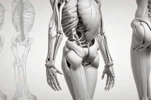
Osteology of the Knee
- The knee is made up of 4 osseous structures: distal femur, proximal tibia, fibula, and patella
- The knee contains 2 distinct joints patellofemoral and tibiofemoral
- Osteology is the Scientific study of bones
Frontal View
- The frontal view of the right knee has the patella reflected to show underlying structures such as:
- Femur (thighbone)
- Patella (underside)
- Trochlea (patellofemoral groove)
- Anterior Cruciate Ligament
- Posterior Cruciate Ligament
- Lateral Femoral Condyle
- Medial Collateral Ligament
- Lateral Meniscus
- Medial Meniscus
- Lateral Collateral Ligament
- Tibial Plateau
- Tibia (shinbone)
- Fibula
- Tibial Tuberosity
Lateral View
- The lateral view of the Patellofemoral Joint features the:
- Femur
- Quadriceps tendon
- Patello-femoral joint
- Patella
- Articular cartilage
- Patella tendon
- Medial Meniscus
- Lateral Meniscus
- Tibia
- Fibula
Distal Femur
- Two condyles are different in geometry and shape, which results in obligatory axial rotation at the knee joint
- The distal femur transmits forces to the tibial plateau
- The distal femur is partially covered by articular surfaces which contain articular cartilages
Convex Distal Femur
- Distal femur is convex in both the frontal and sagittal planes
- It is separated by the intercondylar notch space for cruciate ligaments (ACL, PCL) during flexion and extension
Medial and Lateral Femoral Condyles
- Medial femoral condyle has a Larger A/P dimension than the lateral condyle and is more symmetrical
- Lateral femoral condyle is aligned with the shaft of the femur, and it is the primary transmitter of compressive forces
Lower Leg
- The lower leg contains two osseous structures: the tibia and fibula.
- The tibia is stronger for force transmission
- Lateral and medial condyles are separated by the intercondylar eminences, which serves as a pivot point during axial rotation by locating into the intercondylar notch of the femur
Lateral Tibial Condyle
- Lateral tibial condyle with a facet on posteriolateral aspect for articulation with the fibular head
- Circular in profile when viewed superiorly, slightly hollowed in its central part
Medial Tibial Condyle
- Medial tibial condyle is more oval in outline and larger than lateral tibial condyle
- Concave in all planes for stability
Tibial Condyles
- Lateral and medial tibial condyle are stable platforms upon which the femoral condyles can transmit compressive forces
Knee Joint Motion
- Control of knee joint motion is shared by capsular structures, intra and extra-articular ligaments, joint contours, and muscles & tendons crossing the joint
- Arthrology is the Scientific study of joints
Menisci Structure
- The Menisci is an Intermediary structure to provide shock absorption, lubrication and stabilization
- The Menisci makes the joint surface congruous by providing a deeper fossa (hollow or depression) on the tibial surface
- The Menisci resists translation of femur on tibia by dispersing compressive forces in a radial manner across their structure.
Medial and Lateral Meniscus
- Medial meniscus has a Semicircular shape with wider posterior horn
- Lateral meniscus has a Circular shape
Cruciate Ligaments
- The ACL prevents anterior translation of the tibia on femur and restricts external rotation of the tibia on femur
- The PCL restricts posterior translation of the tibia on the femur and resists hyperextension
Anterior Cruciate Ligament (ACL)
- Attachment is on the tibial plateau anterior to the medial intercondylar eminence and on the posterior aspect of the medial surface of lateral femoral condyle
ACL Information
- ACL is a bundle of fibers rather than a single structure
- Part of the bundle of fibers remain taut (stretched) throughout the flexion range.
Posterior Cruciate Ligament (PCL)
- Attached to tibial plateau in a broad area on the posterior aspect, usually lateral to the M-L bisector of the tibial plateau and is posterior to the ACL attachment on the femur
- Crosses with the ACL
- 2 major PCL bundles -Posteromedial bundle taut in extension -Anterolateral bundle taut in flexion
Primary Stabilizers of the Knee
- For Anterior translation, the primary stabilizers are the: ACL and PCL, and the secondary stabilizers are the: Collateral ligaments and joint capsule
- For the Medial aspect, the primary stabilizer is the: Medial collateral ligament, and the secondary stabilizer is the: ACL
Tibial (Medial) Collateral Ligament
- Robust, broad, and flat structure
- Consists of 3 function units -Superficial medial ligament, referred to as the long fibers -Deep medial ligament, alternatively described as the medial capsular ligament, and is anatomically completely separate from the superficial portion -Posterior oblique ligament
MCL - Medial Collateral Ligament
- Superficial Ligament is below adductor tubercle and has the medial surface of the shaft of tibia
- Deep Fibers have the Medial femoral condyle and Medial tibial plateau
Posterior Oblique Ligament
- Is an Extension of the superficial MCL
- Joins the semimembranosus tendon to the posteromedial aspect of the joint capsule
- Reinforces the area
Functions of the MCL
- All 3 units that constitute of the MCL work in conjunction as the primary medial stabilizer of the knee
- The superficial ligament exerts most influence on stability, against valgus and rotatory stress
- When the knee flexes - The anterior bundle tightens, and the posterior bundle slacken
- The deep fibers of the medial collateral ligament reinforce the action of the superficial ligament
- The posterior oblique fibers tends to support the posterolateral aspect of the knee and the posterior joint capsule
Fibular (Lateral) Collateral Ligament
- The Main ligament supports the capsular structures
- It is attached proximally to the lateral epicondyle of the femur and inserted on the posterior aspect of the fibular head
- No attachments to the menisci/capsular structures
Lateral Collateral Ligament
- Functions on the thin cord-like structure and remains taut during flexion range
- Resists tensile loading and varus angulation
Joint Capsule
- Knee joint is the largest synovial joint in the body
- The joint capsule provides static joint stability
- Muscular and tendinous insertions into the capsule provide further stability
- Synovial joint: the ends of the bones are covered by a joint capsule filled with fluid for movement
Rotations and Translations
- Translation along z-axis, distract/compress at joint surface
- Rotation around z-axis, In / Ext rotation
- Translation along y-axis, Anteroposterior draw
- Rotation around y-axis, Valgus / Varus
- Translation along x-axis, Medial-lateral translation
- Rotation around x-axis, Flexion / Extension
ICOR
- The loci of ICOR can be drawn as a reverse "C" shape when the flexion of the tibia was plotted relative to a femur tibia.
- ICOR stands for Instantaneous center of rotation
Hip Joint
- The hip joint is a ball and socket joint
Hip Osteology
- Ball (femoral head) and socket (acetabulum) in structure
- Acetabular labrum (ring of cartilage) & transverse acetabular ligament increase stability by deepening articulation
Neck-Shaft Angle
- Neck-shaft angle in frontal plane ~125° -> 125 °Coxa valga -< 125 °Coxa vara
Anteversion & Retroversion
- Anteversion refers to the angle between the femoral neck axis and knee axis in the transverse plane (Adult ~12 deg)
- Increases in anteversion when femur rotate internally to keep femoral head in acetabulum
- Increases in retroversion when femur rotates externally to keep femoral head in acetabulum
Ligamentous Structure
- Ligament of the head of femur is responsible for blood supply to femoral head
- Provides stability when hip is adducted
Spanning Ligaments
- Transverse acetabular ligament spans the acetabular notch and together with acetabular labrum deepens and increases stability of the articulation
Stabilizing hip in extension
- Iliofemoral ligament is the Strongest hip ligament, inverted Y shape, where the base attaches to ASIS Major element to stabilize hip in extension when a person is standing in erect position, where the Posterior bands also resists internal rotation
Pubofemoral Ligament
- Runs horizontally from the pubis to the anterior band of the iliofemoral ligament.
- It resists abduction and external rotation.
Ischiofemoral Ligament
- Crosses over the posterior-inferior aspect of the joint capsule and may be ruptured by posterior dislocation of the hip joint
Range of Motion
- Flexion: 100
- Hyperextion: 30
- Abduction: 40
- Adduction: 20
- Internal rotation: 40
- External rotation: 50
Studying That Suits You
Use AI to generate personalized quizzes and flashcards to suit your learning preferences.