Podcast
Questions and Answers
At what stage of embryonic development do the mesonephroi appear?
At what stage of embryonic development do the mesonephroi appear?
- During the 3rd week of gestation
- During the 2nd week of gestation
- Late in the 4th week of gestation (correct)
- Early in the 5th week of gestation
In which anatomical region of the embryo are the mesonephroi located?
In which anatomical region of the embryo are the mesonephroi located?
- Cervical region
- Sacral region
- Lower thoracic region
- Upper lumbar region (correct)
What is the relationship between the mesonephroi and the pronephroi?
What is the relationship between the mesonephroi and the pronephroi?
- The mesonephroi appear before the pronephroi in development
- The mesonephroi form from the remnants of the pronephroi
- The mesonephroi replace the pronephroi entirely
- The mesonephroi develop caudal to the pronephroi (correct)
What characteristics define the mesonephroi?
What characteristics define the mesonephroi?
Which somite range is the mesonephroi associated with during their development?
Which somite range is the mesonephroi associated with during their development?
What could be a possible underlying cause of polyhydramnios in a pregnant woman during the 10th week of gestation?
What could be a possible underlying cause of polyhydramnios in a pregnant woman during the 10th week of gestation?
Which diagnostic method would most likely confirm the suspicion of organ absence in this case of polyhydramnios?
Which diagnostic method would most likely confirm the suspicion of organ absence in this case of polyhydramnios?
In evaluating polyhydramnios, what is the significance of identifying any absent fetal organs during ultrasound?
In evaluating polyhydramnios, what is the significance of identifying any absent fetal organs during ultrasound?
What complication may arise if polyhydramnios due to organ absence is not diagnosed during early gestation?
What complication may arise if polyhydramnios due to organ absence is not diagnosed during early gestation?
What role does maternal fluid management play in cases of polyhydramnios?
What role does maternal fluid management play in cases of polyhydramnios?
Flashcards
Polyhydramnios
Polyhydramnios
An excessive amount of amniotic fluid surrounding the fetus in the womb.
Organ Absence
Organ Absence
The condition of being born without a specific organ.
Ultrasound
Ultrasound
A diagnostic test that uses sound waves to create images of the fetus and surrounding structures.
Fetal Imaging
Fetal Imaging
Signup and view all the flashcards
Amniocentesis
Amniocentesis
Signup and view all the flashcards
Mesonephroi
Mesonephroi
Signup and view all the flashcards
Location of Mesonephroi
Location of Mesonephroi
Signup and view all the flashcards
Somite Association of Mesonephroi
Somite Association of Mesonephroi
Signup and view all the flashcards
Specific Location of Mesonephroi
Specific Location of Mesonephroi
Signup and view all the flashcards
Temporary Nature of Mesonephroi
Temporary Nature of Mesonephroi
Signup and view all the flashcards
Study Notes
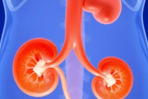
Kidney Development
- The urogenital system originates from the intraembryonic mesoderm.
- After embryonic folding, the intermediate mesoderm moves ventrally, forming the urogenital ridge on each side of the dorsal aorta.
- The part of the urogenital ridge that develops into the urinary system is called the nephrogenic cord.
Intended Learning Outcomes (ILOs)
- Identify the embryological origin of the urinary and genital systems.
- Describe the stages of kidney development.
- Memorize congenital anomalies of the kidney.
Polycystic Kidney Disease
- An autosomal disorder identified by ultrasound at birth.
- Characterized by multiple small cysts in both kidneys.
- Often leads to renal insufficiency.
- Death may occur shortly after birth in severe cases.
Multi-Cystic Dysplastic Kidney Disease (MDK)
- Results from abnormal renal system development.
- Usually has a favorable outcome.
- Death in some cases can happen shortly after birth.
Horseshoe Kidney
- Results from fusion of the caudal ends of both kidneys.
- An interconnecting bridge often forms behind the inferior mesenteric artery.
- Kidneys are typically positioned in the low lumbar region.
Ectopic Pelvic Kidney
- The kidney does not fully ascend during development.
- It's frequently positioned at the pelvic brim.
Renal Agenesis
- Absence of one or both kidneys during development.
- Unilateral agenesis (one kidney missing) often causes few symptoms.
- Bilateral agenesis (both missing) is related to oligohydramnios. (low amniotic fluid)
Kidney Development Stages
- Pronephros: Nonfunctional, transient structures appearing in the fourth week. Appears as cell clusters and tubular structures in the neck region opposing the 7th to 14th somites. The pronephric ducts eventually degenerate.
- Mesonephros: Well-developed, temporary kidneys appearing later in the fourth week, caudal to the pronephros. It appears at a lower level, opposite to the 14th to 28th thoracic and upper lumbar somites. Functions as transient kidneys for about four weeks. Consists of glomeruli and mesonephric tubules that open into the mesonephric ducts. Degenerates during the first trimester.
- Metanephros: Primordia of the permanent kidneys. Appears during the fifth week and begins functioning approximately four weeks later. Develops from two sources: the ureteric bud and the metanephric mass, both of which are mesodermal in origin.
Metanephric Diverticulum Development
- An outgrowth from the mesonephric duct, initiating the ureter and its associated structures.
- The elongating stalk forms the ureter, and the expanded cranial end develops into the renal pelvis.
- Straight collecting tubules repeat branching, forming collecting tubules, major calices, minor calices.
Metanephric Tubules (Nephron Primordia)
- A mass of intermediate mesoderm originating from the caudal nephrogenic cord.
- Metanephric vesicles lengthen into metanephric tubules.
- Tubule proximal ends invaginate to form glomeruli.
- Glomerular filtration begins around the ninth fetal week and increases in rate until the 32nd week.
- Number of nephrons increase after birth.
- The renal corpuscle , proximal and distal convoluted tubules and loop of henle compose a nephron.
- Nephrons become continuous with the collecting tubules.
Fetal Kidneys
- Divided into lobes.
- Lobulation lessens by the end of the first postnatal year.
- Contains 800,000 to 1,000,000 nephrons at term.
- Increase in kidney size post-birth comes from the elongation of the proximal convoluted tubules and interstitial tissue increase.
- Functional maturation happens post-birth.
Positional Changes of Kidneys
- Initially, metanephric kidneys are near each other in the pelvic region ventral to the sacrum.
- Kidneys descend into the abdomen and move further apart during development.
- By the ninth week, the hilum is directed anteromedially.
Changes in Blood Supply of Kidneys
- Initially, renal arteries originate from the common iliac and median sacral arteries.
- As kidneys ascend, they receive blood supply from the distal ends of the aorta.
- Cranial arterial branches from the abdominal aorta supply blood in their permanent form.
- Kidney ascent stops when they contact suprarenal glands in the ninth developmental week.
- Caudal branches undergo involution, and disappear.
Studying That Suits You
Use AI to generate personalized quizzes and flashcards to suit your learning preferences.