Podcast
Questions and Answers
What structure does the internal carotid artery terminate into after supplying blood to the brain?
What structure does the internal carotid artery terminate into after supplying blood to the brain?
- Basilar artery
- Anterior and middle cerebral arteries (correct)
- Vertebral arteries
- Posterior cerebral artery
Which layer of the meninges is also known as the outermost covering of the brain?
Which layer of the meninges is also known as the outermost covering of the brain?
- Arachnoid mater
- Subarachnoid space
- Dura mater (correct)
- Pia mater
Which arteries are given off by the cranial portion of the vertebral artery?
Which arteries are given off by the cranial portion of the vertebral artery?
- Posterior spinal, anterior spinal, and posterior inferior cerebellar (correct)
- Anterior cerebral and middle cerebral
- Pontine and labyrinthine
- Medullary and meningeal
From which structure do the internal carotid arteries originate?
From which structure do the internal carotid arteries originate?
Where do the vertebral arteries enter the cranial cavity?
Where do the vertebral arteries enter the cranial cavity?
Which artery branches from the basilar artery?
Which artery branches from the basilar artery?
What is the role of the carotid sinus in the internal carotid artery?
What is the role of the carotid sinus in the internal carotid artery?
Which two major arteries supply the brain directly?
Which two major arteries supply the brain directly?
What forms when the vertebral arteries merge?
What forms when the vertebral arteries merge?
What additional structures does the internal carotid artery supply before terminating?
What additional structures does the internal carotid artery supply before terminating?
What is the primary site of cerebrospinal fluid (CSF) production?
What is the primary site of cerebrospinal fluid (CSF) production?
How does the total volume of cerebrospinal fluid differ between a pediatric patient and an adult?
How does the total volume of cerebrospinal fluid differ between a pediatric patient and an adult?
Which condition is characterized by excessive accumulation of cerebrospinal fluid?
Which condition is characterized by excessive accumulation of cerebrospinal fluid?
What is the formation rate of cerebrospinal fluid in adults?
What is the formation rate of cerebrospinal fluid in adults?
Which of the following is NOT a method used for diagnosing meningitis?
Which of the following is NOT a method used for diagnosing meningitis?
What is the typical CSF pressure referred to as?
What is the typical CSF pressure referred to as?
Which anatomical structures are involved in the presence of epidural, subdural, and intracerebral hematomas?
Which anatomical structures are involved in the presence of epidural, subdural, and intracerebral hematomas?
What percentage of cerebrospinal fluid is found intracranially in adults?
What percentage of cerebrospinal fluid is found intracranially in adults?
What is the average daily cerebrospinal fluid production rate in neonates?
What is the average daily cerebrospinal fluid production rate in neonates?
What anatomical structure is the falx cerebelli located between?
What anatomical structure is the falx cerebelli located between?
Which of the following correctly describes dural venous sinuses (DVS)?
Which of the following correctly describes dural venous sinuses (DVS)?
What is a true statement about the structure of dural venous sinuses compared to veins?
What is a true statement about the structure of dural venous sinuses compared to veins?
Where are dural venous sinuses primarily located?
Where are dural venous sinuses primarily located?
Which cranial nerve courses through the lateral wall of the cavernous sinus?
Which cranial nerve courses through the lateral wall of the cavernous sinus?
What type of veins do dural venous sinuses receive blood from?
What type of veins do dural venous sinuses receive blood from?
Which of the following statements is correct regarding the internal carotid artery and the cavernous sinus?
Which of the following statements is correct regarding the internal carotid artery and the cavernous sinus?
Which feature is NOT characteristic of dural venous sinuses?
Which feature is NOT characteristic of dural venous sinuses?
What type of tissue primarily composes the dura mater surrounding the dural venous sinuses?
What type of tissue primarily composes the dura mater surrounding the dural venous sinuses?
What is the primary role of the diploic veins in relation to dural venous sinuses?
What is the primary role of the diploic veins in relation to dural venous sinuses?
What is the primary function of the dural septae?
What is the primary function of the dural septae?
Which layer of the meninges is described as the periosteal layer?
Which layer of the meninges is described as the periosteal layer?
The falx cerebri is primarily responsible for separating which part of the brain?
The falx cerebri is primarily responsible for separating which part of the brain?
What does the term 'tentorium cerebelli' refer to?
What does the term 'tentorium cerebelli' refer to?
Which of the following statements is true about the meningeal layer?
Which of the following statements is true about the meningeal layer?
What does 'dura' refer to in the context of the dura mater?
What does 'dura' refer to in the context of the dura mater?
The term 'meninges' collectively refers to which layers?
The term 'meninges' collectively refers to which layers?
What is the significance of the foramen magnum in relation to the meninges?
What is the significance of the foramen magnum in relation to the meninges?
The term 'falx', meaning 'sickle' in Latin, is associated with which structure in the brain?
The term 'falx', meaning 'sickle' in Latin, is associated with which structure in the brain?
What role do the dural venous sinuses play in the brain?
What role do the dural venous sinuses play in the brain?
What is the primary function of the subarachnoid space in relation to the brain and spinal cord?
What is the primary function of the subarachnoid space in relation to the brain and spinal cord?
Which structure is directly involved in the drainage of cerebrospinal fluid from the subarachnoid space?
Which structure is directly involved in the drainage of cerebrospinal fluid from the subarachnoid space?
How does the cerebrospinal fluid (CSF) flow after it drains into the superior sagittal sinus?
How does the cerebrospinal fluid (CSF) flow after it drains into the superior sagittal sinus?
What does the term 'Arachnoid' refer to in the context of the meninges?
What does the term 'Arachnoid' refer to in the context of the meninges?
What is the relationship between the arachnoid mater and the internal jugular vein?
What is the relationship between the arachnoid mater and the internal jugular vein?
What anatomical feature is responsible for the communication of the arachnoid mater with other vascular structures?
What anatomical feature is responsible for the communication of the arachnoid mater with other vascular structures?
Flashcards are hidden until you start studying
Study Notes
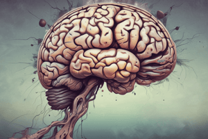
Intracranial Injuries and Protection
- Cerebrospinal fluid (CSF) cushions and nourishes the brain; vital for maintaining intracranial pressure.
- CSF pressure is crucial in diagnosing conditions such as meningitis, bleeding, and multiple sclerosis (MS).
- Hydrocephalus refers to excessive accumulation of CSF, leading to increased pressure.
CSF Production and Volume
- The choroid plexus is the main site for CSF production.
- CSF total volume in newborns is about 5 mL, while adults have approximately 150 mL (50% intracranial, 50% spinal).
- In adults, the formation rate of CSF is about 0.3-0.35 mL/min, totaling 450-750 mL per day.
Blood Supply to the Brain
- Brain supplied by two internal carotid arteries and two vertebral arteries.
- Internal carotid arteries begin at the bifurcation of the common carotid artery and include a dilation known as the carotid sinus.
- They enter the cranial cavity via the carotid canal, eventually branching into anterior and middle cerebral arteries.
- Vertebral arteries enter through the foramen magnum and merge to form the basilar artery, supplying additional arteries to the brain.
Meninges Structure
- The meninges consist of three layers: dura mater, arachnoid mater, and pia mater.
- Dura mater is the toughest and most superficial layer, comprising the periosteal and meningeal layers.
- Dural septae are formed by the separation of the two layers and help restrict brain displacement, similar to a seatbelt.
Dural Venous Sinuses
- Located between the periosteal and meningeal layers of the dura mater, these sinuses contain venous blood and are lined with endothelium.
- They lack valves and tunica media, distinguishing them from regular veins.
- Significant sinuses include the cavernous sinus, located near the pituitary gland, which drains blood from various sources.
Arachnoid Mater
- The middle layer of the meninges, resembling a spider's web, that drapes over brain and spinal cord.
- Contains the subarachnoid space filled with CSF, which provides cushioning.
- Arachnoid granulations (villi) facilitate CSF drainage into the superior sagittal sinus.
CSF Flow and Drainage
- CSF circulates around the brain and spinal cord, providing protection and nourishment.
- Ultimately, all dural venous sinuses drain into the internal jugular vein, facilitating blood return from the brain.
Studying That Suits You
Use AI to generate personalized quizzes and flashcards to suit your learning preferences.