Podcast
Questions and Answers
What is the fundamental process involved in fluorescence?
What is the fundamental process involved in fluorescence?
- Emission of ultraviolet light from all substances
- Emission of high-energy light after absorbing low-energy light
- Absorption of light of higher energy and emission of lower energy (correct)
- Absorption of light of lower energy and emission of higher energy
Which of the following fluorochromes is specifically used for staining DNA?
Which of the following fluorochromes is specifically used for staining DNA?
- Green Fluorescent Protein (GFP)
- Rhodamine
- Calcein-AM
- DAPI (correct)
What is a key application of confocal microscopy in biological research?
What is a key application of confocal microscopy in biological research?
- Studying molecular dynamics and interactions (correct)
- Examining chemical compositions
- Assessing heart rhythms
- Measuring blood pressure
Which application is supported by the technique known as FRET?
Which application is supported by the technique known as FRET?
What does live cell imaging with confocal microscopy primarily reveal?
What does live cell imaging with confocal microscopy primarily reveal?
What is the purpose of using bleaching techniques like FRAP in fluorescence studies?
What is the purpose of using bleaching techniques like FRAP in fluorescence studies?
In confocal microscopy, how does Z-series imaging enhance results?
In confocal microscopy, how does Z-series imaging enhance results?
What is an essential precaution to take when employing confocal microscopy for colocalization studies?
What is an essential precaution to take when employing confocal microscopy for colocalization studies?
Which approach is effective in mitigating the challenges of autofluorescence in immunofluorescence imaging?
Which approach is effective in mitigating the challenges of autofluorescence in immunofluorescence imaging?
Which of the following is a notable advancement in immunofluorescence that enhances live cell imaging?
Which of the following is a notable advancement in immunofluorescence that enhances live cell imaging?
What is a primary limitation of fluorochromes during imaging?
What is a primary limitation of fluorochromes during imaging?
What role do negative controls play in immunofluorescence?
What role do negative controls play in immunofluorescence?
What process allows flow cytometry to analyze thousands of cells per second?
What process allows flow cytometry to analyze thousands of cells per second?
How does forward scatter provide information about cells in flow cytometry?
How does forward scatter provide information about cells in flow cytometry?
Which technique can help minimize the impact of photobleaching in immunofluorescence?
Which technique can help minimize the impact of photobleaching in immunofluorescence?
In flow cytometry, what is the main purpose of laser interaction with cells?
In flow cytometry, what is the main purpose of laser interaction with cells?
What distinguishes immunohistochemistry (IHC) from immunocytochemistry and immunofluorescence?
What distinguishes immunohistochemistry (IHC) from immunocytochemistry and immunofluorescence?
Which of the following processes is NOT part of tissue preparation for IHC?
Which of the following processes is NOT part of tissue preparation for IHC?
Which antibody type used in IHC is characterized by high specificity and is produced by a single clone?
Which antibody type used in IHC is characterized by high specificity and is produced by a single clone?
What is one key advantage of the indirect method in IHC over the direct method?
What is one key advantage of the indirect method in IHC over the direct method?
In which area is IHC NOT commonly applied?
In which area is IHC NOT commonly applied?
What challenge does IHC face when applied to marine sciences?
What challenge does IHC face when applied to marine sciences?
Which of the following practices is crucial for successful IHC?
Which of the following practices is crucial for successful IHC?
Which advanced method increases sensitivity and reduces background noise in IHC?
Which advanced method increases sensitivity and reduces background noise in IHC?
Flashcards
Fluorescence
Fluorescence
A process where a substance absorbs higher-energy light and emits lower-energy light.
Fluorochrome
Fluorochrome
A compound that emits light after absorbing light.
Fluorescent protein
Fluorescent protein
A protein that emits fluorescence.
Confocal microscopy
Confocal microscopy
Signup and view all the flashcards
Live cell imaging
Live cell imaging
Signup and view all the flashcards
FRAP (Fluorescence Recovery After Photobleaching)
FRAP (Fluorescence Recovery After Photobleaching)
Signup and view all the flashcards
FRET (Förster Resonance Energy Transfer)
FRET (Förster Resonance Energy Transfer)
Signup and view all the flashcards
Colocalization studies
Colocalization studies
Signup and view all the flashcards
IHC vs. Immunocytochemistry
IHC vs. Immunocytochemistry
Signup and view all the flashcards
IHC Sample Types
IHC Sample Types
Signup and view all the flashcards
Monoclonal Antibody
Monoclonal Antibody
Signup and view all the flashcards
Polyclonal Antibody
Polyclonal Antibody
Signup and view all the flashcards
IHC Tissue Preparation
IHC Tissue Preparation
Signup and view all the flashcards
Direct IHC Method
Direct IHC Method
Signup and view all the flashcards
Indirect IHC Method
Indirect IHC Method
Signup and view all the flashcards
IHC Applications
IHC Applications
Signup and view all the flashcards
Immunofluorescence Limitations
Immunofluorescence Limitations
Signup and view all the flashcards
Autofluorescence
Autofluorescence
Signup and view all the flashcards
Photobleaching
Photobleaching
Signup and view all the flashcards
Flow Cytometry
Flow Cytometry
Signup and view all the flashcards
Forward Scatter
Forward Scatter
Signup and view all the flashcards
Hydrodynamic Focusing
Hydrodynamic Focusing
Signup and view all the flashcards
Flow Cytometry Controls
Flow Cytometry Controls
Signup and view all the flashcards
Cell Analysis Parameters
Cell Analysis Parameters
Signup and view all the flashcards
Study Notes
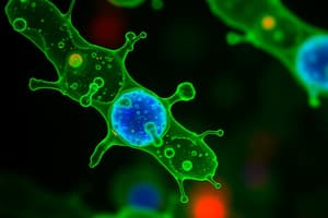
Immunofluorescence (IF) Technique
- A vital technique in cellular and molecular biology
- Visualizes specific intracellular processes and structures precisely
- Combines specific antibodies and fluorescent dyes
- Enhances understanding of biological processes
- Offers valuable applications in research and clinical diagnostics
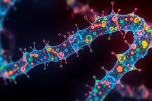
Fluorescence Principles
- Fluorescence occurs when luminescent molecules absorb light at one wavelength and emit it at another
- The light source is removed, fluorescence ceases immediately
- IF utilizes fluorochromes to visualize molecular targets
Immunofluorescence Methods
- Direct Immunofluorescence: Fluorochrome-conjugated primary antibodies bind directly to the antigen. Simpler and faster, but limited sensitivity.
- Indirect Immunofluorescence: Primary antibodies bind to the antigen, secondary antibodies (linked to fluorochromes) bind to the primary. Amplifies the signal, but can introduce cross-reactivity challenges
Multiplexing and Fluorochrome Properties
- IF allows for simultaneous detection of multiple targets (multiplexing)
- Achieved by using fluorochromes with distinct emission spectra
- Important fluorochrome properties include excitation and emission wavelengths, quantum yield, and photostability
- Compatibility with the detection system is crucial; FITC (green) and TRITC (red) are common examples; Alexa Fluor dyes offer better brightness and stability
Applications and Visualization
- Applications span numerous fields, from studying apoptotic markers to tracking specific proteins in tissues (like the mouse cerebellum)
- Offers versatility as a platform
- Coupled with advanced imaging techniques like confocal microscopy, IF allows for higher resolution and dynamic cellular events observation in 3D
Limitations and Controls
- No technique is without challenges; IF is no exception
- Autofluorescence from cellular components (e.g., collagen, lipofuscin) can complicate signal detection
- Photobleaching: Photochemical destruction of fluorophores limits signal longevity. Strategies such as photostable dyes and minimized light exposure help address this limitation
Flow Cytometry
- A process for analyzing cells quickly in a flow
- High capacity (thousands of cells per second)
- Multiple parameters (e.g., 50 ) can be assessed per cell
- Identifies cell subpopulations
- Evaluates cell cycle, phenotype, intracellular/extracellular characteristics, cell counts, apoptosis, calcium flow, cell division.
- Utilizes lasers and hydrodynamic focusing for cell analysis.
- Uses scattering and fluorescent signals for data analysis
- Quantifies using a PMT (photomultiplier tube)
- Biparametric graph analysis shows size and granularity of cells.
Evaluation of Apoptosis
- Uses propidium iodide and annexin V to distinguish viable cells, late apoptosis cells, and apoptotic cells
- Uses fluorescence intensities to determine the stage of apoptosis within a cell population
Phenotyping
- Involves incubating cells with antibodies recognizing specific proteins
- Allows the identification of cell subpopulations (e.g., blood cells) based on their fluorescence emission properties
Confocal Microscopy
- Advanced imaging technology for high lateral and axial resolution microscopic image creation.
- Uses a laser and pinhole to block out-of-focus light to enhance image contrast
- Enables detailed 3D visualization of microscopic structures
- Important for morphological studies, live-cell imaging, and protein interactions.
Hematology: General Principles and Methods
- Preserving Blood Samples. Specific anticoagulants are used relevant to the species and purpose, protecting blood integrity (e.g., EDTA for mammals, Heparin, others).
- Blood collection locations.
- Erythrocyte characteristics depend greatly on the organism type studied.
- Erythrocyte morphology and characteristics often vary significantly; blood cell production.
- Complete Blood Counts (CBC) are vital for diagnosing health conditions, including hemoparasite infections.
- Cellular analysis can range from manual methods (e.g., hemocytometer and staining) to sophisticated automated techniques(e.g., Flow cytometry, Coulters type impedance, automated cell counters).
- Blood smear evaluation under a microscope is an essential technique for assessing cell morphology and quantity.
Immunohistochemistry (IHC)
- A technique relying on the specificity of antigen-antibody interactions
- Different from immunocytochemistry (isolated cells) and immunofluorescence (fluorochromes).
- Can be performed on various sample types (cryopreserved, paraffin-embedded tissues, whole organs).
- Antibody types may be monoclonal (specific to one epitope) or polyclonal (reacting with various epitopes)
- Steps in tissue preparation for IHC (dewaxing, hydration, antigen retrieval, blocking endogenous peroxidases or non-specific reactions).
- Key methods include direct (primary antibody labeled) and indirect methods (primary and labeled secondary antibody).
Additional assays used to measure the effectiveness of a substance or condition
- Genotoxicity tests (Ames Test, chromosomal aberration, sister chromatid exchange, HPRT assay).
- Cytotoxic assays (ATP assays, Caspase activity, Trypan blue, Eosin Y, Propidium Iodide, MTT, XTT).
Studying That Suits You
Use AI to generate personalized quizzes and flashcards to suit your learning preferences.
Related Documents
Description
This quiz covers the essential concepts and methods in immunofluorescence, a key technique in cellular and molecular biology. Learn about direct and indirect methods, the principles of fluorescence, and their applications in research and diagnostics. Enhance your understanding of how this technique visualizes specific biological processes.