Podcast
Questions and Answers
Match the following descriptions with their corresponding immunofluorescence assay type:
Match the following descriptions with their corresponding immunofluorescence assay type:
Detects antigens in a sample using a primary antibody directly conjugated to a fluorescent dye. = Direct Immunofluorescence (DIF) Used in pathology to detect tissue-bound antigens (e.g., autoantibodies in skin or kidney biopsies). = Direct Immunofluorescence (DIF) More versatile, as different primary antibodies can use the same labeled secondary antibody. = Indirect Immunofluorescence Assay (IDIFA) Commonly used for serological tests and autoimmune or infectious disease studies. = Indirect Immunofluorescence Assay (IDIFA)
Match the following terms related to immunofluorescence with their corresponding descriptions:
Match the following terms related to immunofluorescence with their corresponding descriptions:
Fluorescent dyes = Dyes that absorb UV light and emit visible light Immunofluorescence (IF) = A technique visualizing specific antigens by binding fluorescently labelled antibodies Direct Immunofluorescence (DIF) = Uses a single, fluorescently labelled antibody to directly bind to the target antigen Indirect Immunofluorescence Assay (IDIFA) = Detects the presence of specific antibodies in a sample using a secondary, fluorescently labelled antibody
Match the following fluorescent dyes with their corresponding emitted light color:
Match the following fluorescent dyes with their corresponding emitted light color:
Fluorescein isothiocyanate (FITC) = Green light Tetramethylrhodamine = Red light DAPI = Blue light Alexa Fluor 488 = Green light
Match the following steps in Direct Immunofluorescence (DIF) with their corresponding descriptions:
Match the following steps in Direct Immunofluorescence (DIF) with their corresponding descriptions:
Match the following steps in Indirect Immunofluorescence Assay (IDIFA) with their corresponding descriptions:
Match the following steps in Indirect Immunofluorescence Assay (IDIFA) with their corresponding descriptions:
Match the following aspects of immunofluorescence with their corresponding applications:
Match the following aspects of immunofluorescence with their corresponding applications:
Match the following terms related to immunofluorescence with their corresponding advantages:
Match the following terms related to immunofluorescence with their corresponding advantages:
Match the following limitations of immunofluorescence with their corresponding explanations:
Match the following limitations of immunofluorescence with their corresponding explanations:
Match the following techniques related to immunofluorescence with their corresponding uses:
Match the following techniques related to immunofluorescence with their corresponding uses:
Match the following features of immunofluorescence with their corresponding effects:
Match the following features of immunofluorescence with their corresponding effects:
Match the following factors influencing immunofluorescence results with their corresponding effects:
Match the following factors influencing immunofluorescence results with their corresponding effects:
Flashcards
Immunofluorescence (IF)
Immunofluorescence (IF)
A technique that visualizes antigens using antibodies conjugated with fluorescent dyes.
Fluorescence Dyes
Fluorescence Dyes
Substances that absorb UV light and emit visible light.
Fluorescein Isothiocyanate (FITC)
Fluorescein Isothiocyanate (FITC)
A fluorescent dye that emits green light when excited.
Tetramethylrhodamine
Tetramethylrhodamine
Signup and view all the flashcards
Direct Immunofluorescence (DIF)
Direct Immunofluorescence (DIF)
Signup and view all the flashcards
Sample Preparation in DIF
Sample Preparation in DIF
Signup and view all the flashcards
Indirect Immunofluorescence Assay (IDIFA)
Indirect Immunofluorescence Assay (IDIFA)
Signup and view all the flashcards
Antigen Coating
Antigen Coating
Signup and view all the flashcards
Fluorescence Detection
Fluorescence Detection
Signup and view all the flashcards
Positive Reaction Indication
Positive Reaction Indication
Signup and view all the flashcards
Fluorescence Label in DIF
Fluorescence Label in DIF
Signup and view all the flashcards
Fluorescence Label in IDIFA
Fluorescence Label in IDIFA
Signup and view all the flashcards
Specificity of DIF
Specificity of DIF
Signup and view all the flashcards
Specificity of IDIFA
Specificity of IDIFA
Signup and view all the flashcards
Sensitivity of DIF
Sensitivity of DIF
Signup and view all the flashcards
Sensitivity of IDIFA
Sensitivity of IDIFA
Signup and view all the flashcards
Applications of DIF
Applications of DIF
Signup and view all the flashcards
Applications of IDIFA
Applications of IDIFA
Signup and view all the flashcards
Study Notes
Immunofluorescence
- Immunofluorescence is a technique used to visualize specific antigens.
- Antibodies chemically linked to fluorescent dyes bind to the antigen.
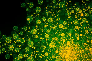
- Fluorochromes absorb short wavelength UV light and emit longer wavelength visible light (e.g., green or red).
- Examples of fluorochromes are fluorescein isothiocyanate (FITC) which emits green light, and tetramethylrhodamine, which emits red light.
- This technique can improve visibility of viral plaques.
Principle of Immunofluorescence (IF)
- IF is based on the specific antigen-antibody interaction.
- A labeled antibody binds to its corresponding antigen.
- When exposed to UV light, the fluorescent dye emits visible light.
- This allows visualization of the antigen-antibody complexes.
Requirements for Immunofluorescence
- Slide: A glass slide is used as a support.
- Tagged Antibody: Antibodies are tagged with a fluorescent dye.
- Fluorescent Microscope: This microscope is equipped to detect the emitted light.
- Specimen: The biological sample containing the antigen needs to be prepared.
Methods of IF Assay
-
Direct IF Assay (DIFA): A primary antibody labeled with a fluorescent dye is used to directly bind to the target antigen.
- Tissue sections/cells are fixed to a slide.
- A fluorescently labelled antibody specific to the target antigen is applied to the sample.
- The antibody binds to the antigen.
- Fluorescence under a microscope indicates a positive result.
-
Indirect IF Assay (IDIFA): A secondary antibody with a fluorescent label is used to bind to the primary antibody which has already bound to a specific antigen.
- A slide or substrate is coated with a specific antigen.
- The serum containing antibodies is applied to the slide.
- If the target antibodies are present, they bind to the antigen.
- A labelled secondary antibody, which binds to the patient’s antibodies, is added.
- Fluorescence under a microscope confirms the presence of specific antibodies.
Results
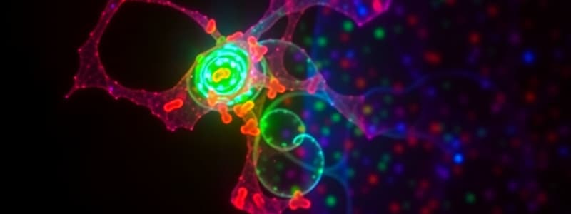
- Confocal images can be used to detect phosphorylated AKT.
- The resulting image shows green fluorescence in cardiomyocytes infected with adenovirus.
Comparison of Direct and Indirect IF Assays
- Direct IF: Detects antigens using a primary antibody conjugated to a fluorescent dye. Faster and cheaper. Lower sensitivity.
- Indirect IF: Detects antibodies using a secondary antibody conjugated to a fluorescent dye. More versatile, higher sensitivity, due to multiple binding events, and greater antibody detection capacity. More time-consuming.
Studying That Suits You
Use AI to generate personalized quizzes and flashcards to suit your learning preferences.