Podcast
Questions and Answers
DIF uses a single-step process where the primary antibody is labeled, while IDIFA uses a two-step process.
DIF uses a single-step process where the primary antibody is labeled, while IDIFA uses a two-step process.
True (A)
DIF is more sensitive than IDIFA due to signal amplification.
DIF is more sensitive than IDIFA due to signal amplification.
False (B)
DIF and IDIFA both utilize fluorescently labeled antibodies.
DIF and IDIFA both utilize fluorescently labeled antibodies.
True (A)
IDIFA is often used in pathology for identifying antigens directly in tissue or cells.
IDIFA is often used in pathology for identifying antigens directly in tissue or cells.
DIF is generally faster and less expensive than IDIFA due to the use of fewer reagents.
DIF is generally faster and less expensive than IDIFA due to the use of fewer reagents.
Tetramethylrhodamine emits green light when exposed to UV radiation.
Tetramethylrhodamine emits green light when exposed to UV radiation.
Direct immunofluorescence (DIF) uses a single, fluorescently labeled antibody to detect antigens.
Direct immunofluorescence (DIF) uses a single, fluorescently labeled antibody to detect antigens.
In indirect immunofluorescence, the fluorescently labeled antibody binds directly to the antigen.
In indirect immunofluorescence, the fluorescently labeled antibody binds directly to the antigen.
Immunofluorescence relies on the interaction between an antibody and an antigen, similar to how a lock and key fit together.
Immunofluorescence relies on the interaction between an antibody and an antigen, similar to how a lock and key fit together.
A positive result in a direct immunofluorescence assay indicates the presence of the target antibody.
A positive result in a direct immunofluorescence assay indicates the presence of the target antibody.
The presence of fluorescence in an indirect immunofluorescence assay indicates the presence of the target antibody in the sample.
The presence of fluorescence in an indirect immunofluorescence assay indicates the presence of the target antibody in the sample.
Immunofluorescence could be used to identify the presence of a specific virus in a cell culture.
Immunofluorescence could be used to identify the presence of a specific virus in a cell culture.
Immunofluorescence is a method that utilizes light emission from a radioactive source.
Immunofluorescence is a method that utilizes light emission from a radioactive source.
Flashcards
Immunofluorescence (IF)
Immunofluorescence (IF)
A technique to visualize antigens using fluorescently labeled antibodies.
Direct Immunofluorescence (DIF)
Direct Immunofluorescence (DIF)
Detects specific antigens using a single fluorescently labeled primary antibody.
Indirect Immunofluorescence Assay (IDIFA)
Indirect Immunofluorescence Assay (IDIFA)
Detects antibodies in a sample using a secondary fluorescently labeled antibody.
Fluorescent Dyes
Fluorescent Dyes
Dyes that absorb UV light and emit visible light (fluorescence).
Signup and view all the flashcards
Fluorescein Isothiocyanate (FITC)
Fluorescein Isothiocyanate (FITC)
A fluorescent dye that emits green light when excited by UV light.
Signup and view all the flashcards
Tetramethylrhodamine
Tetramethylrhodamine
A fluorescent dye that emits red light when excited by UV light.
Signup and view all the flashcards
Sample Preparation
Sample Preparation
The process of fixing tissue sections or cells onto a slide for fluorescence examination.
Signup and view all the flashcards
Fluorescence Detection
Fluorescence Detection
Examining slides under a fluorescence microscope to detect emitted light indicating a positive result.
Signup and view all the flashcards
Sensitivity in DIF vs. IDIFA
Sensitivity in DIF vs. IDIFA
DIF has lower sensitivity due to direct detection; IDIFA has higher sensitivity from signal amplification by the secondary antibody.
Signup and view all the flashcards
Application of DIF
Application of DIF
Used primarily in pathology to detect tissue-bound antigens, like autoantibodies.
Signup and view all the flashcards
Time and Cost Comparison
Time and Cost Comparison
DIF is faster and cheaper due to fewer reagents compared to the more complex IDIFA.
Signup and view all the flashcardsStudy Notes
Immunofluorescence
- Immunofluorescence is a technique used to visualize specific antigens.
- It involves binding a specific antibody, chemically conjugated with a fluorescent dye, to the antigen.
Definition
- Immunofluorescence is a technique that visualizes a specific antigen by binding a specific antibody conjugated with a fluorescent dye.
- Fluorochromes (e.g., fluorescein isothiocyanate (FITC), tetramethylrhodamine) absorb short UV wavelengths and emit longer wavelengths (visible light) such as green or red light.
- This technique aids in making Viral Plaques more visible.
Principle of Immunofluorescence (IF)
- IF is based on specific antigen-antibody interaction.
- A labeled antibody binds to its corresponding antigen.
- UV or specific wavelengths of light cause the fluorescent dye to emit visible light.
- This allows visualization of antigen-antibody complexes.
Principle of the Test (Diagram)
- Cells with specific human antigens are involved.
- Specific antibodies are labeled with a fluorescent dye like FITC.
- The labeled antibodies bind to the target antigens on the cells.
- Complexes of antigens and antibodies are visualized.
Requirements
- A slide
- Antibody tagged with a fluorescent dye.
- A fluorescent microscope.
- The specimen (e.g., tissue sections or cells).
Methods of IF Assay
- Two types of assays exist: direct and indirect.
Direct IF Assay (DIFA)
- A single fluorescent antibody binds directly to the target antigen.
- Sample preparation involves fixing tissues or cells to a slide.
- Applying the fluorescent antibody to the target antigen (if present).
- Looking at the slide under a fluorescence microscope.
- Fluorescence indicates a positive result (antigen presence).
Indirect IF Assay (IDIFA)
- Detects antibodies in a biological sample (e.g., serum).
- A slide/substrate is coated with a specific antigen.
- The patient serum sample is applied to the slide, with antibodies binding to the antigen (if present).
- A secondary antibody, labeled fluorescently, binds to the patient's antibodies.
- Observed with a fluorescence microscope, fluorescence indicates a positive result (antibody presence).
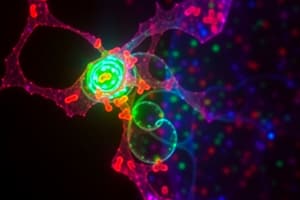
Result
- A confocal image showing phosphorylated AKT (green) in cardiomyocytes infected with adenovirus.
Comparison of Direct and Indirect IF
| Aspect | Direct IF | Indirect IF |
|---|---|---|
| Definition | Detects antigen using primary antibody. | Detects antibodies using primary and secondary antibody. |
| Fluorescence label | Primarily antibody is labeled. | Secondary antibody is labeled. |
| Steps | Single step | Two steps |
| Purpose | Identify antigens in tissue/cells | Detect antibodies in serum |
| Specificity | High, limited to labeled antibody. | More versatile, different primary antibodies can use same secondary antibody. |
| Sensitivity | Lower | Higher due to signal amplification |
| Applications | Pathology (tissue antigens). | Serological tests, autoimmune studies. |
| Time and Cost | Faster, generally cheaper. | Slower, potentially more expensive. |
Studying That Suits You
Use AI to generate personalized quizzes and flashcards to suit your learning preferences.