Podcast
Questions and Answers
What is the most common organ affected by hydatid cysts?
What is the most common organ affected by hydatid cysts?
- Spleen
- Liver (correct)
- Lungs
- Heart
Which symptom is most commonly associated with the rupture of a liver cyst?
Which symptom is most commonly associated with the rupture of a liver cyst?
- Obstructive jaundice (correct)
- Hemoptysis
- Skin rash
- Bronchospasm
Which statement about lung cysts is accurate?
Which statement about lung cysts is accurate?
- They typically require surgical intervention.
- Most cases are detected through symptoms of anaphylaxis.
- Non-complicated lung cysts always show symptoms.
- They are often confused with pulmonary tuberculosis. (correct)
What percentage of patients have hydatid cysts located in the heart or intracranial regions?
What percentage of patients have hydatid cysts located in the heart or intracranial regions?
Which complication can arise from a ruptured lung cyst?
Which complication can arise from a ruptured lung cyst?
Which location has the second highest frequency of hydatid cyst occurrence?
Which location has the second highest frequency of hydatid cyst occurrence?
What can happen when a liver cyst ruptures into the peritoneal cavity?
What can happen when a liver cyst ruptures into the peritoneal cavity?
Which of the following is a common initial symptom of a lung cyst?
Which of the following is a common initial symptom of a lung cyst?
What is the first symptom often associated with cerebral cysts?
What is the first symptom often associated with cerebral cysts?
Which symptom is least likely to be associated with kidney cysts?
Which symptom is least likely to be associated with kidney cysts?
Bone cysts are typically misdiagnosed as which of the following?
Bone cysts are typically misdiagnosed as which of the following?
What is a significant risk associated with heart cysts?
What is a significant risk associated with heart cysts?
Which diagnostic method is preferred for detecting abdominal cysts?
Which diagnostic method is preferred for detecting abdominal cysts?
What classification added a 'cystic lesion' (CL) stage in 1995?
What classification added a 'cystic lesion' (CL) stage in 1995?
What shared feature might pulmonary hydatid cysts and pulmonary tuberculosis exhibit?
What shared feature might pulmonary hydatid cysts and pulmonary tuberculosis exhibit?
What characterizes CE3 transitional cysts?
What characterizes CE3 transitional cysts?
What is a common site for the development of hydatid cysts in humans?
What is a common site for the development of hydatid cysts in humans?
Which animal is primarily responsible for the transmission of Echinococcus granulosus?
Which animal is primarily responsible for the transmission of Echinococcus granulosus?
Which treatment method is specifically limited to hydatid cysts in the liver?
Which treatment method is specifically limited to hydatid cysts in the liver?
What mechanism allows the oncosphere to migrate through the body after infection?
What mechanism allows the oncosphere to migrate through the body after infection?
What type of disease is hydatid disease classified as?
What type of disease is hydatid disease classified as?
What is the role of the definitive hosts in the life cycle of Echinococcus granulosus?
What is the role of the definitive hosts in the life cycle of Echinococcus granulosus?
Which factor contributes to the endemicity of echinococcosis in certain regions?
Which factor contributes to the endemicity of echinococcosis in certain regions?
How does the diagnosis of hydatid disease typically proceed?
How does the diagnosis of hydatid disease typically proceed?
What is the primary reason for the surgical excision of cysticerci before significant intraocular inflammation occurs?
What is the primary reason for the surgical excision of cysticerci before significant intraocular inflammation occurs?
Which of the following is NOT a common treatment for muscular and subcutaneous cysticercosis?
Which of the following is NOT a common treatment for muscular and subcutaneous cysticercosis?
How is sparganosis primarily transmitted to humans?
How is sparganosis primarily transmitted to humans?
Which clinical manifestation is associated with neural coenurosis?
Which clinical manifestation is associated with neural coenurosis?
What is a diagnostic tool commonly used for detecting ocular coenurosis?
What is a diagnostic tool commonly used for detecting ocular coenurosis?
Which of the following treatments can potentially lead to serious toxic endophthalmitis and loss of vision?
Which of the following treatments can potentially lead to serious toxic endophthalmitis and loss of vision?
Which organism causes coenurosis in humans?
Which organism causes coenurosis in humans?
What is the most plausible transmission method for sparganosis when using frogs?
What is the most plausible transmission method for sparganosis when using frogs?
What is the recommended method for assessing the response to treatment for cysts?
What is the recommended method for assessing the response to treatment for cysts?
What indicates a successful outcome after radical surgery for hydatid disease?
What indicates a successful outcome after radical surgery for hydatid disease?
Which of the following strategies has been effective in controlling echinococcosis?
Which of the following strategies has been effective in controlling echinococcosis?
Which organism causes alveolar echinococcosis (AE)?
Which organism causes alveolar echinococcosis (AE)?
How is hydatid disease primarily characterized in humans?
How is hydatid disease primarily characterized in humans?
What role do serologic tests play in defining the outcome of chemotherapy or PAIR?
What role do serologic tests play in defining the outcome of chemotherapy or PAIR?
What preventive measure can help reduce the transmission of hydatid disease to humans?
What preventive measure can help reduce the transmission of hydatid disease to humans?
What is a key aspect of the management strategy for tissue cystodes diseases?
What is a key aspect of the management strategy for tissue cystodes diseases?
What is the primary definitive host for Echinococcus granulosus?
What is the primary definitive host for Echinococcus granulosus?
What is the typical time frame for protoscolices to develop into adult stages in the definitive host?
What is the typical time frame for protoscolices to develop into adult stages in the definitive host?
Which lifecycle stage of Echinococcus granulosus is typically found in the intermediate hosts?
Which lifecycle stage of Echinococcus granulosus is typically found in the intermediate hosts?
What is the nature of the echinococcal cyst in humans?
What is the nature of the echinococcal cyst in humans?
How do intermediate hosts typically acquire Echinococcus granulosus infection?
How do intermediate hosts typically acquire Echinococcus granulosus infection?
Which of the following best describes the habitat of adult Echinococcus granulosus in the definitive host?
Which of the following best describes the habitat of adult Echinococcus granulosus in the definitive host?
What type of reaction surrounds the echinococcal cyst as it develops in tissues?
What type of reaction surrounds the echinococcal cyst as it develops in tissues?
What can be a potential consequence of the growth of hydatid cysts in humans?
What can be a potential consequence of the growth of hydatid cysts in humans?
Flashcards
Echinococcosis
Echinococcosis
A parasitic infection caused by the tapeworm Echinococcus granulosus.
Adult Echinococcus granulosus
Adult Echinococcus granulosus
The stage of the Echinococcus granulosus tapeworm that lives in the intestines of dogs and other canines.
Hydatid cyst
Hydatid cyst
The stage of the Echinococcus granulosus tapeworm that forms cysts in the body of intermediate hosts.
Intermediate host
Intermediate host
Signup and view all the flashcards
Liver
Liver
Signup and view all the flashcards
Cyst development
Cyst development
Signup and view all the flashcards
Chemotherapy for echinococcosis
Chemotherapy for echinococcosis
Signup and view all the flashcards
Surgery for echinococcosis
Surgery for echinococcosis
Signup and view all the flashcards
Raised intracranial pressure
Raised intracranial pressure
Signup and view all the flashcards
Focal epilepsy
Focal epilepsy
Signup and view all the flashcards
Loin pain
Loin pain
Signup and view all the flashcards
Hematuria
Hematuria
Signup and view all the flashcards
Definitive Host
Definitive Host
Signup and view all the flashcards
Pathologic fracture
Pathologic fracture
Signup and view all the flashcards
Oncosphere
Oncosphere
Signup and view all the flashcards
Systemic dissemination
Systemic dissemination
Signup and view all the flashcards
Cardiac tamponade
Cardiac tamponade
Signup and view all the flashcards
Protoscolices
Protoscolices
Signup and view all the flashcards
Oncosphere Migration
Oncosphere Migration
Signup and view all the flashcards
Echinococcal Cyst Development
Echinococcal Cyst Development
Signup and view all the flashcards
Germinal Layer
Germinal Layer
Signup and view all the flashcards
Onset of Symptoms in Hydatid Disease
Onset of Symptoms in Hydatid Disease
Signup and view all the flashcards
Growth Rate of Hydatid Cysts
Growth Rate of Hydatid Cysts
Signup and view all the flashcards
Common Locations for Hydatid Cysts
Common Locations for Hydatid Cysts
Signup and view all the flashcards
Number of Cysts in Hydatid Disease
Number of Cysts in Hydatid Disease
Signup and view all the flashcards
Location of Hydatid Cysts in the Liver
Location of Hydatid Cysts in the Liver
Signup and view all the flashcards
Asymptomatic Hydatid Cysts
Asymptomatic Hydatid Cysts
Signup and view all the flashcards
Infected Hydatid Cysts
Infected Hydatid Cysts
Signup and view all the flashcards
Complications from Rupture of a Hydatid Cyst
Complications from Rupture of a Hydatid Cyst
Signup and view all the flashcards
What causes echinococcosis?
What causes echinococcosis?
Signup and view all the flashcards
Where do hydatid cysts form?
Where do hydatid cysts form?
Signup and view all the flashcards
How is the effectiveness of treatment for echinococcosis assessed?
How is the effectiveness of treatment for echinococcosis assessed?
Signup and view all the flashcards
What are common treatment options for echinococcosis?
What are common treatment options for echinococcosis?
Signup and view all the flashcards
What happens to antibody levels after successful surgical removal of a hydatid cyst?
What happens to antibody levels after successful surgical removal of a hydatid cyst?
Signup and view all the flashcards
What causes alveolar echinococcosis?
What causes alveolar echinococcosis?
Signup and view all the flashcards
What is the most serious feature of alveolar echinococcosis?
What is the most serious feature of alveolar echinococcosis?
Signup and view all the flashcards
How can echinococcosis be prevented?
How can echinococcosis be prevented?
Signup and view all the flashcards
Sparganosis
Sparganosis
Signup and view all the flashcards
How is Sparganosis spread?
How is Sparganosis spread?
Signup and view all the flashcards
What are some symptoms of Sparganosis in subcutaneous tissue?
What are some symptoms of Sparganosis in subcutaneous tissue?
Signup and view all the flashcards
What is the primary treatment for Sparganosis?
What is the primary treatment for Sparganosis?
Signup and view all the flashcards
What is Coenuriasis?
What is Coenuriasis?
Signup and view all the flashcards
How is Coenuriasis spread?
How is Coenuriasis spread?
Signup and view all the flashcards
What are some symptoms of Coenuriasis in the nervous system?
What are some symptoms of Coenuriasis in the nervous system?
Signup and view all the flashcards
What are the treatment options for Coenuriasis?
What are the treatment options for Coenuriasis?
Signup and view all the flashcards
Study Notes
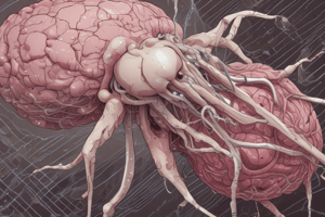
Echinococcosis
- Echinococcosis is a zoonotic parasitic disease, primarily transmitted between dogs and domestic livestock (specifically sheep).
- Humans are accidental intermediate hosts.
- The liver is the most common site of echinococcal (hydatid) cysts, followed by the lungs. Cysts less frequently occur in the spleen, kidneys, heart, and bone.
- Diagnostic methods and effective therapies for cystic hydatid disease (CHD) have been developed in recent years (last 10 years).
- Treatment options include surgery, chemotherapy with anthelmintic agents, or puncture-aspiration-injection-re-aspiration (PAIR).
- PAIR is mostly used for liver cysts
- Despite advancements, echinococcosis remains a significant public health concern in several countries and an emerging/re-emerging problem in others.
Echinococcus Life Cycle
- Adult Echinococcus granulosus worms reside in the small intestines of definitive hosts (dogs and other canines).
- Proglottids release eggs, which are then passed in feces.
- Intermediate hosts (e.g., sheep, goats, swine, cattle, horses, camels, or humans) ingest the eggs.
- Eggs hatch in the small intestine, releasing oncospheres.
- Oncospheres penetrate the intestinal wall and migrate through the circulatory system into organs (especially the liver and lungs).
- Oncospheres develop into cysts that enlarge gradually, forming protoscolices and daughter cysts.
- Definitive hosts become infected by ingesting the cyst-containing organs of intermediate hosts.
- Protoscolices evaginate and attach to the intestinal mucosa.
- Adult stages develop (32-80 days).
Liver Cysts
- Liver cysts are frequently located in the right lobe.
- Typically, patients with hepatic hydatid cysts are asymptomatic, but complications (e.g. abdominal distention, palpable mass, hepatomegaly) arise with cyst enlargement.
- Cysts can become infected with bacteria, potentially mimicking a liver abscess.
- Rupture can occur spontaneously, due to trauma, or during surgery.
- Rupture can result in obstructive jaundice, colic, or bacterial overgrowth.
- Rupture into the peritoneal cavity may lead to secondary peritoneal cysts or peritonitis.
- Rupture can also cause an erythematous rash or anaphylaxis.
Lung Cysts
- Non-complicated lung cysts often go unnoticed until a routine chest X-ray.
- Symptoms can include chest pain, fever, cough, dyspnea (shortness of breath), and hemoptysis (coughing up blood).
- Rupture of cysts can lead to the expulsion of hydatid fluid and/or membranes, potentially leading to bacterial overgrowth and lung abscess.
- Rupture into the lung can result in pneumothorax (collapsed lung) or empyema (pus in the pleural cavity).
- Rupture into the pleural cavity can trigger allergic reactions (e.g., pruritus, urticaria) and, rarely, anaphylaxis.
Other Sites of Infection
- Hydatid cysts can be found in various organs, though the spleen (3-5%) and heart and intracerebral areas (1-1.5%) are other common sites.
- Cysts in these locations can lead to conditions like: raised intracranial pressure, focal epilepsy, loin pain, hematuria, bone fractures or misdiagnosis as tuberculous lesions, and potentially cardiac tamponade.
Diagnosis and Imaging
- Abdominal ultrasonography, CT scans, and often ELISA assays for diagnosis are frequently used.
- The WHO-IWGE classification of cysts (into CE stages) is frequently used.
Treatment
- "Watch and wait" strategy for inactive, consolidated cysts
- Chemotherapy with benzimidazoles (e.g., albendazole, mebendazole), in conjunction or singly for cysts in multiple areas or peritoneal hydatidosis, to improve or cure 2/3 of cases.
- Surgical resection can be necessary for infected large cysts located in strategic areas (e.g., brain, heart) .
- PAIR (percutaneous aspiration of hydatid cysts under ultrasound guidance).
- Protoscolicidal agents (e.g., 95% ethanol or hypertonic saline).
Prevention
- Control measures include periodic treatment of dogs with praziquantel, prohibitions on feeding raw infected viscera to dogs, and inspection of abattoirs to help control echinococcosis. Education is key to modifying human practices.
Cysticercosis
- Human Cysticercosis is infection of human tissues by pork tapeworm larvae ( Taenia solium).
- Acquired through fecal-oral route (ingesting tapeworm eggs).
- Humans ingest eggs and develop cysticerci similarly to how pigs do.
- Cysticercosis can be contracted by ingesting objects or food contaminated with human feces.
- Neurocysticercosis: Epilepsy is a frequent manifestation; focal seizures and secondary generalization (spread) are common. Symptoms can be severe in areas within the brain.
- Ophthalmic cysticercosis: Retina and subretina lesions are frequent; floaters in vitreous or aqueous humor, choroidoretinitis, and retinal detachment are possible.
Sparganosis
- Caused by infection with Sparganum, the second-stage larvae of Spirometra, a particular type of tapeworm.
- Intermediate hosts such as frogs become infected.
- Humans acquire the infection by ingesting contaminated water or consuming raw meats (insects, amphibians, etc).
- Infection is primarily found in East and Southeast Asia.
- Development of adult stages does not occur in the human.
Coenuriasis
- Caused by the larval stage (Taenia multiceps)
- Adult tapeworms reside in the small intestine (generally dogs).
- Eggs are shed in feces, disintegrating to free eggs when ingested by humans.
- Larvae develop into Coenuri.
- Infection is comparatively rare, but reported in the USA, England, France, and Brazil.
Studying That Suits You
Use AI to generate personalized quizzes and flashcards to suit your learning preferences.