Podcast
Questions and Answers
What is the primary function of the platysma muscle?
What is the primary function of the platysma muscle?
- Cover the anterolateral aspect of the neck (correct)
- Support the thyroid gland
- Assist in the movement of the jaw
- Facilitate neck movement
From which embryonic structure does the platysma muscle develop?
From which embryonic structure does the platysma muscle develop?
- Neural crest cells
- Mesenchyme from the 2nd pharyngeal arch (correct)
- Mesodermal layer of the somites
- The 1st pharyngeal arch
Which structure is located deep to the platysma?
Which structure is located deep to the platysma?
- Subclavian artery
- Superficial lymph nodes
- Common carotid artery
- External jugular vein (correct)
What are the three layers of the deep cervical fascia?
What are the three layers of the deep cervical fascia?
What is one of the functions of the deep cervical fascia?
What is one of the functions of the deep cervical fascia?
What does the investing layer of the deep cervical fascia surround?
What does the investing layer of the deep cervical fascia surround?
Which layer of deep cervical fascia forms the carotid sheath?
Which layer of deep cervical fascia forms the carotid sheath?
Which of the following structures does not attach to the investing layer of deep cervical fascia?
Which of the following structures does not attach to the investing layer of deep cervical fascia?
What vital structures are particularly vulnerable in the neck due to lack of bony protection?
What vital structures are particularly vulnerable in the neck due to lack of bony protection?
Which bones contribute to the structure of the neck?
Which bones contribute to the structure of the neck?
What anatomical feature provides flexibility for head positioning in the cervical vertebrae?
What anatomical feature provides flexibility for head positioning in the cervical vertebrae?
What anatomical structures are enclosed by the visceral part of the pretracheal layer?
What anatomical structures are enclosed by the visceral part of the pretracheal layer?
Which layer of fascia blends with the endothoracic fascia inferiorly?
Which layer of fascia blends with the endothoracic fascia inferiorly?
Which statement accurately describes the typical cervical vertebrae?
Which statement accurately describes the typical cervical vertebrae?
What distinguishes the C1 vertebrae from other cervical vertebrae?
What distinguishes the C1 vertebrae from other cervical vertebrae?
What is a key feature of the hyoid bone's greater horns?
What is a key feature of the hyoid bone's greater horns?
Where is the hyoid bone located?
Where is the hyoid bone located?
Where do arterial blood flow and principal venous drainage in the neck lie?
Where do arterial blood flow and principal venous drainage in the neck lie?
What does the carotid sheath contain?
What does the carotid sheath contain?
Which structure passes through the thickening of the pretracheal fascia at the hyoid level?
Which structure passes through the thickening of the pretracheal fascia at the hyoid level?
Where does the prevertebral fascia fuse with the anterior longitudinal ligament?
Where does the prevertebral fascia fuse with the anterior longitudinal ligament?
What is the shape of the superior surface of the vertebral bodies in the typical cervical vertebrae?
What is the shape of the superior surface of the vertebral bodies in the typical cervical vertebrae?
Which of the following statements is true about the cervical vertebrae?
Which of the following statements is true about the cervical vertebrae?
What is the significance of the retropharyngeal space?
What is the significance of the retropharyngeal space?
What identifies the C2 vertebrae compared to C1?
What identifies the C2 vertebrae compared to C1?
How is the hyoid bone unique among the bones in the body?
How is the hyoid bone unique among the bones in the body?
Which of the following muscles is associated with the prevertebral layer of fascia?
Which of the following muscles is associated with the prevertebral layer of fascia?
Which of the following accurately represents the drainage of lymph from the body?
Which of the following accurately represents the drainage of lymph from the body?
How does the carotid sheath relate to other fascial layers?
How does the carotid sheath relate to other fascial layers?
What is true about the spinous process of C7 vertebra?
What is true about the spinous process of C7 vertebra?
What function does the superficial cervical fascia serve in the neck?
What function does the superficial cervical fascia serve in the neck?
How do the transverse processes of the C7 vertebrae compare in size to those of other cervical vertebrae?
How do the transverse processes of the C7 vertebrae compare in size to those of other cervical vertebrae?
What is the primary function of the potential space between the prevertebral layer of deep cervical fascia and the buccopharyngeal fascia?
What is the primary function of the potential space between the prevertebral layer of deep cervical fascia and the buccopharyngeal fascia?
What condition may indicate a malignant tumor elsewhere in the body when associated with enlarged cervical lymph nodes?
What condition may indicate a malignant tumor elsewhere in the body when associated with enlarged cervical lymph nodes?
What is a common consequence of a fractured hyoid bone due to manual strangulation?
What is a common consequence of a fractured hyoid bone due to manual strangulation?
What results from injury to the cervical branch of the facial nerve?
What results from injury to the cervical branch of the facial nerve?
What role does the investing layer of deep cervical fascia play in infection control?
What role does the investing layer of deep cervical fascia play in infection control?
What complication may arise from improper care during surgical dissections of the neck?
What complication may arise from improper care during surgical dissections of the neck?
How does a pretracheal fascia infection typically limit its spread?
How does a pretracheal fascia infection typically limit its spread?
What is a possible consequence of a swollen platysma due to cervical nerve injury?
What is a possible consequence of a swollen platysma due to cervical nerve injury?
Flashcards are hidden until you start studying
Study Notes
Introduction
- Neck is the area between the cranium and the clavicle.
- Neck allows head flexibility and positioning for sense organs.
- Neck has vital structures with limited bony protection.
- Neck serves as a pathway for nerves, the oesophagus and trachea.
- The brachial plexuses of nerves originate in the neck.
- Arterial blood flow to the head and neck.
- Principal venous drainage (jugular veins) lie anterolaterally in the neck.
- Lymph from the head and neck drains into cervical lymph nodes.
- Lymph from the whole body except the superior right quadrant enters the thoracic duct.
Bones of the Neck
- The skeleton of the neck is formed by cervical vertebrae, the hyoid bone, the manubrium of the sternum, and clavicles.
Cervical Vertebrae
- Seven cervical vertebrae form the cervical region, enclosing the spinal cord and meninges.
- Cervical vertebrae support the head and allow flexibility for head positioning.
Typical Cervical Vertebrae (3-6)
- The vertebral body is small and longer from side to side.
- The vertebral foramen is large and triangular.
- Transverse processes are smaller.
- Superior facets of the articular processes are directed superoposteriorly, and the inferior facets are directed inferoposteriorly.
- Spinous processes are short and bifid.
Atypical Cervical Vertebrae (1,2 and 7)
- C1 (atlas) is ring-like, lacking a spinous process or body.
- C2 (axis) has a peg-like dens that projects superiorly from its body.
- C7 has a prominent spinous process that is not bifid.
- C7 has large transverse processes, but its transverse foramina are small.
Hyoid Bone
- It lies in the anterior part of the neck at the level of the C3 vertebra.
- Suspended by muscles connecting it to the mandible, styloid processes, thyroid cartilage, manubrium and scapulae.
- Consists of a body, greater and lesser horns
- It is isolated from the rest of the skeleton.
Structure of the Hyoid Bone
- The body of the hyoid faces anteriorly and is approximately 2.5 cm wide and 1 cm thick.
- The body has an anterior convex surface and a posterior concave surface.
- Each end of its body is united to a greater horn that projects posterosuperiorly and laterally from the body.
- Greater horns are united to the body by fibrocartilage in younger people and usually united by bone in older people.
- Each lesser horn is a small bony projection from the body near its union with the greater horn.
- It is connected to the body by fibrous tissue and sometimes to the greater horn by a synovial joint.
- The lesser horn projects superoposteriorly toward the styloid process and may be partly or completely cartilaginous in some adults.
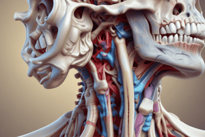
Fascia of the Neck
- Structures in the neck are compartmentalized by layers of deep cervical fascia.
- Subcutaneous tissue of the neck (superficial cervical fascia) is a layer of fatty connective tissue.
- It is usually thinner than in other regions, especially anteriorly.
- It contains cutaneous nerves, blood and lymphatic vessels, superficial lymph nodes, and variable amounts of fat.
- Anterolaterally it contains the platysma.
Platysma
- It is a broad, thin sheet of muscle in the subcutaneous tissue of the neck.
- It develops from a continuous sheet of musculature and is supplied by branches of the facial nerve.
- The external jugular vein and main cutaneous nerves of the neck are deep to the platysma.
- Platysma covers the anterolateral aspect of the neck, arising in the deep fascia covering the deltoid and pectoralis major muscles.
Deep Cervical Fascia
- It consists of three fascial layers: investing, pretracheal, and prevertebral.
- These layers support viscera, muscles, vessels, and deep lymph nodes.
- Deep cervical fascia condenses around the common carotid arteries, internal jugular veins, and vagus nerves to form the carotid sheath.
- These fascial layers form natural cleavage planes for surgery and limit the spread of abscesses.
- The fascial layers allow structures in the neck to move without difficulty.
Investing Layer
- The investing layer of deep cervical fascia surrounds the entire neck deep to the skin and subcutaneous tissue.
- It splits into superficial and deep layers to enclose the trapezius and sternocleidomastoid (SCM) muscles.
- The investing layer attaches superiorly to the superior nuchal line of the occipital bone, mastoid processes, zygomatic arches, inferior border of the mandible, hyoid bone, and spinous processes of the cervical vertebrae.
Pretracheal Layer
- It is limited to the anterior part of the neck.
- It extends inferiorly from the hyoid into the thorax, blending with the fibrous pericardium.
- It includes a muscular part, enclosing the infrahyoid muscles, and a visceral part, enclosing the thyroid gland, trachea, and esophagus.
- It blends laterally with the carotid sheaths.
- A thickening of the pretracheal fascia forms a pulley through which the intermediate tendon of the digastric muscle passes, suspending the hyoid.
Prevertebral Layer
- It forms a tubular sheath for the vertebral column and its associated muscles.
- It is fixed to the cranial base superiorly and blends with the endothoracic fascia peripherally and fuses with the anterior longitudinal ligament centrally at approximately the T3 vertebra.
- The prevertebral fascia extends laterally as the axillary sheath, surrounding the axillary vessels and brachial plexus.
- The cervical parts of the sympathetic trunks are embedded in the prevertebral layer of deep cervical fascia.
Carotid Sheath
- It is a tubular fascial investment extending from the cranial base to the root of the neck.
- It blends with the investing and pretracheal layers anteriorly and with the prevertebral layer posteriorly.
- It communicates freely with the mediastinum and the cranial cavity.
- The carotid sheath contains the common and internal carotid arteries, internal jugular vein, vagus nerve, deep cervical lymph nodes, carotid sinus nerve, and sympathetic nerve fibers.
Retropharyngeal Space
- It is the largest and most important interfascial space in the neck.
- It is a potential space consisting of loose connective tissue between the visceral part of the prevertebral layer and the buccopharyngeal fascia surrounding the pharynx.
- It extends laterally and terminates in the carotid sheath.
- It permits movement of the pharynx, esophagus, larynx, and trachea relative to the vertebral column.
- It is closed superiorly by the cranial base and on each side by the carotid sheath.
Clinical Anatomy
- Cervical pain has various causes, including inflamed lymph nodes, muscle strain or protruding intervertebral discs.
- Enlarged cervical lymph nodes may indicate a malignant tumor.
- Neck connects the head to the trunk, enabling metastasis (e.g., lung cancer).
- A fractured hyoid bone can occur in cases of manual strangulation.
- Depression of the body of the hyoid onto the thyroid cartilage makes swallowing and maintaining separation of the alimentary and respiratory tracts difficult.
- A fractured hyoid bone can result in aspiration pneumonia.
Paralysis of the Platysma
- It is due to injury to the cervical branch of the facial nerve.
- Causes the skin to fall away from the neck in slack folds.
- Surgeons must carefully preserve the cervical branch of the facial nerve.
- Surgeons must carefully suture the skin and edges of the platysma to avoid broad, ugly scars.
Deep Cervical Fascia Infections
- The investing layer of deep cervical fascia prevents the spread of abscesses.
- Infections between the investing layer and the muscular part of the pretracheal fascia will usually not spread beyond the superior edge of the manubrium.
Studying That Suits You
Use AI to generate personalized quizzes and flashcards to suit your learning preferences.