Podcast
Questions and Answers
Which of the following best describes the primary function of the fascial layers in the neck?
Which of the following best describes the primary function of the fascial layers in the neck?
- To lubricate the area between skeletal structures in the neck.
- To allow for the unobstructed passage of blood vessels and nerves within the neck.
- To separate and compartmentalize different neck structures, guiding the spread of infections. (correct)
- To provide a direct attachment point for muscles, facilitating movement.
What distinguishes the superficial fascia of the neck from the deeper fascial layers?
What distinguishes the superficial fascia of the neck from the deeper fascial layers?
- The superficial fascia contains the platysma muscle and superficial veins, which are not found in the deeper layers. (correct)
- The superficial fascia has a uniform thickness throughout the neck, unlike deeper layers.
- The superficial fascia primarily provides skeletal muscle support, whereas deeper layers offer more protection to the nerves.
- The superficial fascia contains only muscle tissue, unlike the deeper layers which contain primarily support tissue.
What is the anatomical term for the border formed by the trapezius and sternocleidomastoid muscles?
What is the anatomical term for the border formed by the trapezius and sternocleidomastoid muscles?
- The anterior border of the posterior triangle. (correct)
- The posterior border of the anterior triangle.
- The anterior border of the anterior triangle.
- The posterior border of the posterior triangle.
If a doctor were to make an incision in the anterior triangle of the neck, they should be aware of which of the following?
If a doctor were to make an incision in the anterior triangle of the neck, they should be aware of which of the following?
Which of these lists only contains contents of the posterior triangle?
Which of these lists only contains contents of the posterior triangle?
Which anatomical landmark is MOST relevant when defining the anterior triangle?
Which anatomical landmark is MOST relevant when defining the anterior triangle?
Which of the following is MOST characteristic of the muscular triangle of the neck?
Which of the following is MOST characteristic of the muscular triangle of the neck?
Which anatomical structure demarcates the superior posterior border of the neck?
Which anatomical structure demarcates the superior posterior border of the neck?
What is the primary role of the neck?
What is the primary role of the neck?
Which specific characteristic is unique to the cervical vertebrae (C2-C7) compared to other vertebral regions?
Which specific characteristic is unique to the cervical vertebrae (C2-C7) compared to other vertebral regions?
What distinguishes the atlas (C1) from the rest of the cervical vertebrae?
What distinguishes the atlas (C1) from the rest of the cervical vertebrae?
Besides the passage of nerve bundles, which major structures pass through the neck as it connects the head and rest of the body?
Besides the passage of nerve bundles, which major structures pass through the neck as it connects the head and rest of the body?
What bone feature is included in the posterior superior border of the neck?
What bone feature is included in the posterior superior border of the neck?
Where does the anterior inferior border of the neck extend from?
Where does the anterior inferior border of the neck extend from?
Which of the following accurately describes the articulation between C1 and the occipital bone?
Which of the following accurately describes the articulation between C1 and the occipital bone?
The neck transitions between the head and the thorax. What aspects of the neck allow this transition?
The neck transitions between the head and the thorax. What aspects of the neck allow this transition?
The geniohyoid muscle originates from which specific location?
The geniohyoid muscle originates from which specific location?
Which nerve provides innervation to the geniohyoid muscle?
Which nerve provides innervation to the geniohyoid muscle?
When the hyoid bone is fixed, what action does the geniohyoid muscle produce?
When the hyoid bone is fixed, what action does the geniohyoid muscle produce?
Which best describes the spatial relationship between the facial artery and vein as they pass through the submandibular triangle?
Which best describes the spatial relationship between the facial artery and vein as they pass through the submandibular triangle?
At what vertebral level does the common carotid artery typically bifurcate into the internal and external carotid arteries?
At what vertebral level does the common carotid artery typically bifurcate into the internal and external carotid arteries?
Which nerve innervates the platysma muscle?
Which nerve innervates the platysma muscle?
Where does the external jugular vein typically form?
Where does the external jugular vein typically form?
What is the most accurate description of the relationship of the external jugular vein to the sternocleidomastoid (SCM) muscle?
What is the most accurate description of the relationship of the external jugular vein to the sternocleidomastoid (SCM) muscle?
Which layer of the deep cervical fascia does the external jugular vein pierce before emptying into the subclavian vein?
Which layer of the deep cervical fascia does the external jugular vein pierce before emptying into the subclavian vein?
The anterior jugular veins communicate through which structure?
The anterior jugular veins communicate through which structure?
What forms the anterior border of the posterior triangle of the neck?
What forms the anterior border of the posterior triangle of the neck?
Which of the following muscles does not form a boundary of the posterior triangle?
Which of the following muscles does not form a boundary of the posterior triangle?
The sternocleidomastoid muscle is innervated by what nerve for its motor function?
The sternocleidomastoid muscle is innervated by what nerve for its motor function?
Where does the sternal head of the sternocleidomastoid muscle originate?
Where does the sternal head of the sternocleidomastoid muscle originate?
The deep boundary (floor) of the posterior triangle consists of which of the following?
The deep boundary (floor) of the posterior triangle consists of which of the following?
Which of the following muscles is NOT directly involved in forming a boundary of the submental or submandibular triangles?
Which of the following muscles is NOT directly involved in forming a boundary of the submental or submandibular triangles?
What is the primary function of the suprahyoid muscles when the mandible is in a fixed position?
What is the primary function of the suprahyoid muscles when the mandible is in a fixed position?
Which nerve innervates the posterior belly of the digastric muscle?
Which nerve innervates the posterior belly of the digastric muscle?
The stylohyoid muscle inserts onto the hyoid bone in which specific location, relative to the digastric muscle's intermediate tendon?
The stylohyoid muscle inserts onto the hyoid bone in which specific location, relative to the digastric muscle's intermediate tendon?
What is the origin of the mylohyoid muscle?
What is the origin of the mylohyoid muscle?
Which of the following best describes the spatial relationship between the mylohyoid muscle and the anterior belly of the digastric muscle?
Which of the following best describes the spatial relationship between the mylohyoid muscle and the anterior belly of the digastric muscle?
Which nerve provides the innervation for the mylohyoid muscle?
Which nerve provides the innervation for the mylohyoid muscle?
Which muscle supports and elevates the floor of the mouth, as well as elevates the hyoid bone?
Which muscle supports and elevates the floor of the mouth, as well as elevates the hyoid bone?
What is the primary function of the digastric muscle when the hyoid bone is fixed?
What is the primary function of the digastric muscle when the hyoid bone is fixed?
Which of the following describes the action of the stylohyoid muscle during swallowing?
Which of the following describes the action of the stylohyoid muscle during swallowing?
Flashcards
What are the borders of the neck?
What are the borders of the neck?
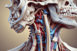
The neck region is bordered by the base of the skull superiorly, the clavicles inferiorly, and the anterior and posterior midline structures of the neck.
What are the functions of the neck?
What are the functions of the neck?
The neck plays a crucial role in supporting the head, enabling movement, and providing passage for vital structures such as airways, blood vessels, and nerves.
Describe the skeletal framework of the neck.
Describe the skeletal framework of the neck.
The skeletal framework of the neck comprises the cervical vertebrae (C1-C7), the hyoid bone, and the mandible. It provides structural support, attachments for muscles, and protection for crucial structures.
What are fascial compartments/spaces, and why are they clinically significant?
What are fascial compartments/spaces, and why are they clinically significant?
Signup and view all the flashcards
What is the Platysma muscle?
What is the Platysma muscle?
Signup and view all the flashcards
What are the superficial veins of the neck?
What are the superficial veins of the neck?
Signup and view all the flashcards
What are the boundaries and contents of the posterior triangle of the neck?
What are the boundaries and contents of the posterior triangle of the neck?
Signup and view all the flashcards
Neck: Region of transition
Neck: Region of transition
Signup and view all the flashcards
Superior Neck Border
Superior Neck Border
Signup and view all the flashcards
Inferior Neck Border
Inferior Neck Border
Signup and view all the flashcards
Cervical Vertebrae
Cervical Vertebrae
Signup and view all the flashcards
Atlas (C1) Vertebra
Atlas (C1) Vertebra
Signup and view all the flashcards
Axis (C2) Vertebra
Axis (C2) Vertebra
Signup and view all the flashcards
Features of Cervical Vertebrae
Features of Cervical Vertebrae
Signup and view all the flashcards
Foramen Transversarium
Foramen Transversarium
Signup and view all the flashcards
Articulation of C1 and C2
Articulation of C1 and C2
Signup and view all the flashcards
Platysma
Platysma
Signup and view all the flashcards
External jugular vein
External jugular vein
Signup and view all the flashcards
Anterior jugular vein
Anterior jugular vein
Signup and view all the flashcards
Posterior triangle
Posterior triangle
Signup and view all the flashcards
Prevertebral fascia
Prevertebral fascia
Signup and view all the flashcards
Sternocleidomastoid muscle (SCM)
Sternocleidomastoid muscle (SCM)
Signup and view all the flashcards
Omoclavicular triangle
Omoclavicular triangle
Signup and view all the flashcards
Occipital triangle
Occipital triangle
Signup and view all the flashcards
Accessory nerve (CN XI)
Accessory nerve (CN XI)
Signup and view all the flashcards
Proprioception
Proprioception
Signup and view all the flashcards
What is the action of the digastric muscle?
What is the action of the digastric muscle?
Signup and view all the flashcards
What is the sub mandibular gland?
What is the sub mandibular gland?
Signup and view all the flashcards
What is the carotid sheath?
What is the carotid sheath?
Signup and view all the flashcards
Where does the common carotid artery bifurcate?
Where does the common carotid artery bifurcate?
Signup and view all the flashcards
What are the contents of the carotid sheath?
What are the contents of the carotid sheath?
Signup and view all the flashcards
Submental Triangle
Submental Triangle
Signup and view all the flashcards
Submandibular Triangle
Submandibular Triangle
Signup and view all the flashcards
What is the Digastric Muscle?
What is the Digastric Muscle?
Signup and view all the flashcards
What are the Origins and Insertions of the Digastric Muscle?
What are the Origins and Insertions of the Digastric Muscle?
Signup and view all the flashcards
What are the Nerve Innervations of the Digastric Muscle?
What are the Nerve Innervations of the Digastric Muscle?
Signup and view all the flashcards
What is the Stylohyoid Muscle?
What is the Stylohyoid Muscle?
Signup and view all the flashcards
What is the Mylohyoid Muscle?
What is the Mylohyoid Muscle?
Signup and view all the flashcards
What is the Geniohyoid Muscle?
What is the Geniohyoid Muscle?
Signup and view all the flashcards
What is the Overall Function of the Suprahyoid Muscles?
What is the Overall Function of the Suprahyoid Muscles?
Signup and view all the flashcards
What are the Suprahyoid Muscles?
What are the Suprahyoid Muscles?
Signup and view all the flashcards
Study Notes
Organization & Neck Triangles
- The neck is a transition zone between the head and thorax
- Anatomical terminology is crucial for describing neck structures and functions
- The skeletal framework defines the spaces and compartments in the neck area
- Fascial layers organize the neck into compartments, impacting clinical significance
- Superficial fascia contains structures like the platysma muscle, affecting mouth movements and skin tension
- The posterior and anterior triangles divide the neck region, with varying muscle content
Subdivisions of the Neck Regions
- Posterior Triangle is defined by the posterior border of SCM, anterior border of trapezius, and inferior clavicle
- Muscles forming the posterior triangle include the SCM and Trapezius.
- Anterior Triangle subdivided further, including the submandibular, submental, and muscular triangles
- Key anatomical landmarks include the hyoid bone and mandible
Structures in the Superficial Fascia
- Platysma muscle, a large, thin sheet muscle, connects the thorax and mandible
- Veins like external jugular, described as passing posterior to mandible angle and crossing the SCM
- Location and innervation of the platysma and superficial veins are key
Nerves in the Neck
- Cutaneous branches of the cervical plexus provide sensory input to the skin
- Important nerves include the lesser occipital, great auricular, and supraclavicular nerves
- The accessory nerve (CN XI) innervates the SCM and trapezius muscles
- Clinical significance of nerve locations, crucial for potential damage from trauma or surgery
Lymphatic Drainage of the Neck
- The superficial lymph nodes include occipital, mastoid (retroauricular), preauricular/parotid, submandibular, and submental nodes
- Deep cervical lymph nodes are along the internal jugular vein
Bones of the Neck
- Cervical vertebrae form the framework, characterized by small bodies, bifid spinous processes, and transverse foramina for vessels
- C1, the atlas, does not have a body and joins with the occipital bone, offering a unique articulation with C2
- Hyoid bone is a unique part of the neck, supporting soft tissue structures without direct bony connections to other bones
Studying That Suits You
Use AI to generate personalized quizzes and flashcards to suit your learning preferences.