Podcast
Questions and Answers
What is an electrode?
What is an electrode?
A piece of metal that conducts potentials from the skin to the electrocardiograph.
How many leads are in a standard ECG (Electrocardiogram)?
How many leads are in a standard ECG (Electrocardiogram)?
- 12 (correct)
- 6
- 3
- 9
Bipolar limb leads measure potential difference between specific points on the limbs, such as Lead I measures potential difference between left arm (VL) & Rt.Arm which is __ - __.
Bipolar limb leads measure potential difference between specific points on the limbs, such as Lead I measures potential difference between left arm (VL) & Rt.Arm which is __ - __.
VL - VR
Unipolar chest leads measure the potential at certain points on the chest facing the heart.
Unipolar chest leads measure the potential at certain points on the chest facing the heart.
What is the function of coagulase in Staphylococci?
What is the function of coagulase in Staphylococci?
Which organisms produce catalase enzyme that breaks down hydrogen peroxide?
Which organisms produce catalase enzyme that breaks down hydrogen peroxide?
Streptococci are classified based on their hemolysis patterns on blood agar.
Streptococci are classified based on their hemolysis patterns on blood agar.
_______ ________ causes a swelling of the capsule in S.pneumoniae in the Quellung reaction.
_______ ________ causes a swelling of the capsule in S.pneumoniae in the Quellung reaction.
Match the following components with their functions in ABP measurement:
Match the following components with their functions in ABP measurement:
Flashcards are hidden until you start studying
Study Notes
Human Anatomy and Embryology
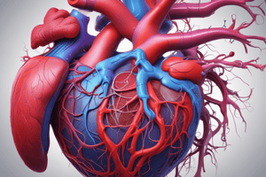
- Right Ventricle
- Located in the heart
- Right Atrium
- Located in the heart
- Tricuspid valve
- Located between the right atrium and ventricle
- Left atrium
- Located in the heart
- Coronary sinus
- Located in the heart
- Inferior vena cava
- Located in the heart
- Descending Thoracic Aorta
- Located near the heart
- Oesophagus
- Located near the heart
- Right coronary artery
- Located near the heart
- Posterior interventricular artery
- Located near the heart
- Pulmonary valve
- Located between the right ventricle and pulmonary artery
- Papillary muscle
- Located in the right ventricle
- Anterior interventricular artery
- Located near the heart
- Apex
- Located at the bottom of the heart
- Superior sternal angle
- Located near the heart
- Brachiocephalic artery
- Located near the heart
- Superior vena cava
- Located near the heart
- Azygos vein
- Located near the heart
- Thoracic duct
- Located near the heart
- Arch of azygos
- Located near the heart
- Arch of aorta
- Located near the heart
Cell Biology
- Wall of heart
- Consists of cardiac muscle fibers
- Endomysium: a layer of connective tissue surrounding cardiac muscle fibers
- Intercalated disc: a gap between cardiac muscle fibers allowing for electrical conduction
- Central oval nuclei: found in cardiac muscle fibers
- Wall of large artery (Aorta)
- Consists of three layers: tunica adventitia, tunica media, and tunica intima
- Tunica adventitia: outer layer of connective tissue
- Tunica media: middle layer of smooth muscle and elastic fibers
- Tunica intima: inner layer of endothelial cells
- Wall of IVC (Inferior Vena Cava)
- Consists of three layers: tunica adventitia, tunica media, and tunica intima
- Medium-sized artery and vein
- Wall consists of three layers: tunica adventitia, tunica media, and tunica intima
Laboratory Diagnosis
- Staphylococci
- General characteristics: coagulase positive or negative, catalase positive
- Examples: S. aureus, S. epidermidis, S. saprophyticus
- Microbiology Laboratory steps to identify Staphylococcus aureus
-
- Selective media: Mannitol salt agar
-
- Identification of colonies: Gram stain, biochemical reactions
-
- Catalase test: differentiates Staphylococci from Streptococci
-
- Coagulase test: differentiates S. aureus from S. epidermidis and S. saprophyticus
-
- Gram-positive cocci
- Streptococci: long chains, non-spore forming, non-motile, capsule of hyaluronic acid
- Examples: S. pyogenes, S. viridans, S. pneumoniae
- Microbiology Laboratory diagnosis of S. pyogenes
-
- Sample: according to clinical presentation
-
- Direct film: morphology
-
- Culture: enriched media, optimum temperature 37°C
-
- Identification: morphology, biochemical reactions, bacitracin sensitivity test
-
- Serology: specific identification of S. pyogenes
-
- Microbiology Laboratory diagnosis of S. pneumoniae
-
- Specimens: sputum
-
- Direct film: Gram-positive diplococci
-
- Culture: enriched media, optimum temperature 37°C
-
- Identification: morphology, biochemical reactions, fermentation of inulin, bile solubility, sensitivity to optochin
-
Blood Pressure
- Arterial blood pressure (ABP)
- Lateral force exerted by the moving column of blood on the lateral wall of arteries
- Normally oscillating during the cardiac cycle
- Systolic blood pressure (SBP)
- Maximum pressure created inside the arteries during ventricular systole
- Normal range: 90-140 mm Hg at rest
- Diastolic blood pressure (DBP)
- Minimum pressure remains inside the arteries at the end of ventricular diastole
- Normal range: 60-90 mm Hg at rest
- Pulse pressure
- Difference between systolic and diastolic BP
- Normal range: 40 mm Hg at rest
- Manometers
- Mercury manometers
- Aneroid manometers
- Digital electronic manometers
- Measurement of ABP in man
- Using a mercury sphygmomanometer
- Technique: many precautions, including patient position, arm support, and cuff size
Electrocardiogram (ECG)
- Definition: graphic representation of the cardiac electrical activity
- Electrode: a piece of metal that conducts potentials from the skin to the electrocardiograph
- Lead: a particular arrangement of each two electrodes
- Types of leads
- Bipolar leads (3)
- Unipolar leads (9)
- Unipolar limb leads (3)
- Unipolar chest leads (6)
- ECG recording
- Standard ECG: 12-lead ECG
- Additional leads may be used in special situations
Studying That Suits You
Use AI to generate personalized quizzes and flashcards to suit your learning preferences.


