Podcast
Questions and Answers
What anatomical feature is indicated by a line that separates the atria from the ventricles?
What anatomical feature is indicated by a line that separates the atria from the ventricles?
- Interatrial sulcus
- Coronary sinus
- Atrioventricular groove (correct)
- Interventricular groove
In which anatomical location can the apex of the heart be found?
In which anatomical location can the apex of the heart be found?
- Left 4th intercostal space
- Right 5th intercostal space
- Left 5th intercostal space (correct)
- Right 4th intercostal space
Which groove is responsible for separating the right atrium from the right ventricle?
Which groove is responsible for separating the right atrium from the right ventricle?
- Anterior interventricular sulcus
- Right atrioventricular groove (correct)
- Posterior interventricular sulcus
- Left atrioventricular groove
What separates the two ventricles from each other?
What separates the two ventricles from each other?
Which component lies above and behind the ventricles in the heart?
Which component lies above and behind the ventricles in the heart?
Which feature of the heart is described by a line that is slightly convex to the right?
Which feature of the heart is described by a line that is slightly convex to the right?
What connects the left atrium to the left ventricle?
What connects the left atrium to the left ventricle?
What is the primary function of the grooves or sulci of the heart?
What is the primary function of the grooves or sulci of the heart?
What is the average weight of the heart in males?
What is the average weight of the heart in males?
Which of the following best describes the anatomical position of the heart?
Which of the following best describes the anatomical position of the heart?
How does the size of the heart compare to common objects?
How does the size of the heart compare to common objects?
In which location does one-third of the heart lie?
In which location does one-third of the heart lie?
What anatomical landmarks define the upper border of the heart?
What anatomical landmarks define the upper border of the heart?
How much of the heart is located to the left of the median plane?
How much of the heart is located to the left of the median plane?
What is the relationship of the heart's location to the diaphragm?
What is the relationship of the heart's location to the diaphragm?
Which structure is NOT a landmark for the cardiac surface anatomy?
Which structure is NOT a landmark for the cardiac surface anatomy?
What percentage of the heart lies to the right side of the median plane?
What percentage of the heart lies to the right side of the median plane?
Which anatomical region contains the heart?
Which anatomical region contains the heart?
What is the primary function of the ductus venosus in fetal circulation? (review)
What is the primary function of the ductus venosus in fetal circulation? (review)
What happens to blood from the placenta once it enters the right atrium during fetal circulation?
What happens to blood from the placenta once it enters the right atrium during fetal circulation?
Which structure opens at the upper end of the smooth posterior part of the right atrium?
Which structure opens at the upper end of the smooth posterior part of the right atrium?
What component is located in the rough anterior part of the right atrium?
What component is located in the rough anterior part of the right atrium?
During fetal life, why does most blood in the pulmonary trunk bypass the lungs? (review)
During fetal life, why does most blood in the pulmonary trunk bypass the lungs? (review)
What distinguishes the tricuspid valve from the bicuspid valve?
What distinguishes the tricuspid valve from the bicuspid valve?
What is the function of the Eustachian valve in the inferior venacava? (review)
What is the function of the Eustachian valve in the inferior venacava? (review)
In which part of the right atrium are the Thebesian vein openings found? (review)
In which part of the right atrium are the Thebesian vein openings found? (review)
What is the oxygen saturation level in the umbilical arteries during fetal circulation?
What is the oxygen saturation level in the umbilical arteries during fetal circulation?
What separates the smooth posterior part from the rough anterior part in the right atrium? (review)
What separates the smooth posterior part from the rough anterior part in the right atrium? (review)
Where does the coronary sinus open in relation to the inferior venacava's opening?
Where does the coronary sinus open in relation to the inferior venacava's opening?
Which structure represents the smallest veins draining into the heart?
Which structure represents the smallest veins draining into the heart?
What characterizes the pectinate part of the right atrium?
What characterizes the pectinate part of the right atrium?
Which of the following structures is NOT located in the smooth posterior part of the right atrium?
Which of the following structures is NOT located in the smooth posterior part of the right atrium?
What is the role of the papillary muscles within the heart?
What is the role of the papillary muscles within the heart?
Which wall of the left atrium features the fossa lunata? (review)
Which wall of the left atrium features the fossa lunata? (review)
Which structure separates the right and left ventricles?
Which structure separates the right and left ventricles?
What type of orifice does the left atrium pump blood into?
What type of orifice does the left atrium pump blood into?
Which structure is specifically responsible for guarding the aortic orifice?
Which structure is specifically responsible for guarding the aortic orifice?
How many pairs of valves are present in the heart?
How many pairs of valves are present in the heart?
Which papillary muscle is located on the septum side of the left ventricle?
Which papillary muscle is located on the septum side of the left ventricle?
What general feature does the inferior wall of the left ventricle possess? (review)
What general feature does the inferior wall of the left ventricle possess? (review)
Which of the following veins does the left atrium receive blood from?
Which of the following veins does the left atrium receive blood from?
Which muscle structure aids in preventing blood regurgitation within the heart?
Which muscle structure aids in preventing blood regurgitation within the heart?
Flashcards
Heart Location
Heart Location
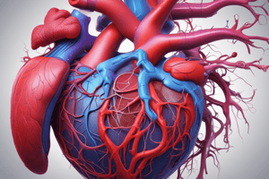
The heart is a conical, muscular organ situated in the middle mediastinum, within the pericardium, superior to the diaphragm, and left of the midline.
Heart Size
Heart Size
The average human heart is about the size of a fist, weighing around 300g for males and 250g for females.
Heart Chambers
Heart Chambers
The heart has four chambers: two atria (right and left) and two ventricles (right and left).
Atria Location
Atria Location
Signup and view all the flashcards
Atrioventricular Groove
Atrioventricular Groove
Signup and view all the flashcards
Interventricular Groove
Interventricular Groove
Signup and view all the flashcards
Right Atrium Parts
Right Atrium Parts
Signup and view all the flashcards
Right Atrium Openings
Right Atrium Openings
Signup and view all the flashcards
Left Atrium Function
Left Atrium Function
Signup and view all the flashcards
Left Ventricle Function
Left Ventricle Function
Signup and view all the flashcards
Interventricular Septum
Interventricular Septum
Signup and view all the flashcards
Tricuspid Valve
Tricuspid Valve
Signup and view all the flashcards
Bicuspid Valve
Bicuspid Valve
Signup and view all the flashcards
Semilunar Valves
Semilunar Valves
Signup and view all the flashcards
Fetal Circulation
Fetal Circulation
Signup and view all the flashcards
Foramen Ovale
Foramen Ovale
Signup and view all the flashcards
Ductus Arteriosus
Ductus Arteriosus
Signup and view all the flashcards
Superior Vena Cava
Superior Vena Cava
Signup and view all the flashcards
Inferior Vena Cava
Inferior Vena Cava
Signup and view all the flashcards
Study Notes
Location and Position of the Heart
- Conical muscular organ located in the middle mediastinum within the pericardium
- Superior surface of the diaphragm, left of the midline
- Anterior to the vertebral column, posterior to the sternum
- Weight- 300g in males, 250g in females
- Size- Approximately the size of your fist
Surface Anatomy of the Heart
- Upper border: straight line joining the lower border of the 2nd left costal cartilage to the upper border of the 3rd right costal cartilage
- Lower border: straight line joining the lower border of the 6th right costal cartilage to the superior venacava
Chambers of the Heart
- 4 Chambers: Two atria (right atrium & left atrium) and Two ventricles (right & left)
- The atria lie above and behind the ventricles
Grooves/Sulci of the Heart
- Atrioventricular groove/Coronary sulcus (right & left): on the surface of the heart, separates the atrium from the ventricles
- Interventricular groove/Sulcus (anterior & posterior): separates the two ventricles from each other
- Interatrial groove/Sulcus: separates the two atria from each other
Right Atrium
- Divided into three parts: smooth posterior part (sinus venarum), rough anterior part (pectinate part including the auricle), interatrial septum
- Smooth Posterior Part: shows the openings of: superior venacava, inferior venacava, coronary sinus, thebesian veins
- Inferior Venacava Opening: guarded by a valve of inferior venacava (Eustachian valve)
- Coronary Sinus Opening: guarded by valve of coronary sinus
- Rough Anterior Part: includes the crista terminalis and musculi pectinati
Left Atrium
- Receives oxygenated blood from the lungs through four pulmonary veins
- Pumps it to the left ventricle through left atrioventricular or bicuspid or mitral orifice
- Features: septal wall shows fossa lunata, receives four pulmonary veins and few vena cordis minimi
Left Ventricle
- Features: The interior is divisible into two parts: lower rough part with trabeculae carneae, upper smooth part or aortic vestibule
- Interior shows two orifices: left atrioventricular/bicuspid/ mitral orifice, aortic orifice
- Has two well developed papillary muscles
Interventricular Septum
- Separates the right and left ventricle
- Upper thin membranous part and lower thick muscular part
Valves of the Heart
- Maintain the unidirectional flow of blood
- Two pairs of valves: atrioventricular valves, semilunar valves
- Right atrioventricular valve: tricuspid valve (3 cusps)
- Left atrioventricular valve: bicuspid valve (2 cusps)
- Semilunar valves: aortic and pulmonary valves (both with 3 semilunar cusps)
Fetal Circulation
- Blood from the placenta, about 80% saturated with oxygen, returns to the fetus by way of the umbilical vein
- Most of this blood flows through the ductus venosus directly into the inferior vena cava, bypassing the liver
- Blood from the inferior vena cava and superior vena cava enters the right atrium
- Blood is guided past the foramen ovale into the left atrium
- Blood in the right ventricle flows through the ductus arteriosus into the descending aorta, bypassing the pulmonary circulation
- Blood then flows toward the placenta by way of the two umbilical arteries
Studying That Suits You
Use AI to generate personalized quizzes and flashcards to suit your learning preferences.


