Podcast
Questions and Answers
What fluid fills the cochlear duct?
What fluid fills the cochlear duct?
- Cerebrospinal fluid
- Interstitial fluid
- Perilymph
- Endolymph (correct)
Which part of the basilar membrane is most responsive to high-frequency vibrations?
Which part of the basilar membrane is most responsive to high-frequency vibrations?
- The apex
- The portion nearest the oval window (correct)
- The portion nearest the helicotrema
- The middle portion
What are the hair-like projections located at the apical ends of hair cells called?
What are the hair-like projections located at the apical ends of hair cells called?
- Stereocilia (correct)
- Cilia
- Flagella
- Microtubules
What structure connects the tip of each stereocilium to the side of the next taller stereocilium within a hair bundle?
What structure connects the tip of each stereocilium to the side of the next taller stereocilium within a hair bundle?
Where do the axons of the optic tract primarily synapse?
Where do the axons of the optic tract primarily synapse?
What is the function of optic radiations?
What is the function of optic radiations?
What type of ion channel is opened when stereocilia bend?
What type of ion channel is opened when stereocilia bend?
Which part of the visual field projects to the left side of the brain?
Which part of the visual field projects to the left side of the brain?
What is a key characteristic of binocular vision, based on the figure?
What is a key characteristic of binocular vision, based on the figure?
If a lesion occurred in the optic tracts, what structure would be most directly affected in terms of signal transmission?
If a lesion occurred in the optic tracts, what structure would be most directly affected in terms of signal transmission?
What is the direct effect of light on rhodopsin in rod cells?
What is the direct effect of light on rhodopsin in rod cells?
What is the immediate consequence of cGMP being converted to GMP in a rod cell?
What is the immediate consequence of cGMP being converted to GMP in a rod cell?
What is the role of transducin in the phototransduction cascade?
What is the role of transducin in the phototransduction cascade?
What causes a rod cell to hyperpolarize?
What causes a rod cell to hyperpolarize?
Which event directly follows the reduction of glutamate release in a rod cell after exposure to light?
Which event directly follows the reduction of glutamate release in a rod cell after exposure to light?
Why does the eye take time to adapt when transitioning from a dark to a bright environment?
Why does the eye take time to adapt when transitioning from a dark to a bright environment?
In dark conditions, what changes occur in the rod cells?
In dark conditions, what changes occur in the rod cells?
What is the primary function of the choroid in relation to vision?
What is the primary function of the choroid in relation to vision?
What is the primary function of the pupils during light and dark adaptation?
What is the primary function of the pupils during light and dark adaptation?
Which photoreceptor is primarily responsible for color vision and visual acuity?
Which photoreceptor is primarily responsible for color vision and visual acuity?
Where are rods primarily located in the retina?
Where are rods primarily located in the retina?
Which photopigment is found in rods?
Which photopigment is found in rods?
Which of the following best describes the shape of rods and cones?
Which of the following best describes the shape of rods and cones?
Which structure is located in the anterior compartment of the eye?
Which structure is located in the anterior compartment of the eye?
What is the primary function of the aqueous humor?
What is the primary function of the aqueous humor?
In what conditions do rods primarily function?
In what conditions do rods primarily function?
Where are cones most numerous in the retina?
Where are cones most numerous in the retina?
Where is the posterior chamber located?
Where is the posterior chamber located?
What is the function of the pigment cell layer in the retina as illustrated?
What is the function of the pigment cell layer in the retina as illustrated?
What is the function of the vitreous humor?
What is the function of the vitreous humor?
Which cell type acts as the first stage of light processing after photoreceptors?
Which cell type acts as the first stage of light processing after photoreceptors?
Where does the aqueous humor return to the venous circulation?
Where does the aqueous humor return to the venous circulation?
What is the direction of light through the retina, as shown in the diagram?
What is the direction of light through the retina, as shown in the diagram?
Which of these is NOT a function of the anterior compartment?
Which of these is NOT a function of the anterior compartment?
Glaucoma is characterized primarily by:
Glaucoma is characterized primarily by:
Which of the following structures is part of the fibrous tunic of the eye?
Which of the following structures is part of the fibrous tunic of the eye?
Which structure is NOT directly filled with perilymph?
Which structure is NOT directly filled with perilymph?
The basilar membrane forms the wall of which structure?
The basilar membrane forms the wall of which structure?
Which of these is the space between the vestibular and basilar membranes?
Which of these is the space between the vestibular and basilar membranes?
Where are the stereocilia located?
Where are the stereocilia located?
What is the function of the endosteum of the inner ear?
What is the function of the endosteum of the inner ear?
Which structure is located within the cochlear duct?
Which structure is located within the cochlear duct?
Which structure within the inner ear is bone?
Which structure within the inner ear is bone?
Which is the correct sequence of structures, starting from the scala vestibuli?
Which is the correct sequence of structures, starting from the scala vestibuli?
What is the main function of the round window?
What is the main function of the round window?
Which of the following structures is NOT part of the membranous labyrinth?
Which of the following structures is NOT part of the membranous labyrinth?
Flashcards
Cornea
Cornea
The transparent, dome-shaped front part of the eye that helps focus light.
Iris
Iris
The colored part of the eye that controls the size of the pupil.
Pupil
Pupil
The black opening in the center of the iris that allows light to enter the eye.
Lens
Lens
Signup and view all the flashcards
Retina
Retina
Signup and view all the flashcards
Aqueous Humor
Aqueous Humor
Signup and view all the flashcards
Vitreous Humor
Vitreous Humor
Signup and view all the flashcards
Glaucoma
Glaucoma
Signup and view all the flashcards
Optic Tracts
Optic Tracts
Signup and view all the flashcards
Lateral Geniculate Nuclei
Lateral Geniculate Nuclei
Signup and view all the flashcards
Optic Radiations
Optic Radiations
Signup and view all the flashcards
Visual Cortex
Visual Cortex
Signup and view all the flashcards
Binocular Vision
Binocular Vision
Signup and view all the flashcards
Rods
Rods
Signup and view all the flashcards
Rhodopsin
Rhodopsin
Signup and view all the flashcards
Iodopsin
Iodopsin
Signup and view all the flashcards
Photoreceptor layer
Photoreceptor layer
Signup and view all the flashcards
Pigmented layer
Pigmented layer
Signup and view all the flashcards
Fovea centralis
Fovea centralis
Signup and view all the flashcards
Outer plexiform layer
Outer plexiform layer
Signup and view all the flashcards
Inner plexiform layer
Inner plexiform layer
Signup and view all the flashcards
Ganglionic layer
Ganglionic layer
Signup and view all the flashcards
Endolymph
Endolymph
Signup and view all the flashcards
Basilar Membrane
Basilar Membrane
Signup and view all the flashcards
Base of the Basilar Membrane
Base of the Basilar Membrane
Signup and view all the flashcards
Apex of the Basilar Membrane
Apex of the Basilar Membrane
Signup and view all the flashcards
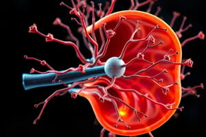
Stereocilia
Stereocilia
Signup and view all the flashcards
What does Transducin do?
What does Transducin do?
Signup and view all the flashcards
How is the signaling pathway activated?
How is the signaling pathway activated?
Signup and view all the flashcards
What is the role of cGMP in the rod cell?
What is the role of cGMP in the rod cell?
Signup and view all the flashcards
What happens when cGMP is broken down?
What happens when cGMP is broken down?
Signup and view all the flashcards
What is the result of the reduction in sodium entry?
What is the result of the reduction in sodium entry?
Signup and view all the flashcards
What is the effect of the hyperpolarized state on glutamate release?
What is the effect of the hyperpolarized state on glutamate release?
Signup and view all the flashcards
What is light adaptation?
What is light adaptation?
Signup and view all the flashcards
What is pupil adaptation?
What is pupil adaptation?
Signup and view all the flashcards
Inner Ear
Inner Ear
Signup and view all the flashcards
Membranous Labyrinth
Membranous Labyrinth
Signup and view all the flashcards
Bony Labyrinth
Bony Labyrinth
Signup and view all the flashcards
Cochlea
Cochlea
Signup and view all the flashcards
Semicircular Canals
Semicircular Canals
Signup and view all the flashcards
Vestibule
Vestibule
Signup and view all the flashcards
Oval Window
Oval Window
Signup and view all the flashcards
Round Window
Round Window
Signup and view all the flashcards
Scala Vestibuli, Scala Tympani, Cochlear Duct
Scala Vestibuli, Scala Tympani, Cochlear Duct
Signup and view all the flashcards
Perilymph & Endolymph
Perilymph & Endolymph
Signup and view all the flashcards
Study Notes
Chapter 15 Outline
- Chapter 15 focuses on the lecture outline for Anatomy and Physiology, Eleventh Edition
- Separate PowerPoint slides provide figures and tables without notes and animations.
- The copyright for the content is held by McGraw Hill Education.
Special Senses
- Olfaction
- Taste
- Visual system
- Hearing and balance
15.1 Olfaction
- Seven primary odors are now recognized, yet people can distinguish around 4000 different odors.
- Olfactory hairs (cilia) of olfactory neurons are embedded in mucus.
- Odorants dissolve in mucus, binding to receptors. The mechanism is unknown, but cilia depolarize triggering action potentials in olfactory neurons.
- A single receptor can respond to multiple odors.
- Olfactory epithelium regenerates; olfactory neurons are replaced by basal cells every two months.
15.2 Taste
- Papillae are bumps on the tongue.
- Taste buds house supporting cells surrounding taste (gustatory) cells.
- Taste cells have microvilli (gustatory hairs) extending into taste pores.
- Taste cells are replaced about every 10 days.
- Taste is closely associated with smell.
Taste Types
- Sour: Most sensitive receptors are on the lateral aspects of the tongue.
- Salty: Most sensitivity with sweet, triggered by Na+ and other metal ions.
- Bitter: Highest sensitivity receptors on posterior tongue, triggered by alkaloids (often toxins).
- Sweet: Sensitivity triggered by sugars, some carbohydrates, and some proteins
- Umami (Glutamate): Scattered sensitivity caused by amino acids.
15.3 Visual System
- Eyebrows: Shade and inhibit sweat.
- Eyelids (palpebrae): have conjunctiva, palpebral fissure, and canthi (medial/lateral).
- Eyelashes: double/triple row with ciliary glands (modified sweat glands) and Meibomian glands (produce sebum).
- Conjunctiva: thin transparent mucous membrane, including palpebral and bulbar conjunctiva.
- Layers of eyelid tissues include a tarsal plate to maintain shape.
Anatomy of the Eye
- Three Tunics: fibrous, vascular, and nervous.
- Fibrous tunic: sclera and cornea
- Vascular tunic: choroid, ciliary body, and iris
- Nervous tunic: retina
- Optic nerve carries information, connecting the eye to the brain.
Lacrimal Apparatus
- Tears are produced in the lacrimal gland.
- Tears exit through several lacrimal ducts
- Tears pass over the surface of the eye.
- Tears enter lacrimal canaliculi.
- Tears are carried through the lacrimal sac to the nasolacrimal duct.
- Tears enter the nasal cavity.
Extrinsic Eye Muscles
- Various extrinsic muscles control eye movement. These include the Superior/Inferior Rectus, Superior/Inferior Oblique, and the Lateral/Medial Rectus muscles.
Chambers of the Eye
- Anterior compartment: anterior to the lens, occupied by the aqueous humor.
- Anterior chamber: between the cornea and iris.
- Posterior chamber: between the iris and the lens.
- Aqueous humor: helps maintain intraocular pressure; delivers nutrients. Refracts light. Ciliary process produces aqueous humor.
- Vitreous humor: posterior to lens. Jelly-like vitreous humor helps maintain intraocular pressure and holds the lens and retina in place; it refracts light.
- Glaucomatous: increased intraocular pressure.
15.4 Hearing and Balance
- External ear: Auricle, external acoustic meatus, tympanic membrane.
- Middle ear: air-filled space, auditory ossicles (malleus, incus, stapes), eustachian tube.
- Inner ear: vestibular complex (vestibule, semicircular canals; responsible for equilibrium), and cochlea (responsible for hearing).
Inner Ear
- Labyrinths (bony and membranous)
- Lymph filled: endolymph (within membranous labyrinth) and perilymph (space between bony and membranous labyrinth)
- Oval/round windows
- Cochlear chambers (scala vestibuli, scala media, scala tympani)
- Spiral organ/Organ of Corti: contains hair cells and associated nerves.
Auditory Function
- Vibrations produce sound waves
- Volume/loudness relates to wave amplitude.
- Pitch relates to wave frequency.
- Timbre relates to resonance quality or overtones.
Muscles of the Middle Ear
- Tensor tympani: inserts on the malleus and innervated by cranial nerve V.
- Stapedius: inserts on the stapes and innervated by cranial nerve VII. (Attenuation reflex muscles that contract during loud noises preventing damage during vibrations.)
Effect of Sound Waves on Cochlear Structures/Points Along the Basilar Membrane
- Sound waves cause the tympanic membrane, malleus, incus, and stapes to vibrate.
- Vibrations of the stapes and oval window cause vibrations in the perilymph of Scala Vestibuli.
- Wave vibrations in perilymph propagate through scala tympani to the round window and dampened.
Sensitivity of Hearing
- The basilar membrane is important in pitch discrimination because it responds to different frequencies at different points of the cochlea.
- Inner hair cells receive efferent inputs, causing them to change in length to enhance or diminish certain auditory inputs.
- Outer hair cell responses to frequency range differences is important for tuning.
15.5 Effects of Aging on the Special Senses
- Slight loss in ability to detect odors
- Decreased sense of taste
- Lens's of eyes lose flexibility/accommodation.
- Development of cataracts, macular degeneration, glaucoma, and diabetic retinopathy
- Decline in visual acuity and color perception.
Hearing and Balance Conditions
- Conductive hearing loss (pathology in external/middle ear)
- Sensorineural hearing loss (pathology of inner ear/cochlear nerve)
- Otosclerosis (bones of middle ear fuse)
- Tinnitus (phantom sound)
- Otitis media (middle ear infection)
- Inner ear infections
- Motion sickness (conflict of inner ear/visual/balance cues)
- Meniere disease (conflicts in inner ear/balance, fluid abnormality)
Balance
- Static labyrinth: utricle and saccule
- Evaluates position of head relative to gravity
- Detects linear acceleration/deceleration (as in a car)
- Kinetic labyrinth: semicircular canals
- Evaluates head movement in three-dimensional space
Static Labyrinth
- Utricle has macula oriented parallel to skull base.
- Saccula has macula oriented perpendicular to skull base.
- Macula: Specialized epithelium with stereocilia (microvilli) and kinocilium (embedded in gelatinous).
- Otoliths (weighted structures) influence action potential rate based on gravity.
- Patterns of stimulation provide information on head position and acceleration.
Function of Vestibule in Maintaining Balance
- Endolymph in utricle, gelatinous mass, and otoliths determine displacement in response of head/body movement.
- Vestibular nerve fibers transmit action potentials from the macula depending on stimulation.
Kinetic Labyrinth
- Three semicircular canals filled with Endolymph: transverse, coronal, and sagittal planes
- Movement initiates an action potential (most intense when the rate of head movement changes)
- Movement changes translate into changes in action potential rate.
Neuronal Pathways for Balance
- Axons from vestibular ganglion pass vestibular to the vestibular nucleus.
- Vestibular nuclei influence postural muscles.
- Vestibular nuclei to other motor nuclei (oculomotor, trochlear, abducens) influence extrinsic eye muscles.
- Thalamic neurons to vestibular area of the cortex.
Studying That Suits You
Use AI to generate personalized quizzes and flashcards to suit your learning preferences.