Podcast
Questions and Answers
Which type of epithelial cells primarily covers the intestinal villi?
Which type of epithelial cells primarily covers the intestinal villi?
- Stratified squamous cells
- Simple cuboidal cells
- Simple columnar absorptive cells (correct)
- Transitional epithelium
What is a prominent type of gland found in the duodenum that secretes alkaline mucus?
What is a prominent type of gland found in the duodenum that secretes alkaline mucus?
- Parotid gland
- Brunner's gland (correct)
- Submandibular gland
- Sublingual gland
Which part of the large intestine is characterized by the absence of Peyer's patches?
Which part of the large intestine is characterized by the absence of Peyer's patches?
- Ascending colon (correct)
- Cecum
- Transverse colon
- Ileum
What differentiates the muscularis mucosa of the large intestine from that of the small intestine?
What differentiates the muscularis mucosa of the large intestine from that of the small intestine?
How does the lamina propria of the large intestine differ from that of the small intestine?
How does the lamina propria of the large intestine differ from that of the small intestine?
What type of glands is characterized by having both fundic and pyloric regions?
What type of glands is characterized by having both fundic and pyloric regions?
Which structure is associated with the absorptive surface of the duodenum?
Which structure is associated with the absorptive surface of the duodenum?
What is a significant feature of the villi in the large intestine?
What is a significant feature of the villi in the large intestine?
What type of villi is found in the ileum that is associated with Payer's patches?
What type of villi is found in the ileum that is associated with Payer's patches?
Which gland primarily does not have any villi in its structural design?
Which gland primarily does not have any villi in its structural design?
Which type of goblet cell distribution is least prominent in the duodenum?
Which type of goblet cell distribution is least prominent in the duodenum?
How do the villi in the small intestine differ from those in the large intestine?
How do the villi in the small intestine differ from those in the large intestine?
What structural feature is primarily lacking in the large intestine compared to the small intestine?
What structural feature is primarily lacking in the large intestine compared to the small intestine?
Which type of salivary gland is located beneath the mandible?
Which type of salivary gland is located beneath the mandible?
What percentage of pancreatic cells are beta cells, which are responsible for insulin production?
What percentage of pancreatic cells are beta cells, which are responsible for insulin production?
What type of blood supply delivers oxygen-rich blood to the liver?
What type of blood supply delivers oxygen-rich blood to the liver?
Which cells in the liver are specialized macrophages involved in immune response?
Which cells in the liver are specialized macrophages involved in immune response?
Which of the following is NOT a function associated with hepatocytes?
Which of the following is NOT a function associated with hepatocytes?
What structural feature do hepatocytes have that projects into the space of Disse?
What structural feature do hepatocytes have that projects into the space of Disse?
Which cellular organelle is abundant in hepatocytes for the purpose of detoxification?
Which cellular organelle is abundant in hepatocytes for the purpose of detoxification?
Which type of blood is primarily rich in nutrients but low in oxygen as it enters the liver?
Which type of blood is primarily rich in nutrients but low in oxygen as it enters the liver?
What is the primary function of the muscularis mucosa in the digestive tract?
What is the primary function of the muscularis mucosa in the digestive tract?
Which cells in the stomach are responsible for secreting hydrochloric acid?
Which cells in the stomach are responsible for secreting hydrochloric acid?
What type of epithelial cells primarily make up the mucosa of the stomach?
What type of epithelial cells primarily make up the mucosa of the stomach?
In which region of the stomach would you find pyloric glands?
In which region of the stomach would you find pyloric glands?
Which layer of the digestive tract wall contains loose connective tissue and mucous glands in the duodenum and esophagus?
Which layer of the digestive tract wall contains loose connective tissue and mucous glands in the duodenum and esophagus?
What are the three layers composing the musculosa of the stomach?
What are the three layers composing the musculosa of the stomach?
Which part of the small intestine is primarily responsible for the complete digestion of food?
Which part of the small intestine is primarily responsible for the complete digestion of food?
Which of the following correctly describes the serosa of the digestive tract?
Which of the following correctly describes the serosa of the digestive tract?
Flashcards
Oral cavity
Oral cavity
The initial part of the digestive tract, responsible for chewing and mechanical breakdown of food.
Mucosa
Mucosa
A layer of the digestive tract wall that contains epithelial lining, lamina propria, and muscularis mucosa.
Submucosa
Submucosa
A layer of the digestive tract wall consisting of loose connective tissue that contains blood vessels and nerves.
Musculosa
Musculosa
Signup and view all the flashcards
Serosa
Serosa
Signup and view all the flashcards
Cardiac region of the stomach
Cardiac region of the stomach
Signup and view all the flashcards
Fundus and body of the stomach
Fundus and body of the stomach
Signup and view all the flashcards
Pylorus of the stomach
Pylorus of the stomach
Signup and view all the flashcards
Enterocytes
Enterocytes
Signup and view all the flashcards
Goblet cells
Goblet cells
Signup and view all the flashcards
Enteroendocrine cells
Enteroendocrine cells
Signup and view all the flashcards
Serosa of the large intestine
Serosa of the large intestine
Signup and view all the flashcards
Taeniae coli
Taeniae coli
Signup and view all the flashcards
Gastric glands
Gastric glands
Signup and view all the flashcards
Fundic glands
Fundic glands
Signup and view all the flashcards
Pyloric glands
Pyloric glands
Signup and view all the flashcards
Duodenal villi
Duodenal villi
Signup and view all the flashcards
Finger-like villi
Finger-like villi
Signup and view all the flashcards
Payer's patches
Payer's patches
Signup and view all the flashcards
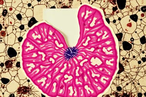
Crypts of Lieberkuhn
Crypts of Lieberkuhn
Signup and view all the flashcards
What is the Parotid gland?
What is the Parotid gland?
Signup and view all the flashcards
What do Beta cells produce?
What do Beta cells produce?
Signup and view all the flashcards
What are hepatocytes?
What are hepatocytes?
Signup and view all the flashcards
What are microvilli on hepatocytes?
What are microvilli on hepatocytes?
Signup and view all the flashcards
What are Kupffer cells?
What are Kupffer cells?
Signup and view all the flashcards
What is the space of Disse?
What is the space of Disse?
Signup and view all the flashcards
What is the primary function of the liver?
What is the primary function of the liver?
Signup and view all the flashcards
How is the liver supplied with blood?
How is the liver supplied with blood?
Signup and view all the flashcards
Study Notes
Digestive System Overview
- The digestive system is a complex process with multiple organs working together to break down food and absorb nutrients.
- It involves two main parts: the digestive tract and accessory organs.
Digestive Tract
- The digestive tract is a long muscular tube beginning at the mouth and ending at the anus.
- It includes the oral cavity, oropharynx, esophagus, stomach, small intestine, large intestine, rectum, and anal canal.
- The structure of the digestive tract wall has four layers: mucosa, submucosa, muscularis, and serosa (or adventitia).
Accessory Organs
- Salivary glands, pancreas, liver, and gall bladder are accessory organs supporting digestion.
- They produce secretions (like enzymes) that aid in the breakdown, neutralization, and absorption of food.
Oesophagus
- A long, muscular tube extending from the pharynx to the stomach.
- Its primary function is to transport food from the mouth to the stomach.
- The wall of the esophagus is composed of stratified non-keratinized squamous epithelium.
Stomach
- Divided into cardiac, fundus/body, and pyloric regions.
- Histologically, the stomach has three layers of muscle: inner oblique, middle circular, and outer longitudinal.
- The mucosa contains rugae and pits with specialized cells secreting mucus to protect the stomach from its own acidity.
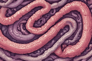
Small Intestine
- Responsible for the completion of digestion and absorption of nutrients.
- Composed of three segments: duodenum, jejunum, and ileum.
- The mucosal lining of the small intestine has villi and crypts for efficient absorption.
Large Intestine
- Concentrates leftover material from digestion, primarily absorbs water and salts.
- Segments include cecum, ascending colon, transverse colon, descending colon, sigmoid colon, and rectum.
Salivary Glands
- Produce saliva in the oral cavity.
- Saliva aids in lubrication, digestion, and immunity.
- Divided into major (parotid, submandibular, sublingual) and minor salivary glands.
Pancreas
- A mixed exocrine and endocrine gland.
- Exocrine function: produces pancreatic juice (enzymes) for digestion.
- Endocrine function: produces hormones (such as insulin and glucagon) regulating blood sugar.
- Contains acini (exocrine) and islets of Langerhans cells (endocrine)
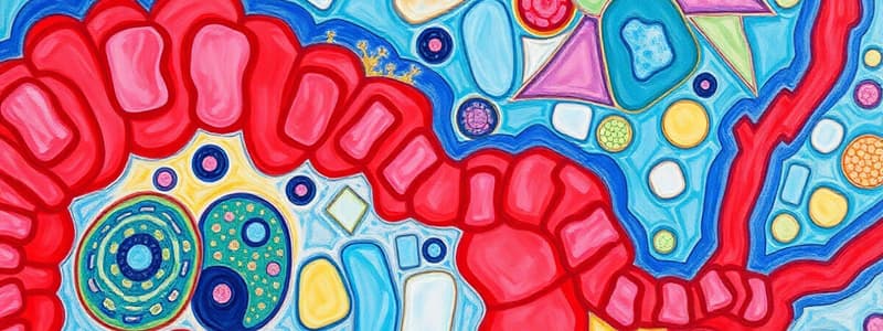
Liver
- The body's largest metabolic organ.
- Involved in a wide range of metabolic activities, including bile production and the detoxification of substances.
- Blood supply to the liver comes primarily from the portal vein and hepatic artery.
- Hepatocytes are the major functional cells in the liver, responsible for many metabolic functions.
Hepatic Lobules
- The liver is organized into lobules, each containing a central vein, surrounded by radiating hepatocyte cords, and thin connective tissue.
- Portal tracts contain the branches of the hepatic portal vein, hepatic artery, and bile ducts.
- The space of Disse (perisinusoidal space) is a space between the hepatocytes and the blood sinusoids containing blood plasma and lipocytes.
Von Kupffer Cells
- Phagocytic cells lining the liver sinusoids, removing microbes and cellular debris.
Studying That Suits You
Use AI to generate personalized quizzes and flashcards to suit your learning preferences.