Podcast
Questions and Answers
Which of the following statements is true regarding the characteristics of muscular tissue?
Which of the following statements is true regarding the characteristics of muscular tissue?
- Muscle fibers are primarily classified based on their color.
- Sarcoplasm is the main structural component of muscle tissue.
- Muscle fibers exhibit specialized development for movement through contractility. (correct)
- All muscle fibers are striated and involuntary.
What type of muscle fiber is characterized by being striated and voluntary?
What type of muscle fiber is characterized by being striated and voluntary?
- Smooth muscle
- Elastic muscle
- Cardiac muscle
- Skeletal muscle (correct)
Which of the following accurately describes the cell membrane and cytoplasm of a muscle fiber?
Which of the following accurately describes the cell membrane and cytoplasm of a muscle fiber?
- The cell membrane of a muscle fiber is termed sarcolemma, while the cytoplasm is known as sarcoplasm. (correct)
- Mitochondria and myofibrils are called sarcolemma.
- Sarcoplasm is specifically referred to as smooth endoplasmic reticulum.
- Sarcoplasmic reticulum serves as the cytoplasm in muscle fibers.
Where in the body would you primarily find smooth muscle tissue?
Where in the body would you primarily find smooth muscle tissue?
Which muscle type functions involuntarily and is striated in appearance?
Which muscle type functions involuntarily and is striated in appearance?
What is the primary function of the T-triad in skeletal muscle fibers?
What is the primary function of the T-triad in skeletal muscle fibers?
Which structural feature is characteristic of a sarcomere?
Which structural feature is characteristic of a sarcomere?
What best describes the I band in the striated pattern of skeletal muscle?
What best describes the I band in the striated pattern of skeletal muscle?
What cellular feature distinguishes skeletal muscle fibers from other types of muscle fibers?
What cellular feature distinguishes skeletal muscle fibers from other types of muscle fibers?
Which component is NOT found in the sarcoplasm of skeletal muscle fibers?
Which component is NOT found in the sarcoplasm of skeletal muscle fibers?
Flashcards
Muscle fibers
Muscle fibers
Muscle tissue is composed of elongated cells called muscle fibers.
Sarcolemma, sarcoplasm, sarcoplasmic reticulum, sarcosomes
Sarcolemma, sarcoplasm, sarcoplasmic reticulum, sarcosomes
The cell membrane of a muscle fiber is called the sarcolemma, the cytoplasm is called sarcoplasm, the endoplasmic reticulum is called sarcoplasmic reticulum and the mitochondria are called sarcosomes.
Classification of muscle fibers
Classification of muscle fibers
Muscle fibers are classified based on their structure (striated or smooth) and their function (voluntary or involuntary).
Skeletal Muscle
Skeletal Muscle
Signup and view all the flashcards
Location of Skeletal Muscle
Location of Skeletal Muscle
Signup and view all the flashcards
Skeletal muscle fiber
Skeletal muscle fiber
Signup and view all the flashcards
Sarcomere
Sarcomere
Signup and view all the flashcards
Z-line
Z-line
Signup and view all the flashcards
A-band
A-band
Signup and view all the flashcards
I-band
I-band
Signup and view all the flashcards
Study Notes
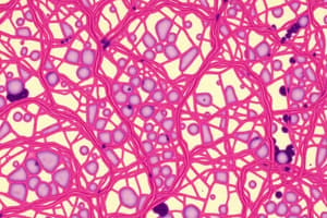
Histology of Muscular Tissue
- Muscle tissue is specialized for producing movement and displays significant contractility
- Muscle fibers are categorized based on structural (striated or non-striated) and functional (voluntary or involuntary) characteristics
- Muscle cells are elongated, narrow structures called muscle fibers
- The cell membrane of a muscle fiber is the sarcolemma
- The cytoplasm is called sarcoplasm
- Smooth endoplasmic reticulum is called sarcoplasmic reticulum
- Mitochondria are called sarcosomes
Types of Muscle Tissue
- There are three main types of muscle fibers: skeletal, cardiac, and smooth
- Skeletal muscle is striated and voluntary, connected to the skeleton
- Cardiac muscle is striated and involuntary, found in the heart
- Smooth muscle is non-striated and involuntary, found in the walls of internal organs and blood vessels
Skeletal Muscle
- Skeletal muscle fibers are long, cylindrical, and multinucleated
- They have a large diameter, typically up to 0.1 mm
- They are non-branching (except in some specific areas like the face and tongue)
- The cytoplasm (sarcoplasm) is acidophilic and contains transverse striations due to myofibrils
- Nuclei are flattened and situated peripherally under the sarcolemma
- Light microscopy (LM) reveals alternating dark (A) and light (I) bands
- The dark Z line bisects the light I band
- The pale H zone bisects the dark A band
- The dark M line bisects the H zone
- The distance between two Z lines is called a sarcomere
- A sarcomere represents the contractile unit
Myofibrils
- Myofibrils are thread-like structures within the sarcoplasm, running parallel to the fiber's long axis
- They show alternating dark (A) and light (I) bands
- Each I band is bisected by a dark Z line
- Each A band is bisected by a pale H zone
- The H zone is bisected by a dark M line
- The region between two Z lines forms the sarcomere
- Myofibrils show the regular arrangement responsible for transverse striations seen under LM
- The sarcomere is the main contractile unit
Electron Microscopy (EM) of Skeletal Muscle Fiber
- The sarcoplasm contains myofibrils
- The sarcoplasm also contains plenty of mitochondria arranged peripherally
- The sarcoplasm contains myoglobin and glycogen
- Transverse triad (T-triad): consists of T-tubules and sarcoplasmic reticulum (sER)
T-tubules, Sarcoplasmic Reticulum (and T-triad)
- T-tubules are invaginations of the sarcolemma (cell membrane) into the sarcoplasm
- They encircle the myofibrils at the junction of A and I bands
- Terminal cisternae of the sarcoplasmic reticulum (sER) surround T-tubules
- One T-tubule + 2 terminal cisternae constitutes the T-triad
- The T-triad plays a critical role in depolarization and calcium ion release during muscle contraction
Myofilaments
- Myofilaments are the two major types of proteins (thick and thin) within the sarcomere, responsible for contraction
- Thick filaments (myosin) are located within the A band, primarily made of myosin molecules
- Thin filaments (actin) are attached to the Z line and extend into the I band and part of the A band, made of actin and associated proteins like tropomyosin and troponin
- Actin is a polymer of globular G-actin molecules wrapped around each other
Molecular Structure of Myofilaments (Actin)
- Formed of two chains of F-actin filaments (polymer of globular G actin)
- Tropomyosin is a 40 nm long coil of polypeptide chains located in the groove between the two twisted actin strands
- Troponin is a complex of three subunits: TnT (attaches to tropomyosin), TnC (binds Ca2+), and TnI (binds to actin)
Molecular Structure of Myofilaments (Myosin)
- Formed of 200-300 myosin II molecules
- Each myosin molecule has a head and a tail
- Composed of two heavy chains and four light chains
Sliding Filament Theory of Muscle Contraction
- Muscle contraction involves the sliding of actin filaments over myosin filaments
- An impulse triggers the release of calcium ions from the sarcoplasmic reticulum
- Calcium ions bind to troponin, altering its conformation
- The altered troponin shifts tropomyosin, exposing myosin-binding sites on actin
- Myosin heads bind to actin, pulling actin filaments towards the M line, shortening the sarcomere
- ATP hydrolysis powers the cycling process of the myosin-actin interactions
Myoglobin
- Pigment protein (red to brown) in muscle fibers
- Provides oxygen to muscle
Connective Tissue Coverings of Skeletal Muscle
- Epimysium: Covers the whole muscle
- Perimysium: Surrounds bundles of muscle fibers
- Endomysium: Surrounds individual muscle fibers
Regeneration of Skeletal Muscle
- Skeletal muscle fibers regenerate through regenerative cells called satellite cells
- Satellite cells are located between the sarcolemma and basement membrane of skeletal muscle fibers
- They differentiate into myoblasts and fuse to form new skeletal muscle fibers
Smooth Muscle
- Smooth muscle is non-striated and involuntary
- Located in the walls of internal organs and blood vessels
- Smooth muscle fibers are short, spindle-shaped, single-nucleated, and lack transverse striations (sarcomeres)
- They lack T-tubules and have less-developed sarcoplasmic reticulum, using caveolae (invaginations of the sarcolemma) for impulse transmission
- Smooth muscle fibers regenerate through mitosis
Studying That Suits You
Use AI to generate personalized quizzes and flashcards to suit your learning preferences.